|
|
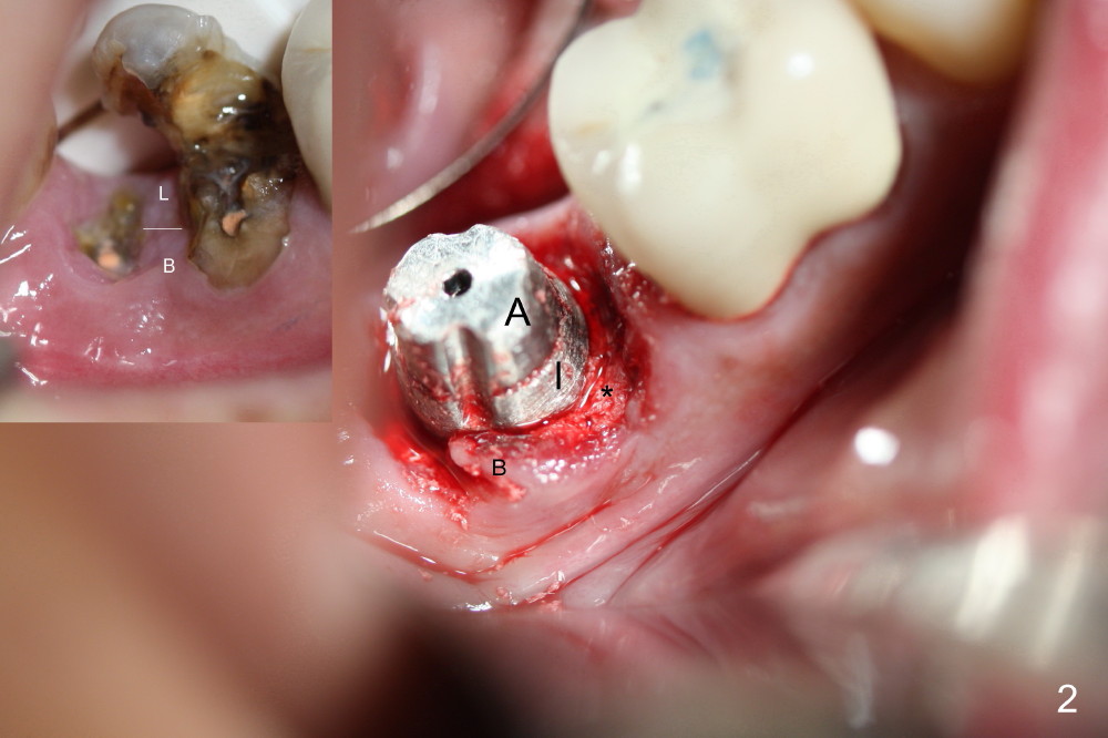
Fig.2 insert (upper left corner): preop image, showing a piece of gingival tissue between the mesial and distal roots. When the roots are extracted, the gingival tissue is torn into the buccal (B) and lingual (L) portions.
When the 7x14 mm gingiva-level implant (I) and the 6x3 mm abutment are placed, the buccal portion of the gingival tissue is pushed distobuccally (Fig.2 B). Bone graft is placed in the peri-implant gap, especially mesial and distal (*).
Xin Wei, DDS, PhD, MS 1st edition 12/28/2014, last revision 12/31/2014