|
|
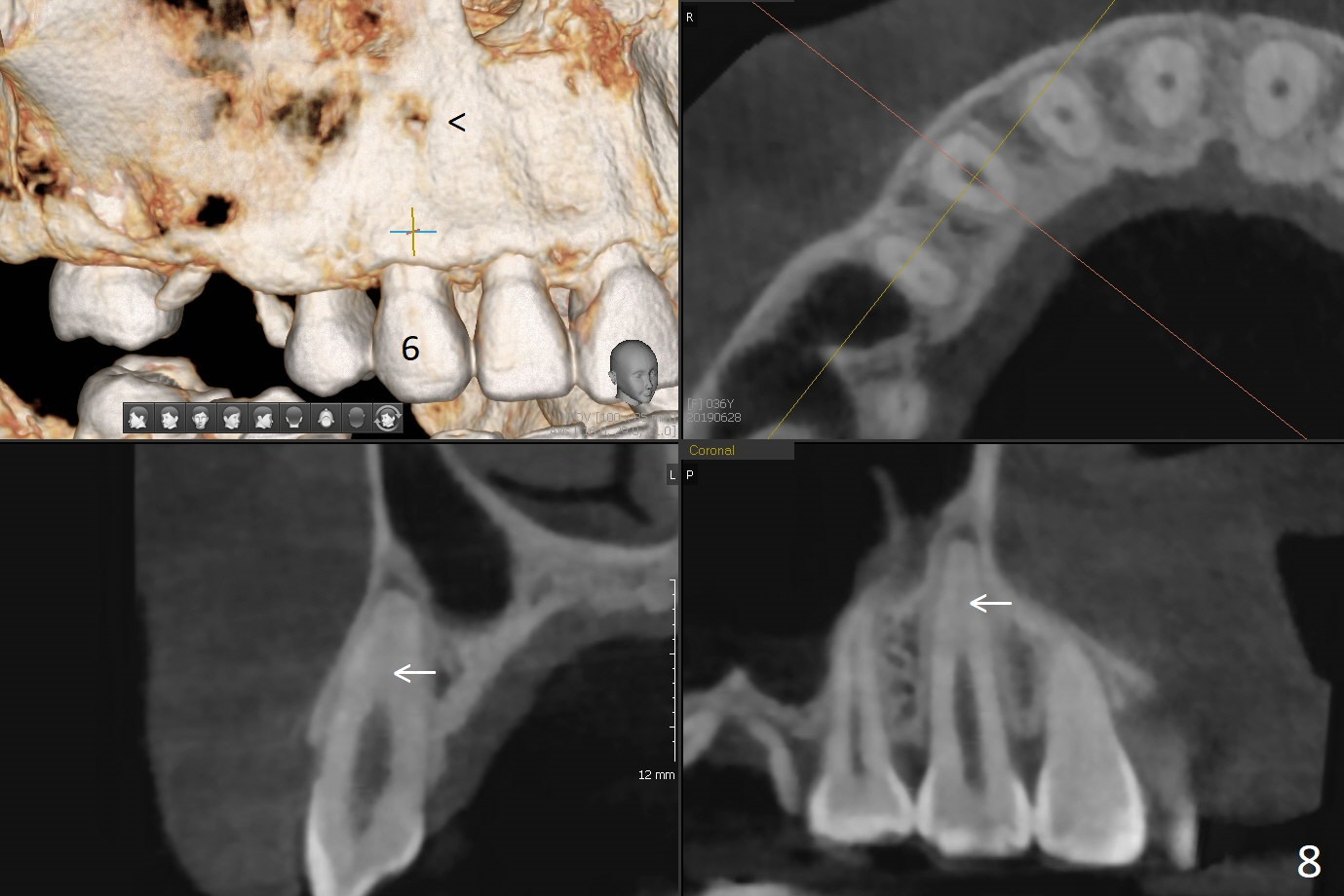
The patient is concerned about the discolored upper right canine, which should be associated with orthodontics 20 years ago. The apical canal is obliterated (Fig.8 arrow) with periapical radiolucency (arrowhead).
Xin Wei, DDS, PhD, MS 1st edition 06/27/2019, last revision 01/25/2020