|
|
|
|
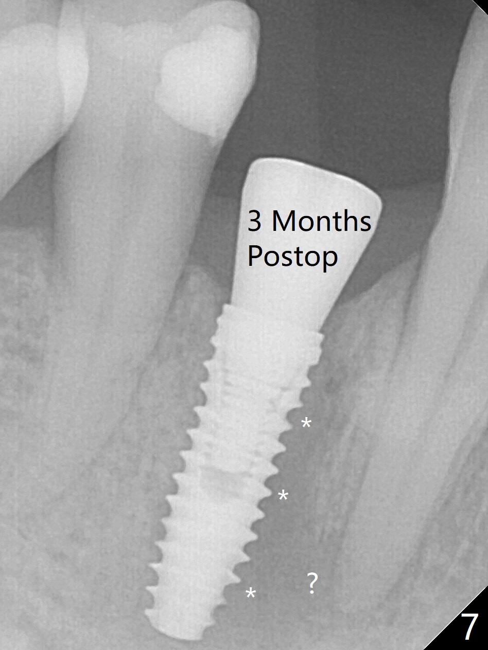
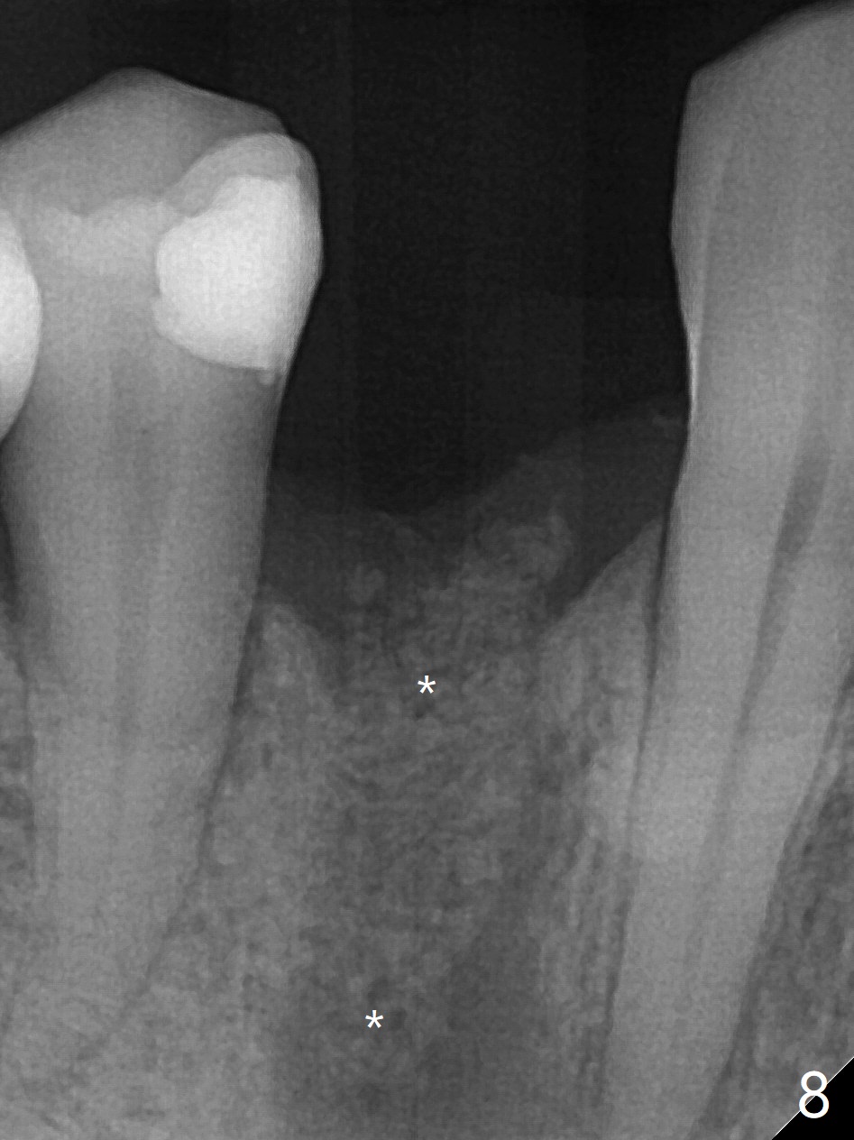
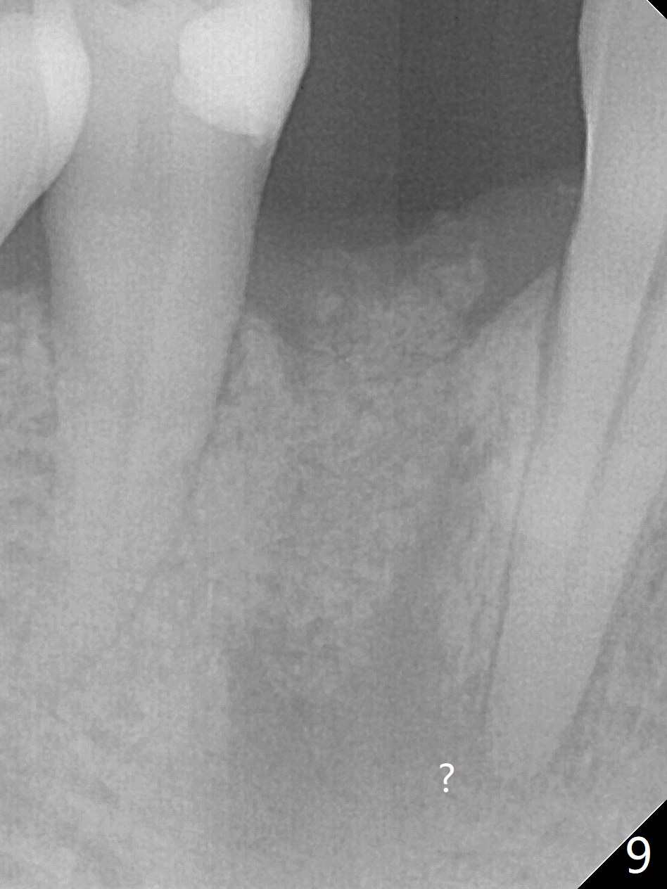
Fig.2 shows the site of #28 immediately before bone expansion and after use of 2.2x11.5 mm drill and small-scaled ridge split (^). The bone is so hard that the bone expansion is minimal (Fig.5) with autogenous bone graft and implant placement (Fig.4). Because of inability to masticate on the left (#18 root fracture with infection), the patient wants to restore #28 and 31 implants 3 months postop (Fig.6,7). The implant at #28 is removed while the healing abutment is being un-torqued without noting bone loss mesially (Fig.7 *). Bone graft is placed (Fig.8 (*),9). Pay attention radiolucency next the apex of the neighboring tooth (Fig.9 ?) before re-placement of an implant. Hard Bone Last Next Density 4-6 Xin Wei, DDS, PhD, MS 1st edition 11/06/2019, last revision 05/23/2020