|
|
|
|
|
|
Implant Placement 3 Months Post
Socket Preservation
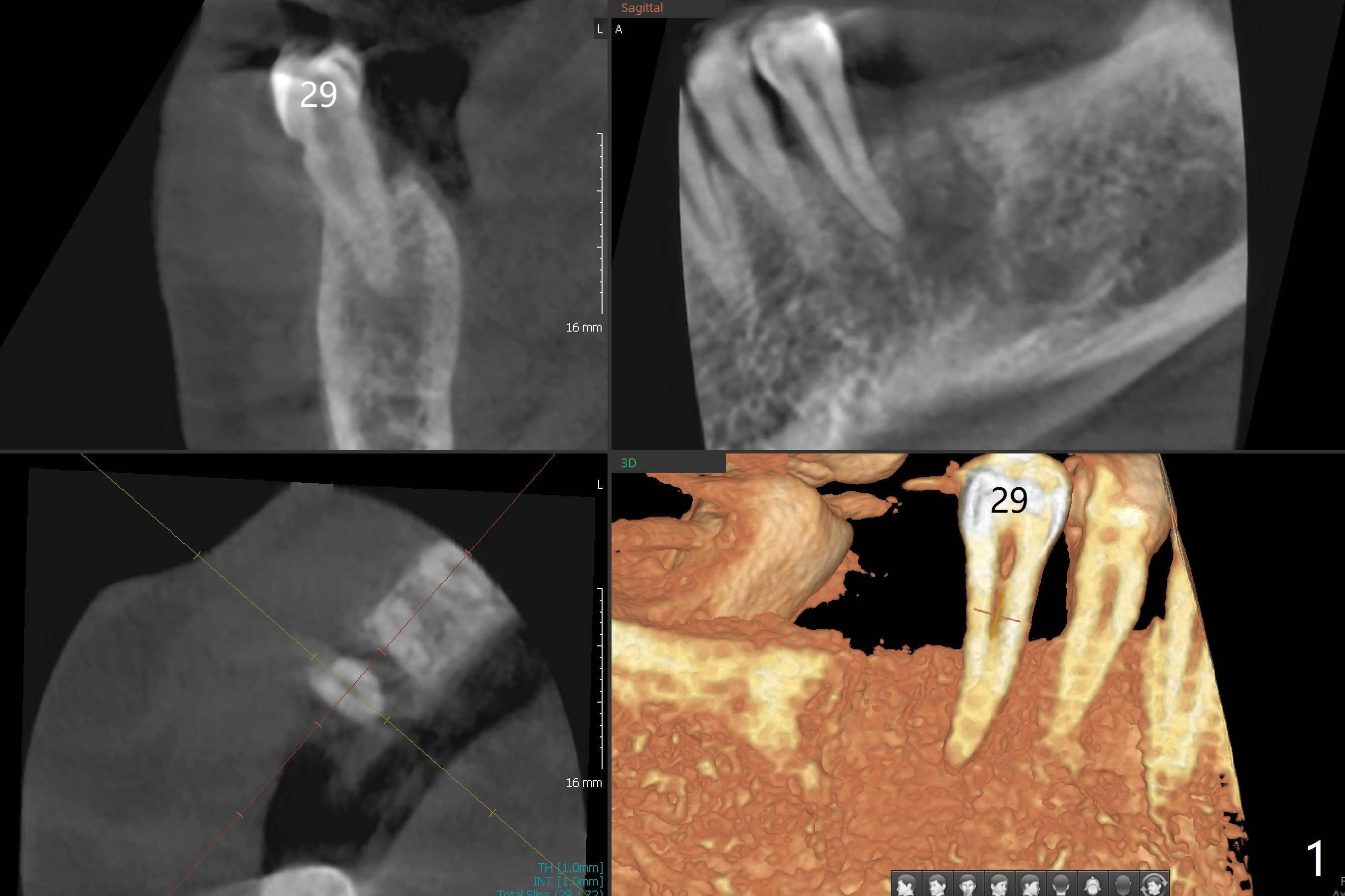
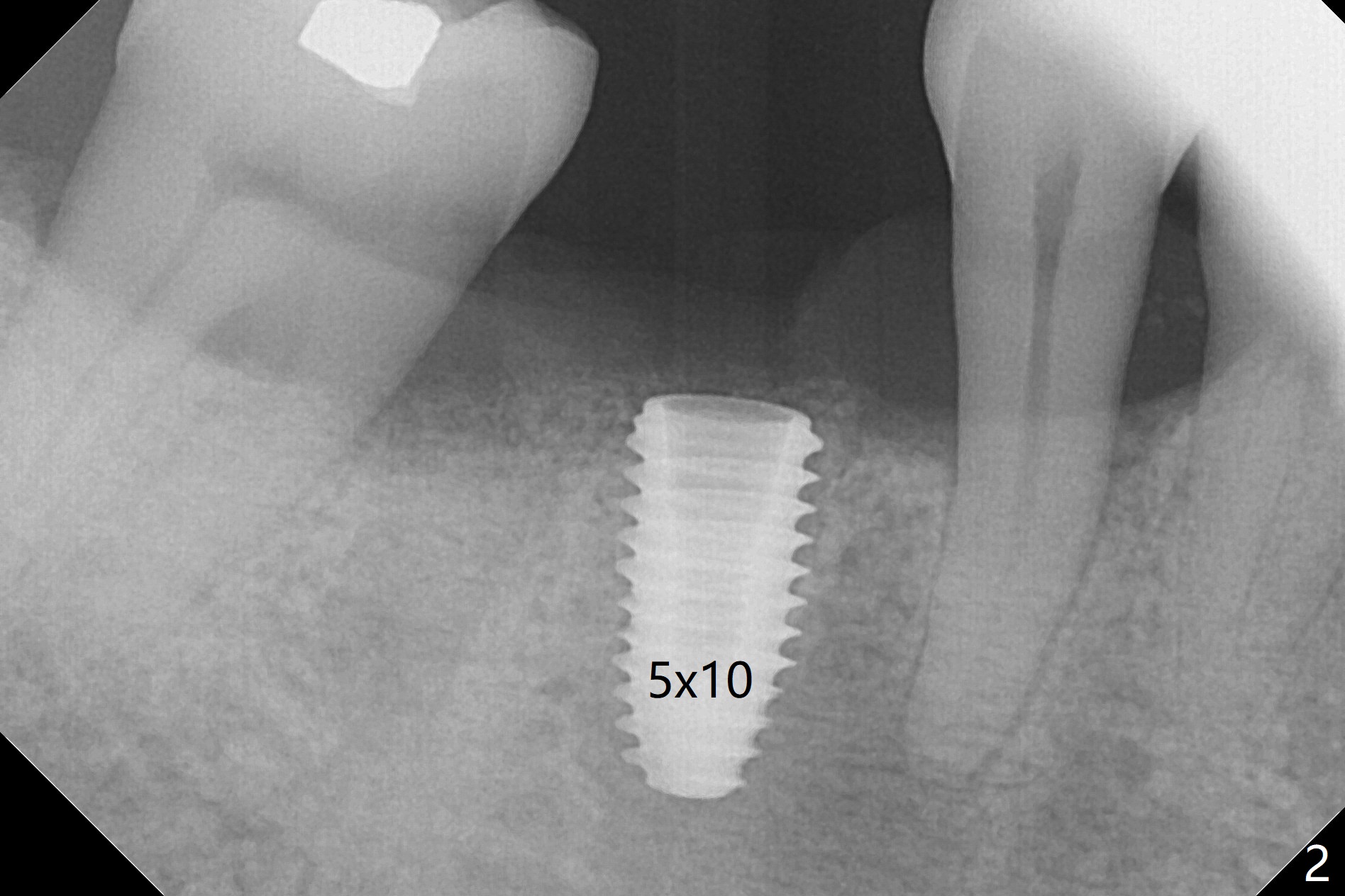
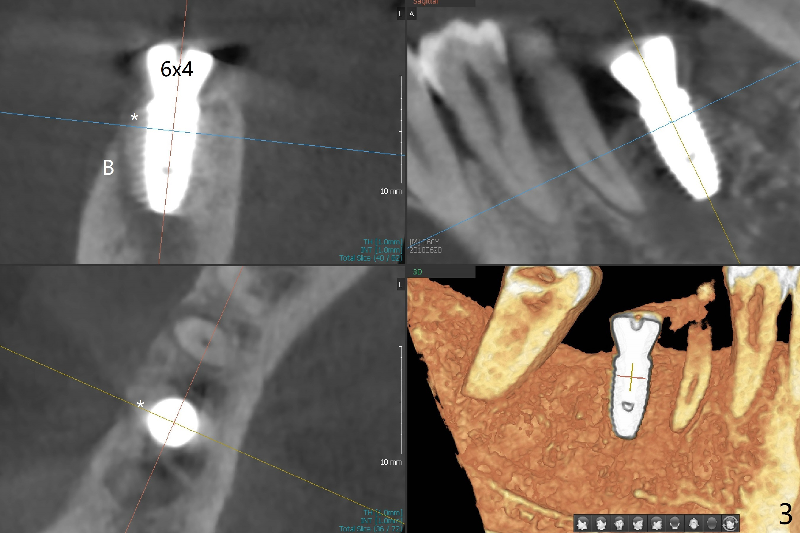
There are two questions before guided surgery at #30. First, is implant placement 3 months post socket preservation too early? Will the periapical radiolucency between #29 and #30 affect osteointegration? Reanalysis of CT >2 months post socket preservation reveals that the lamina dura of the tooth #29 is apparently intact (Fig.1). Immediately preop clinical exam shows no active infection. The patient is reluctant to accept RCT at #29. A 5x10 mm implant is placed slightly subcrestal buccally (which is the lowest, Fig.2). Immediately postop CT (5x5cm field of view) demonstrates that the implant is covered by graft bone (Fig.3 *) buccally (B). In fact the autogenous bone harvested from osteotomy is inserted between the 6x4 mm healing abutment and the buccal gingiva.
Hi Dr. WeiIt seems like apical cyst occurred due to endodontic pulp necrosis. We cannot identify which one is the origin for that it could be #29 or #30 or even may be both. In order to place implant on #30 position, make sure not to touch the cyst and when you do RCT to the #29 then the size of cyst will be minimized. However for the best result, extract #30 first, remove the cyst with curette first and then place implant. Jennifer
Hi, Jennifer: Thanks for the info. It appears that Dr. Heo is as good a dentist as his implants. If he visits USA, I may want to see him and listen to his lecture. After careful clinical evaluation, I have placed the implant with intention to watch. In fact, the osteotomy does not appear to communicate with the apical lesion. Thanks again.
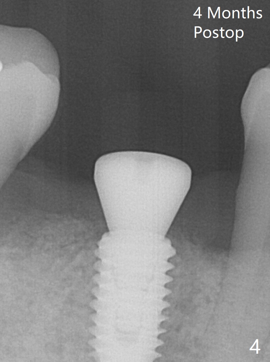
Minimal bone loss is observed 4 months postop (Fig.4).
Return to
Lower
Molar Immediate Implant, Armaments
Xin Wei, DDS, PhD, MS 1st edition 06/28/2018, last revision 11/01/2018