|
|
|
|
||
|
|
|
|
|
|
|
|
|
|
||
1st IBS Guided Surgery
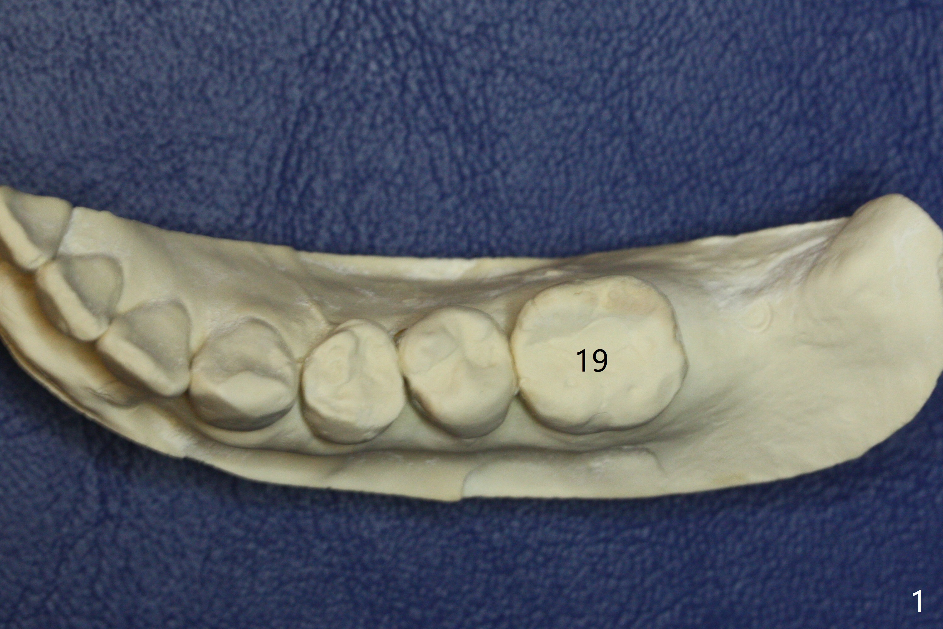
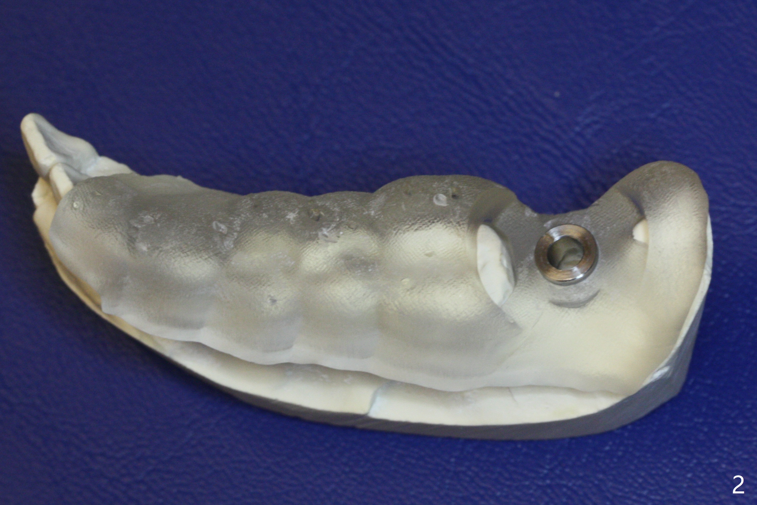
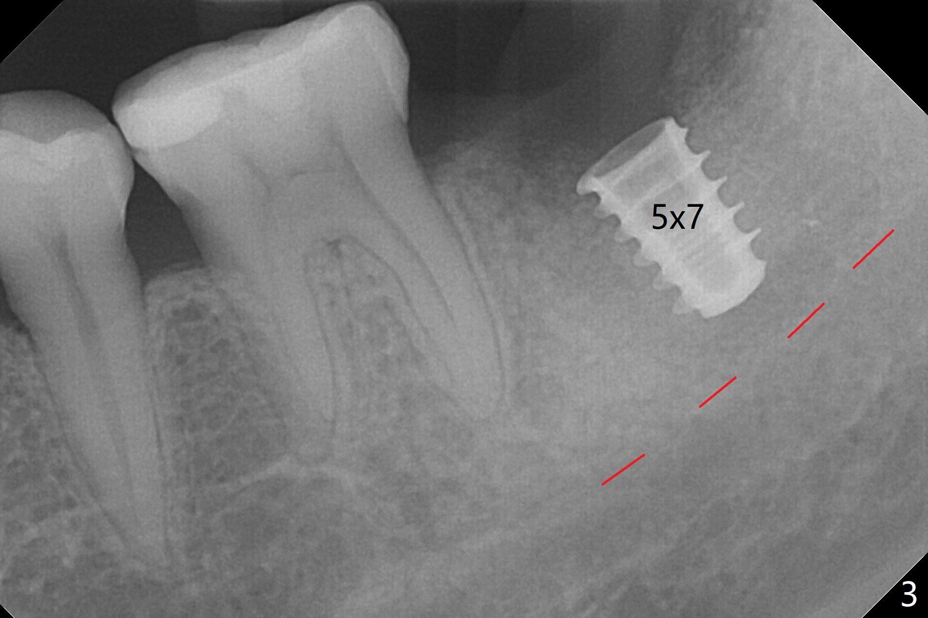
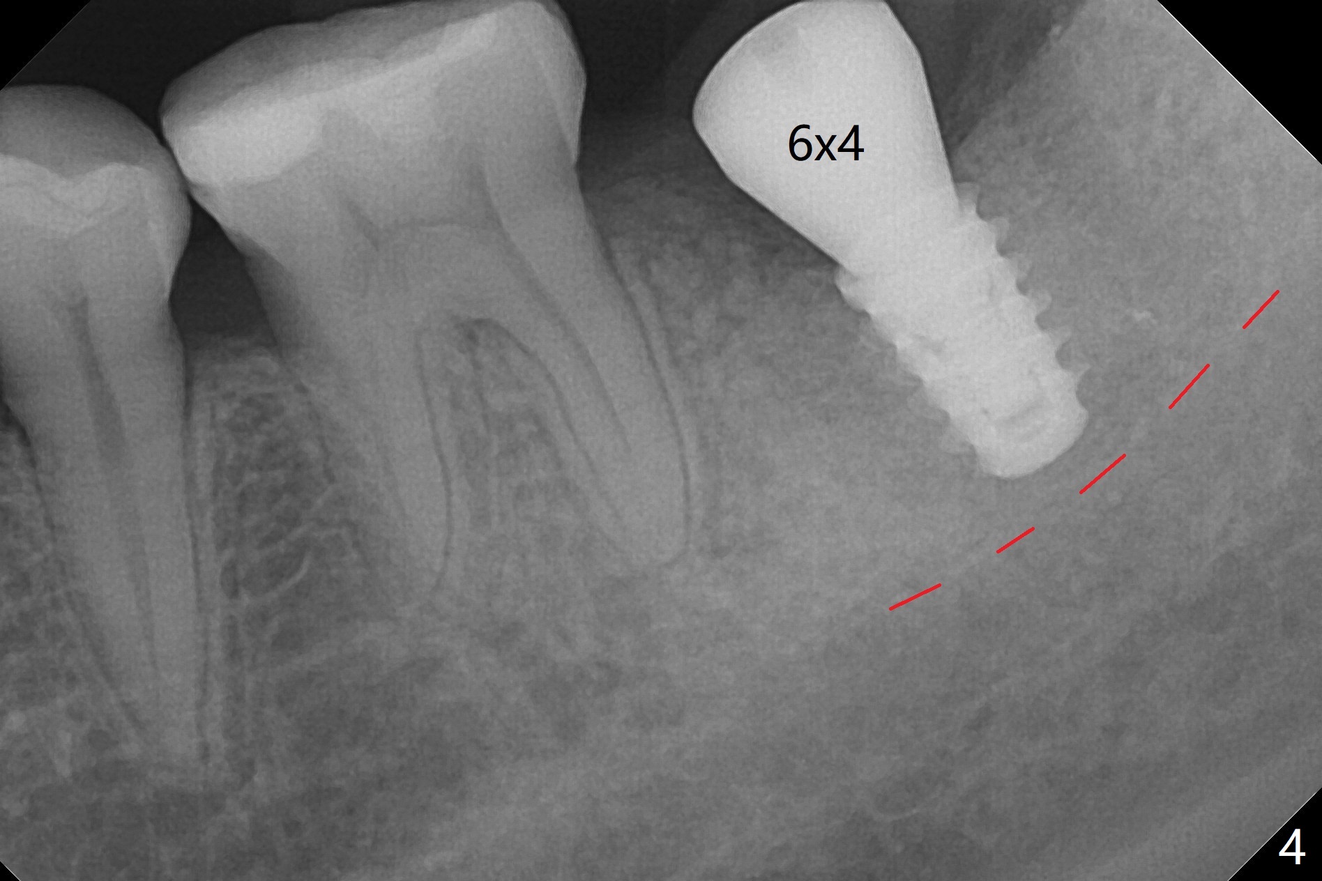
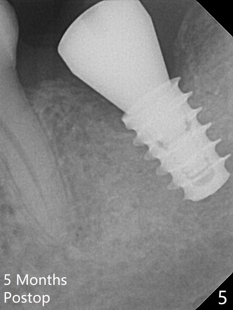
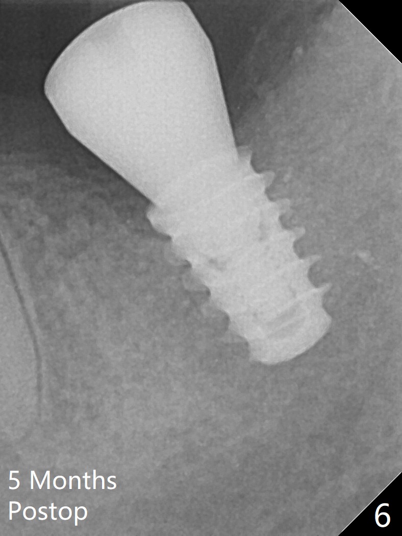
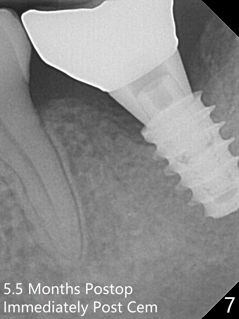
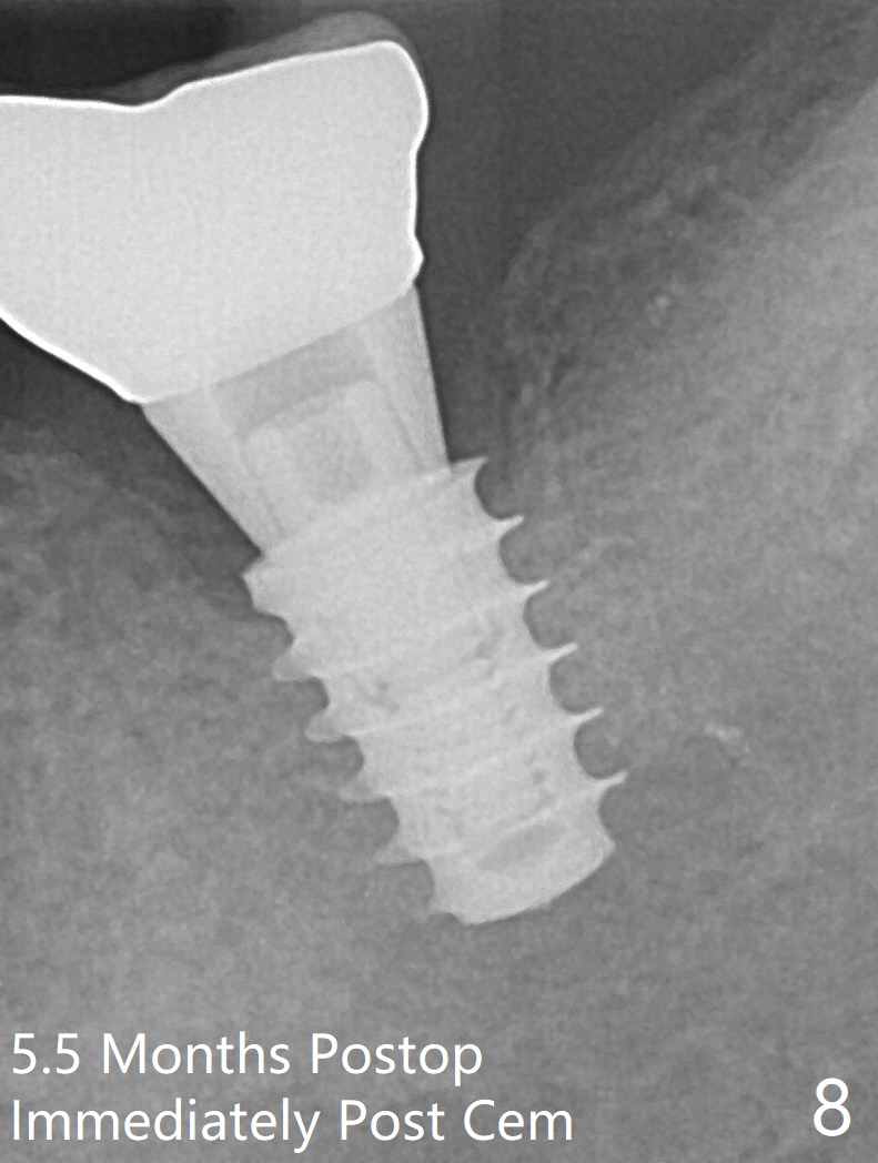
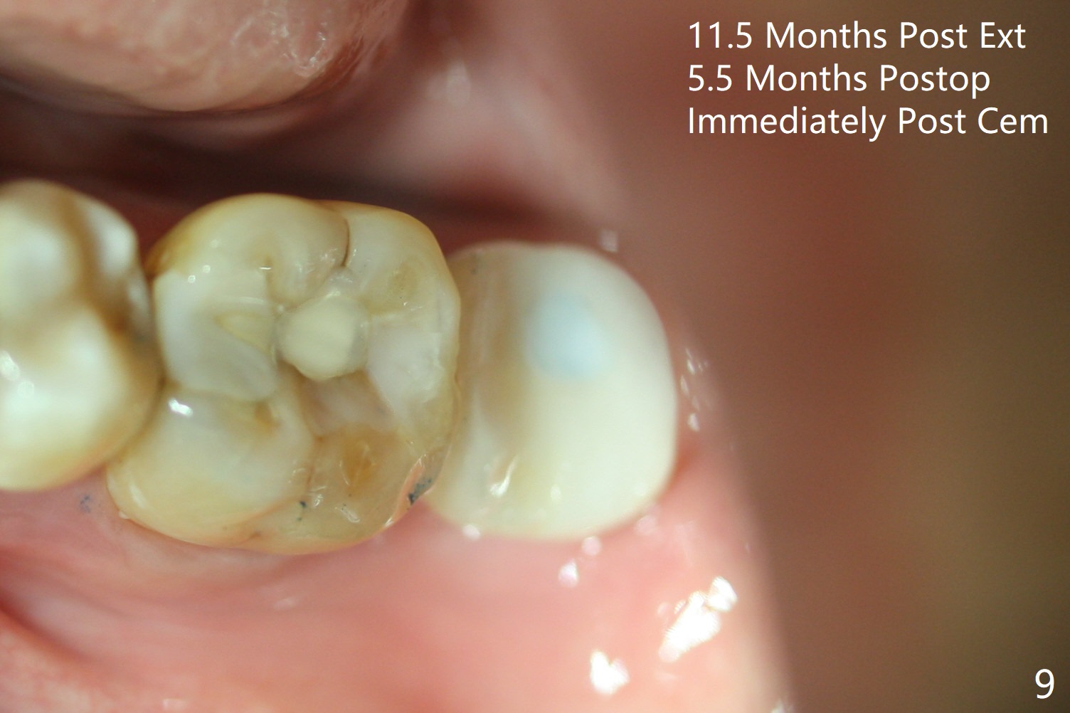
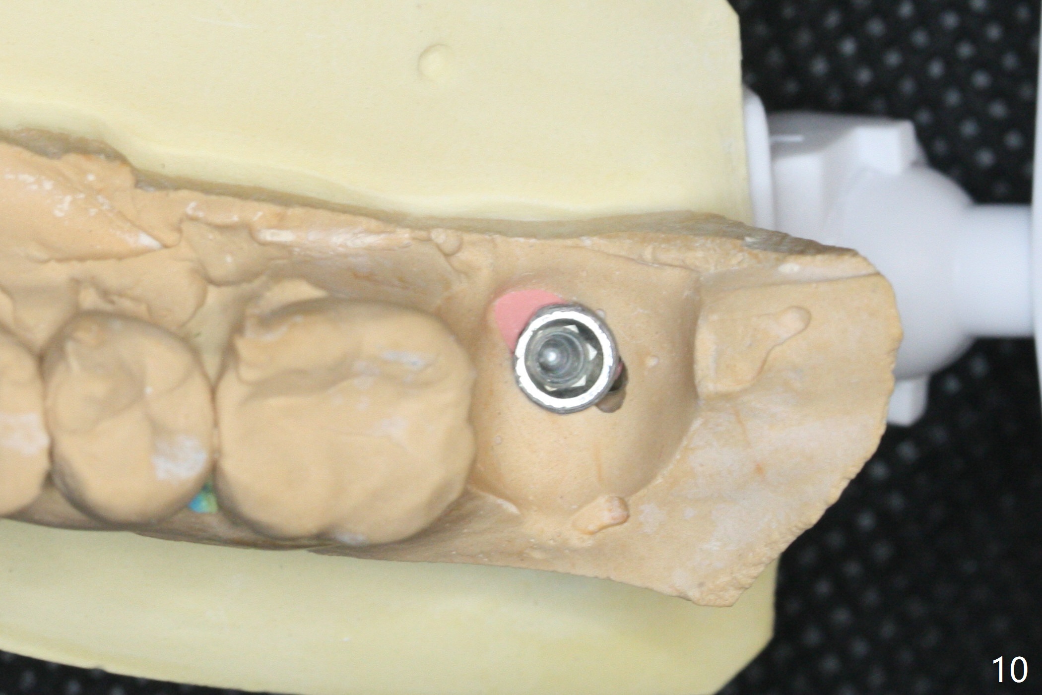
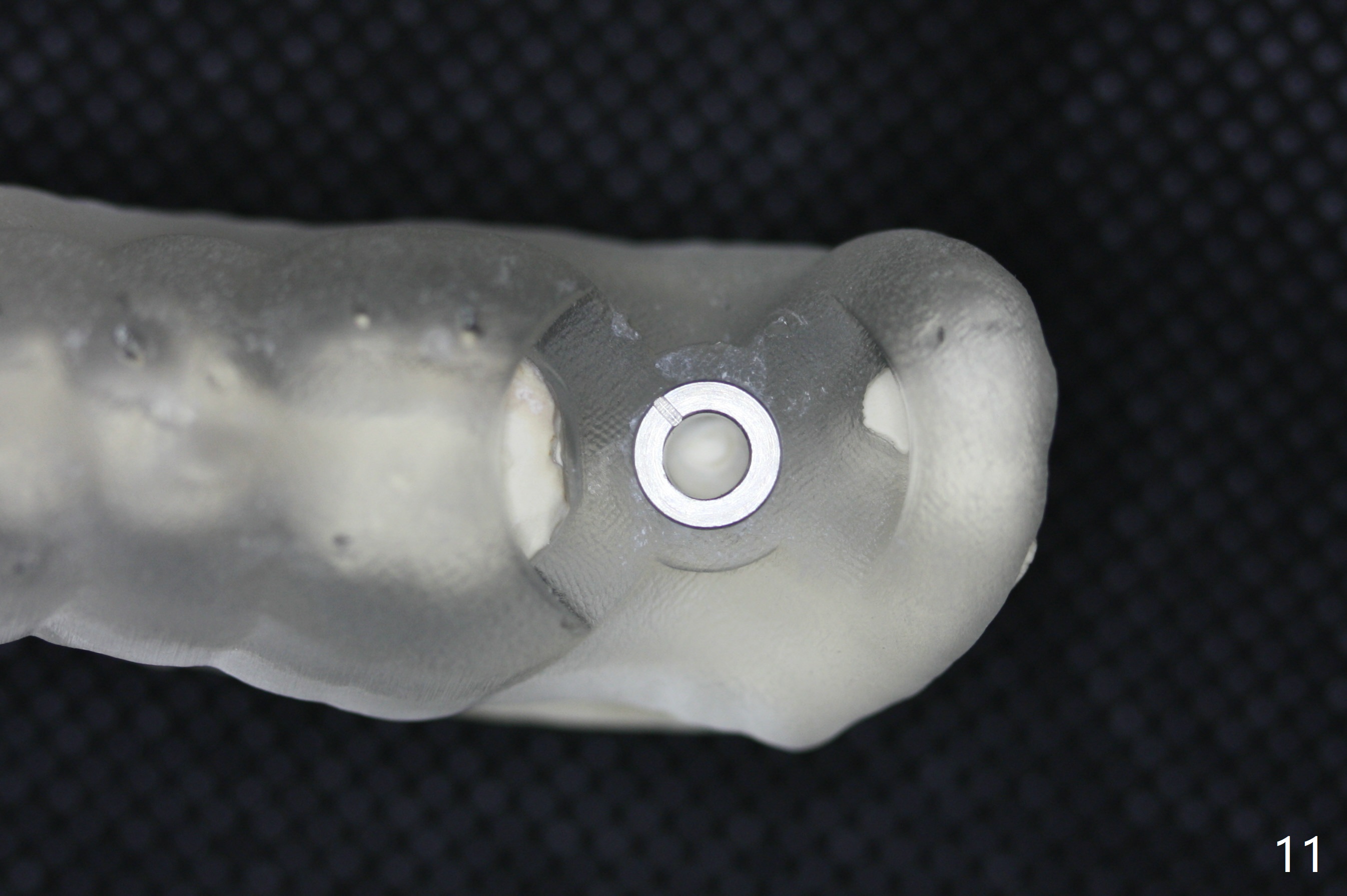
Six months post socket preservation at #18 (with moderate atrophy, Fig.1), a simplified surgical guide (Fig.2 with metal sleeve of 5 mm height and 2.93 mm diameter) determines initial osteotomy with 3.3 mm Magic Drill (MD), followed by 4.8 mm MD for 9 mm (gingival level) free hand. Since a 5x7 mm IBS implant is placed incompletely and in low stability (Fig.3 (in the graft bone)), a 4.3 mm MD is used for ~1 mm deeper. The implant is reseated to more satisfactory level (Fig.4: ~ 1 mm from the upper border of the Inferior Alveolar Canal (red dashed line)). The fearful patient is extremely pleased with quickness of the procedure as compared to that at #30 free hand. The wound heals normally 2 weeks postop. When she finishes the follow up appointment, she voluntarily talks to another patient who is hesitant about implant treatment. Impression is taken 5 months postop (Fig.5,6). There is no gap between the crown and abutment using abutcoping technique (Fig.7,8). The crown at #18 looks low probably related to long termed edentulism (Fig.9). The access hole is lingual (Fig.9), because the implant was placed lingual (Fig.10) due to use of a partial guide (Fig.11).
Return to Lower Molar Immediate Implant, Armaments 6th Meeting Xin Wei, DDS, PhD, MS 1st edition 04/16/2018, last revision 10/06/2018