|
|
|
|
|
|
|
|
|
|
|
|
|
|
|
|
|
|
|||
Bone Graft or Implant Post Extrusion M
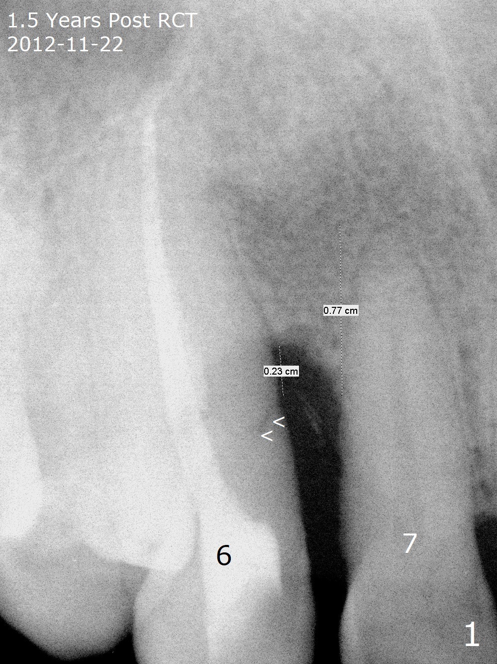
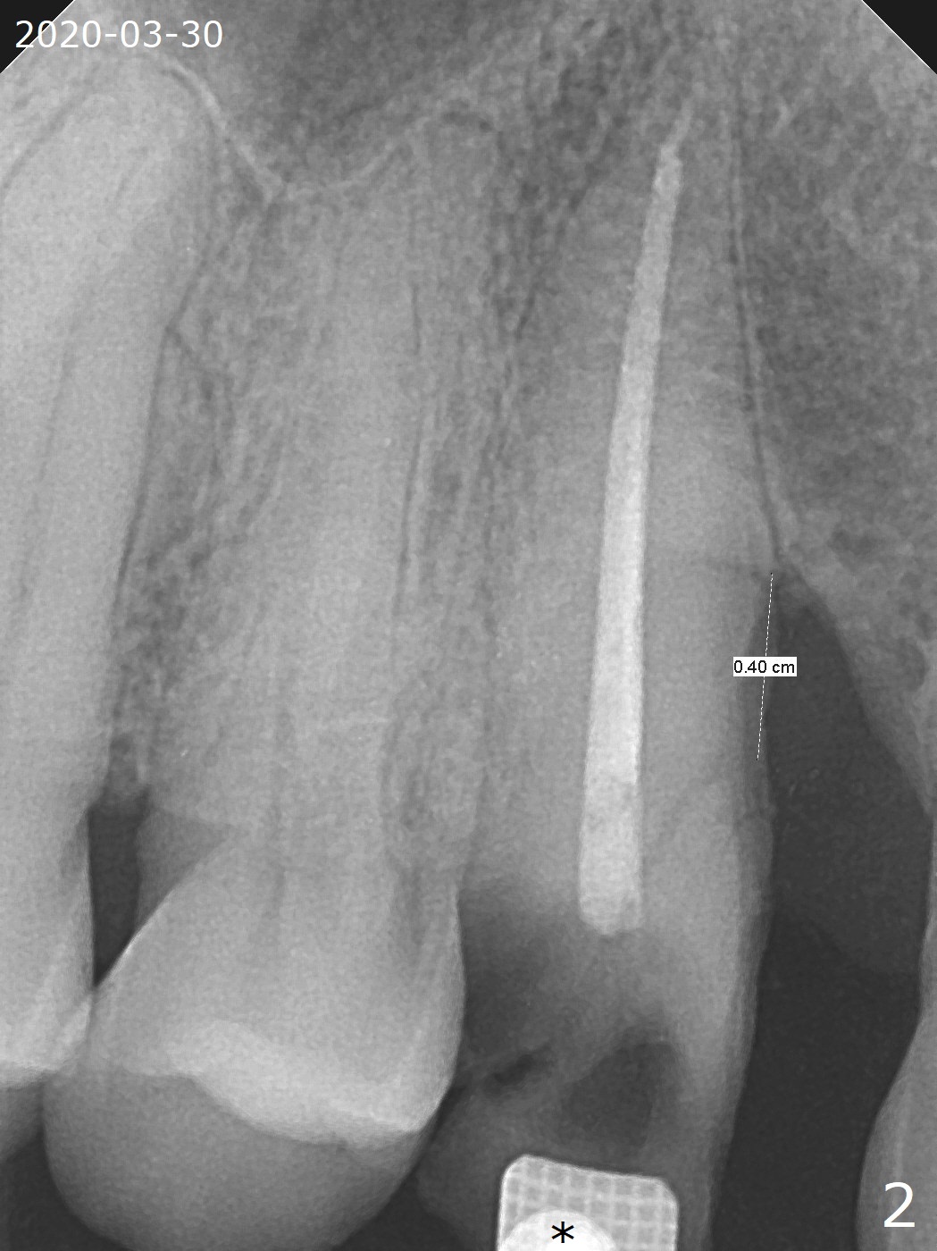
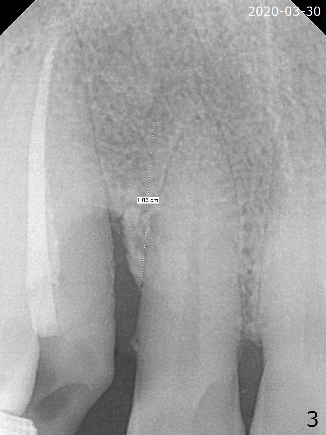
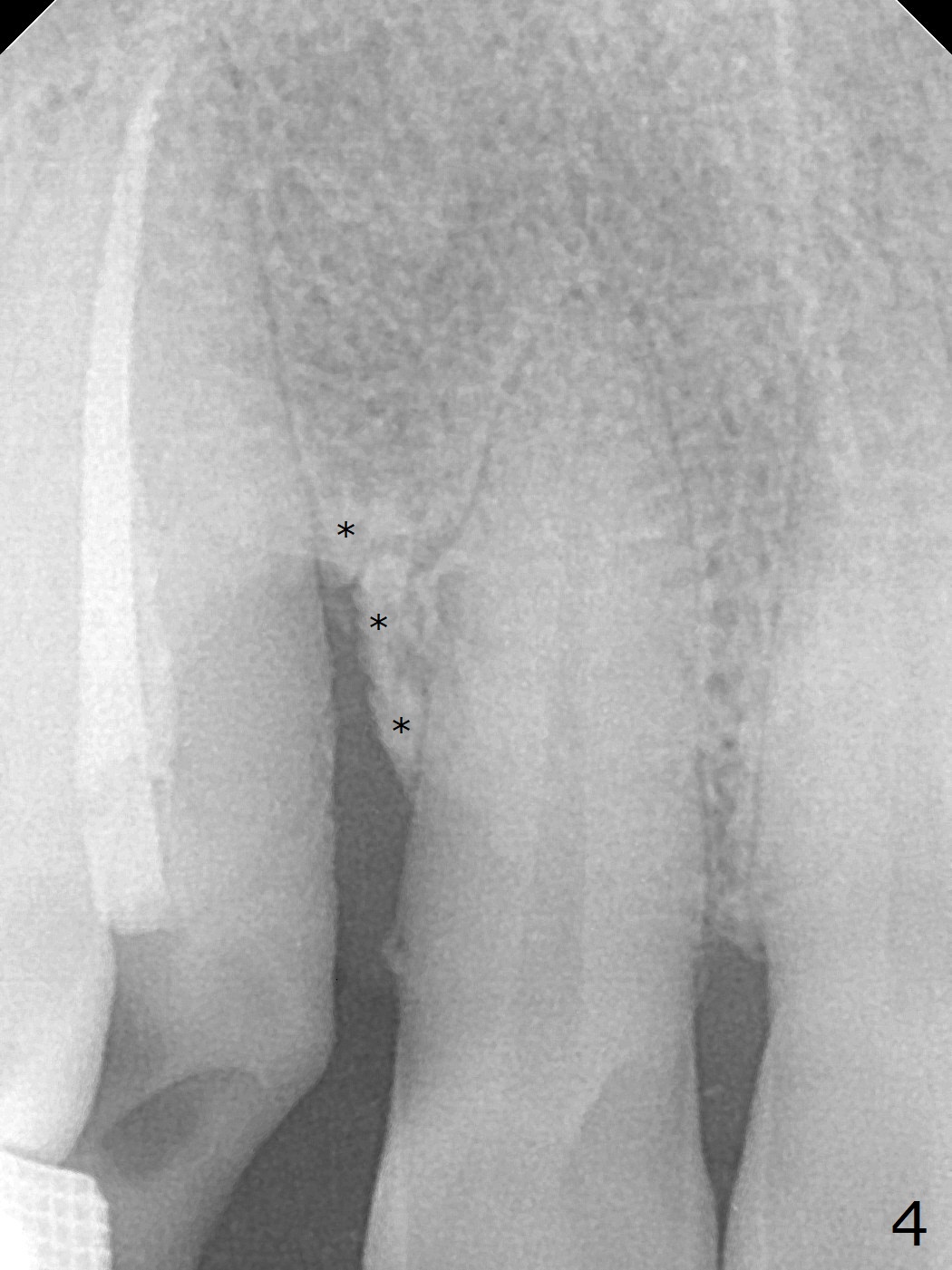
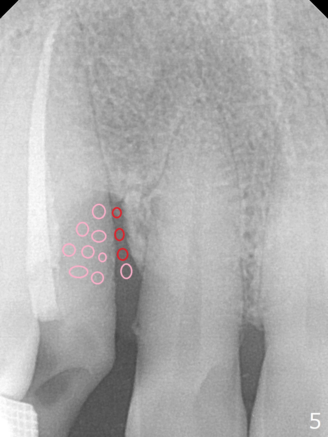
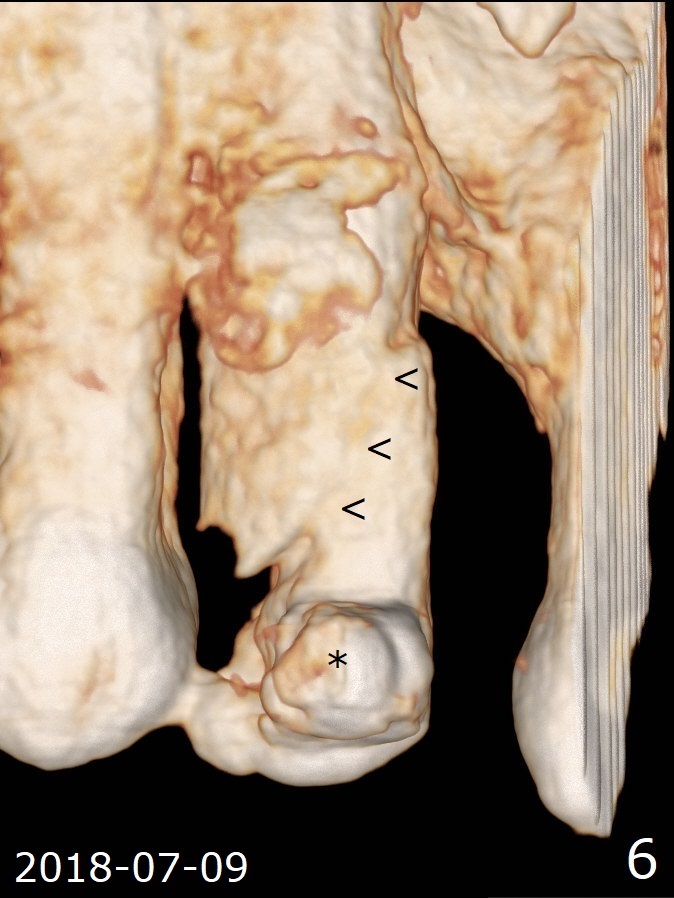
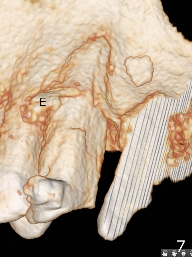
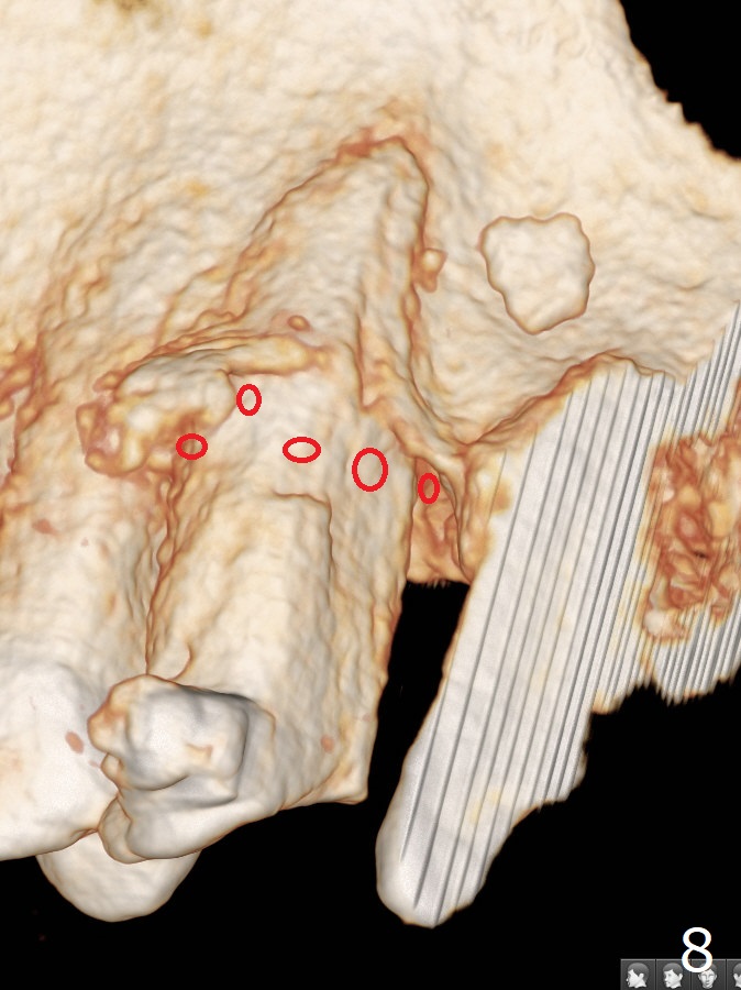
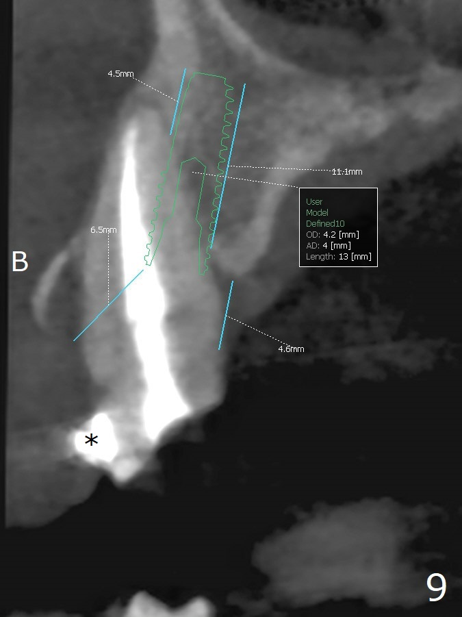
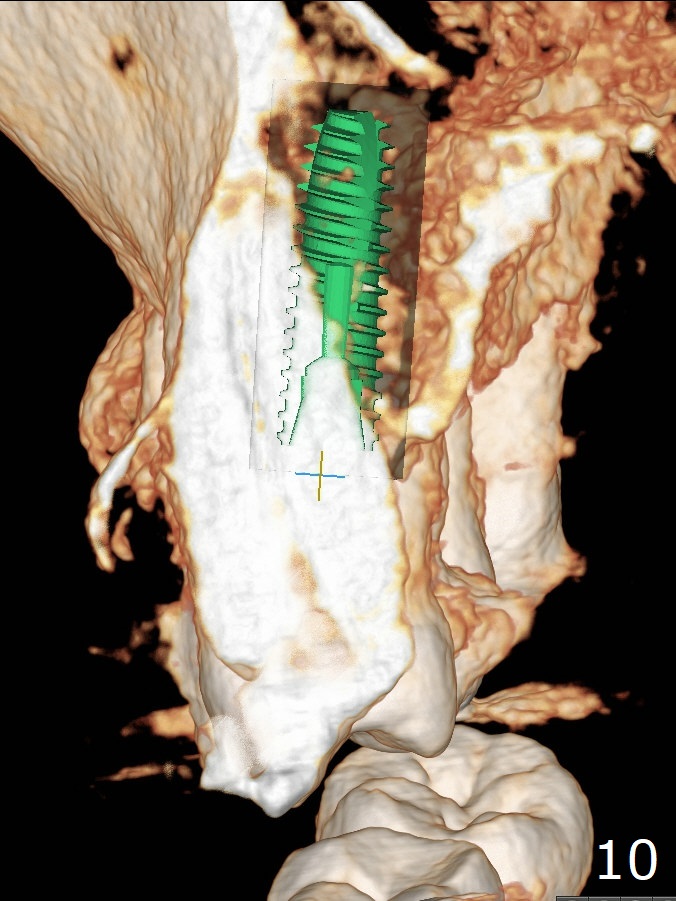
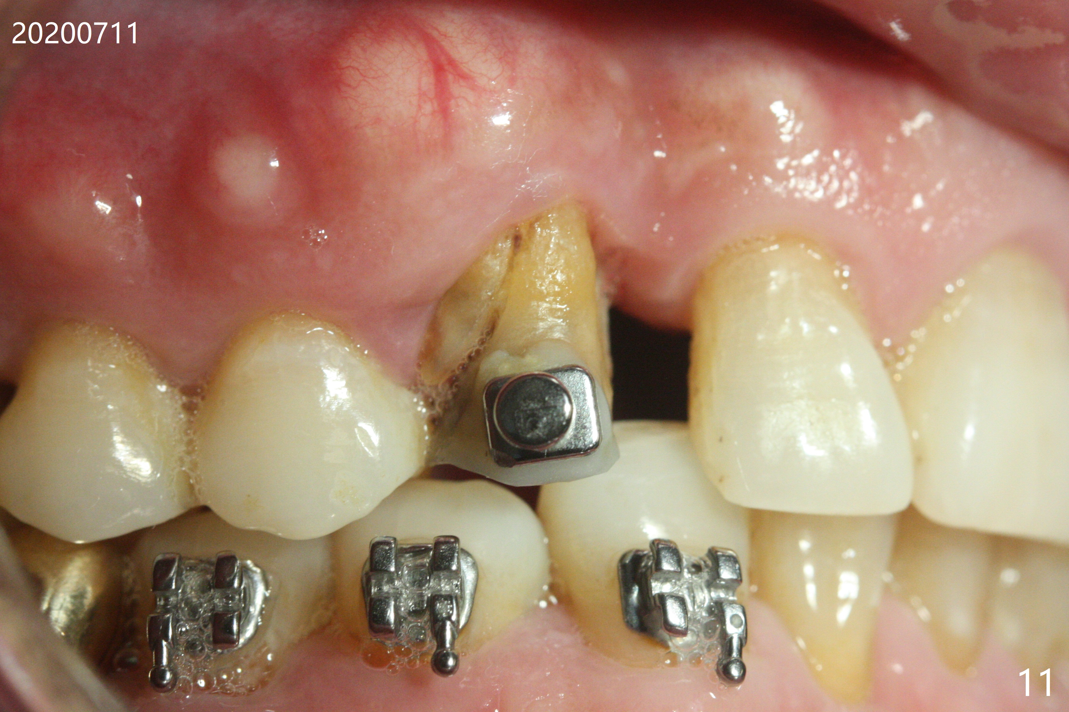
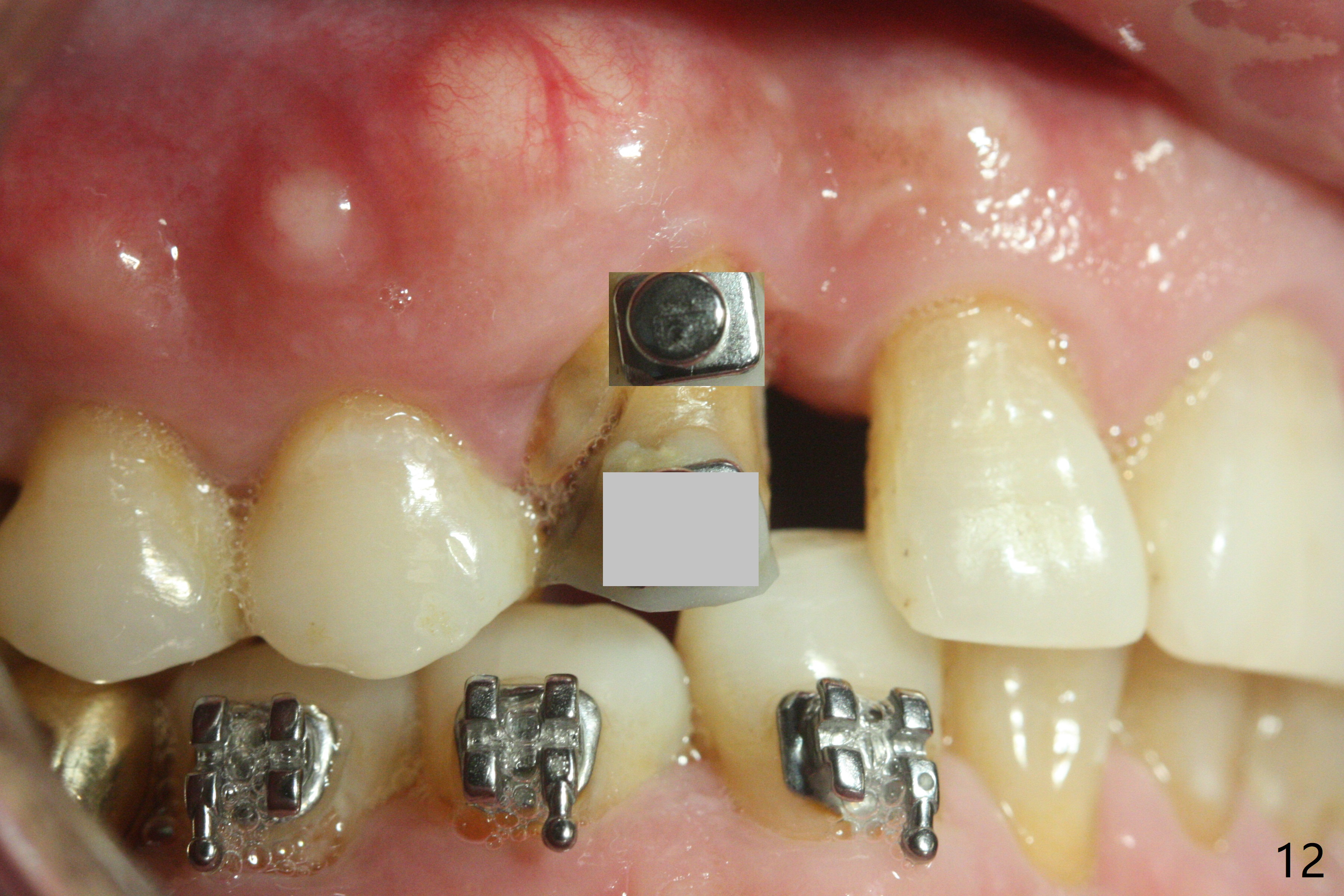
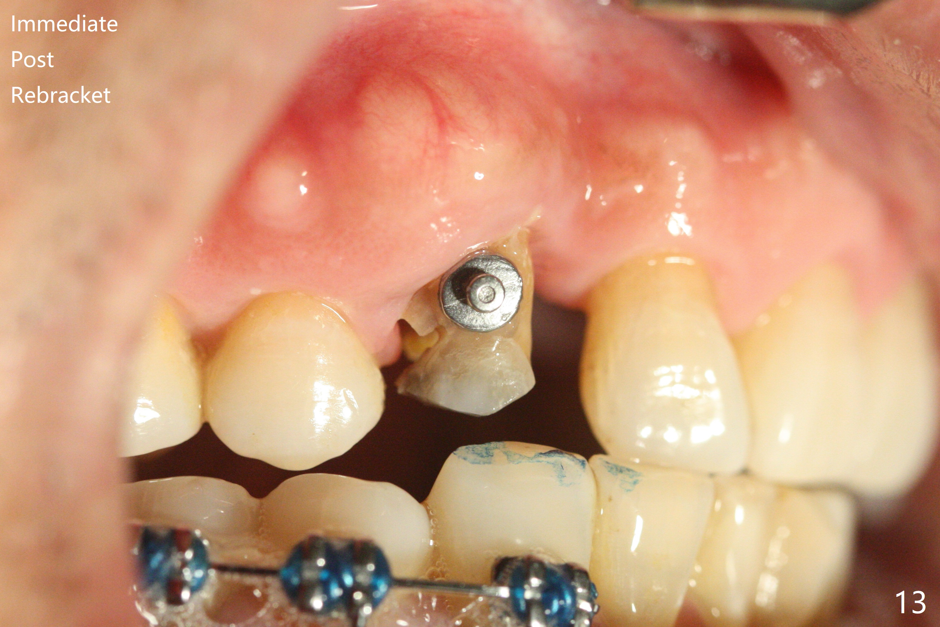
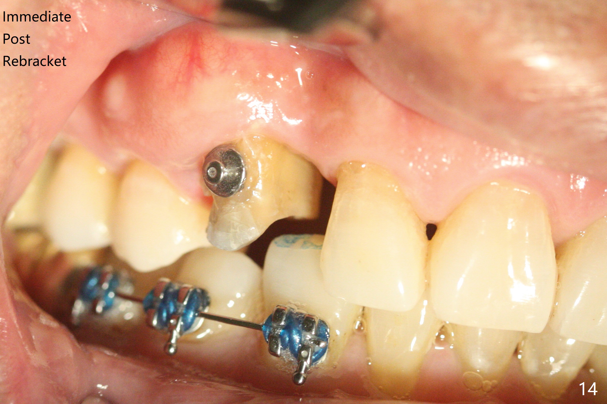
A 62-year-old man had traumatic root fracture at #6 in his teen. The tooth remained asymptomatic until his fifties. Following root canal therapy (Fig.1), the tooth is orthodontically extruded (~ 5 years, Fig.2 (*: bracket)) with apparent disappearance of the infection. The bone distal to #7 seems to increase in height (Fig.3, as compared to Fig.1) and in density (Fig.4). Bone graft could be placed for regeneration with PRF or GEM21S (Fig.5 red (between #6 and 7), pink (buccal to #7 or coronal to the fracture line) circles). With extrusion, the oblique fracture line is more than half or two third supragingival (Fig.6). In spite of severe bone loss, exostosis is present (Fig.7 (mesiobuccal view) E) so that bone graft could be placed palatal to it (Fig.8 red). In case the tooth is non-salvageable, immediate implant will be placed with guide (Fig.9,10). Move lingual button as apical as possible (Fig.12) and make occlusal clearance. Continue extrusion until all of the crack is exposed without deep pocket.
Return to
No Deviation
Clindamycin Metronidazole
No Antibiotic
Ortho Cases
Plug
Professionals
Shield
Waterlase
19
Xin Wei, DDS, PhD, MS 1st edition
03/30/2020, last revision
07/27/2020