,%202.5x12(2).jpg)
,%202.5x14(4).jpg)
|
|
|
|
|||
|
|
|
|
|
|
|
|
|
|
|
|
||
|
|
|
|
|
|
|
|
|
|
||||
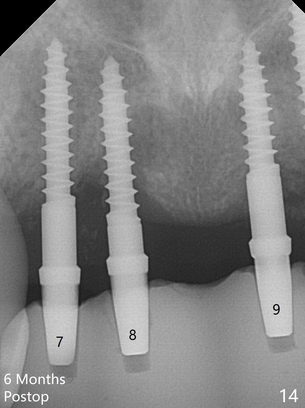
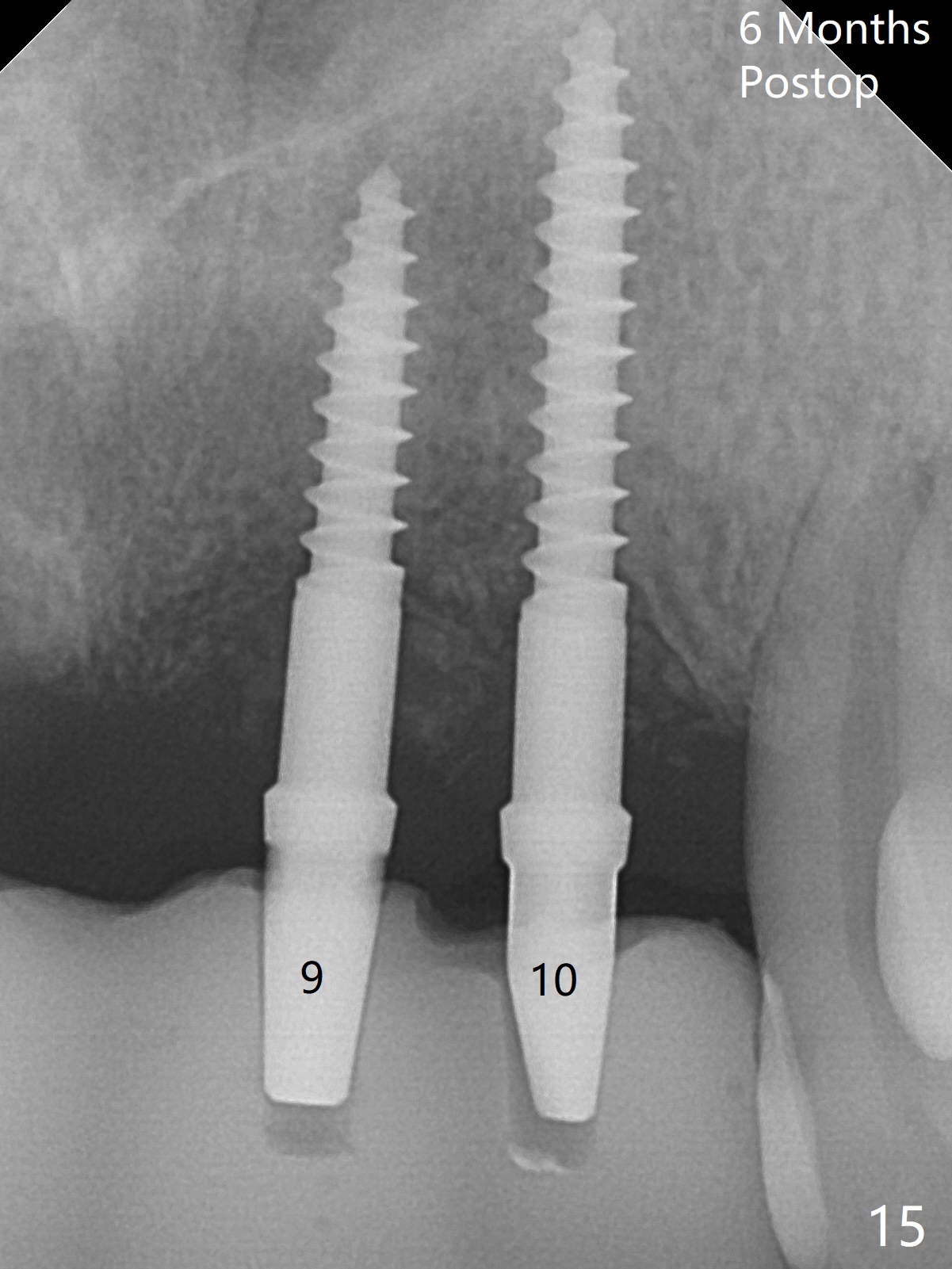
2.5 mm 1-Piece Implants
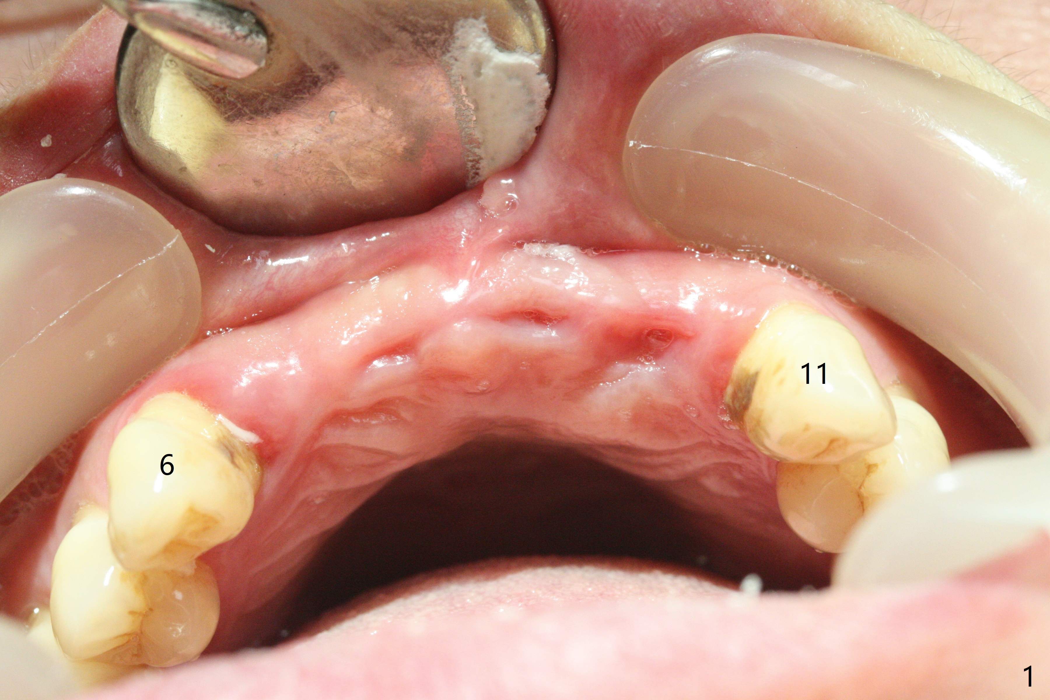
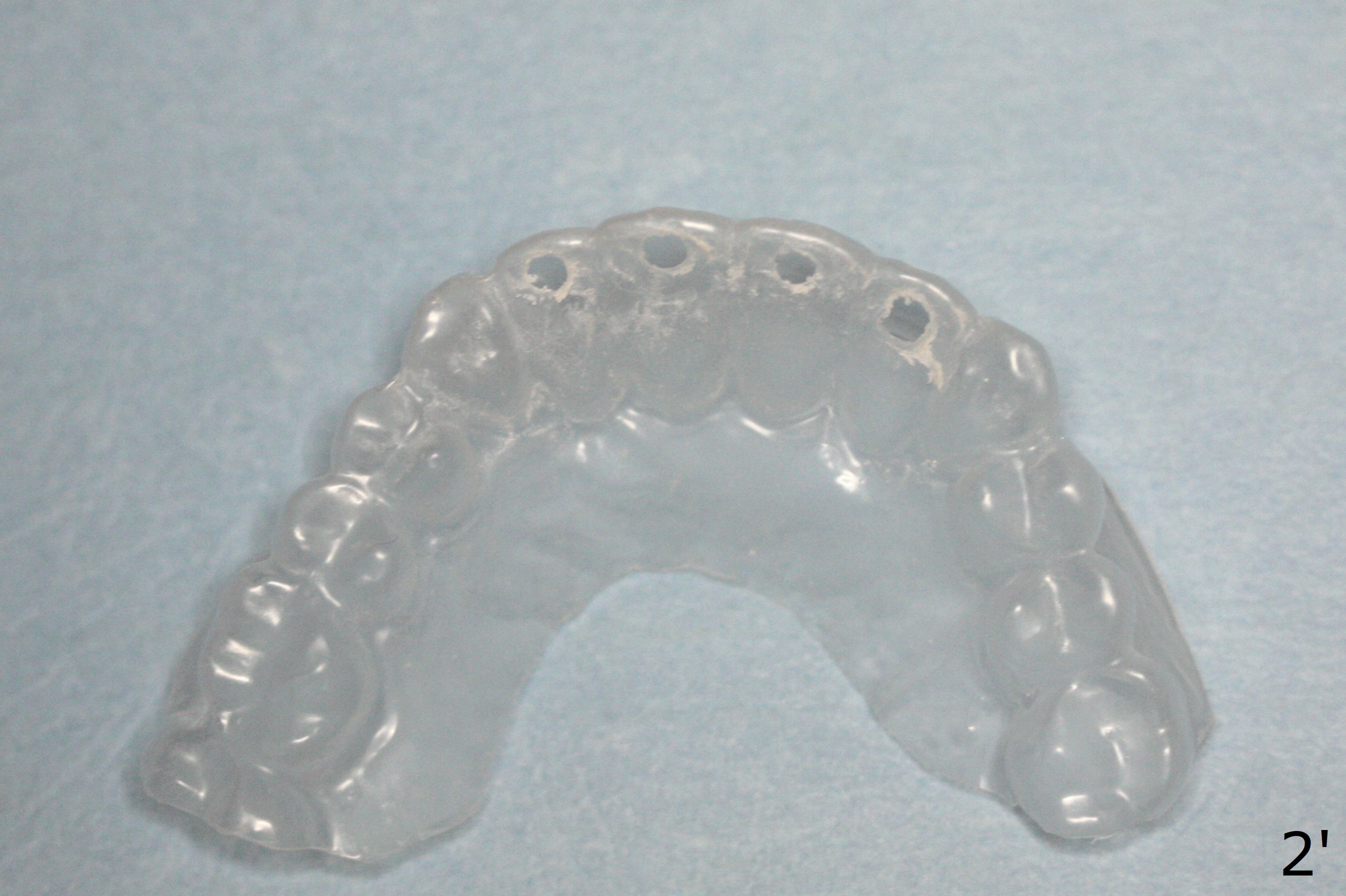
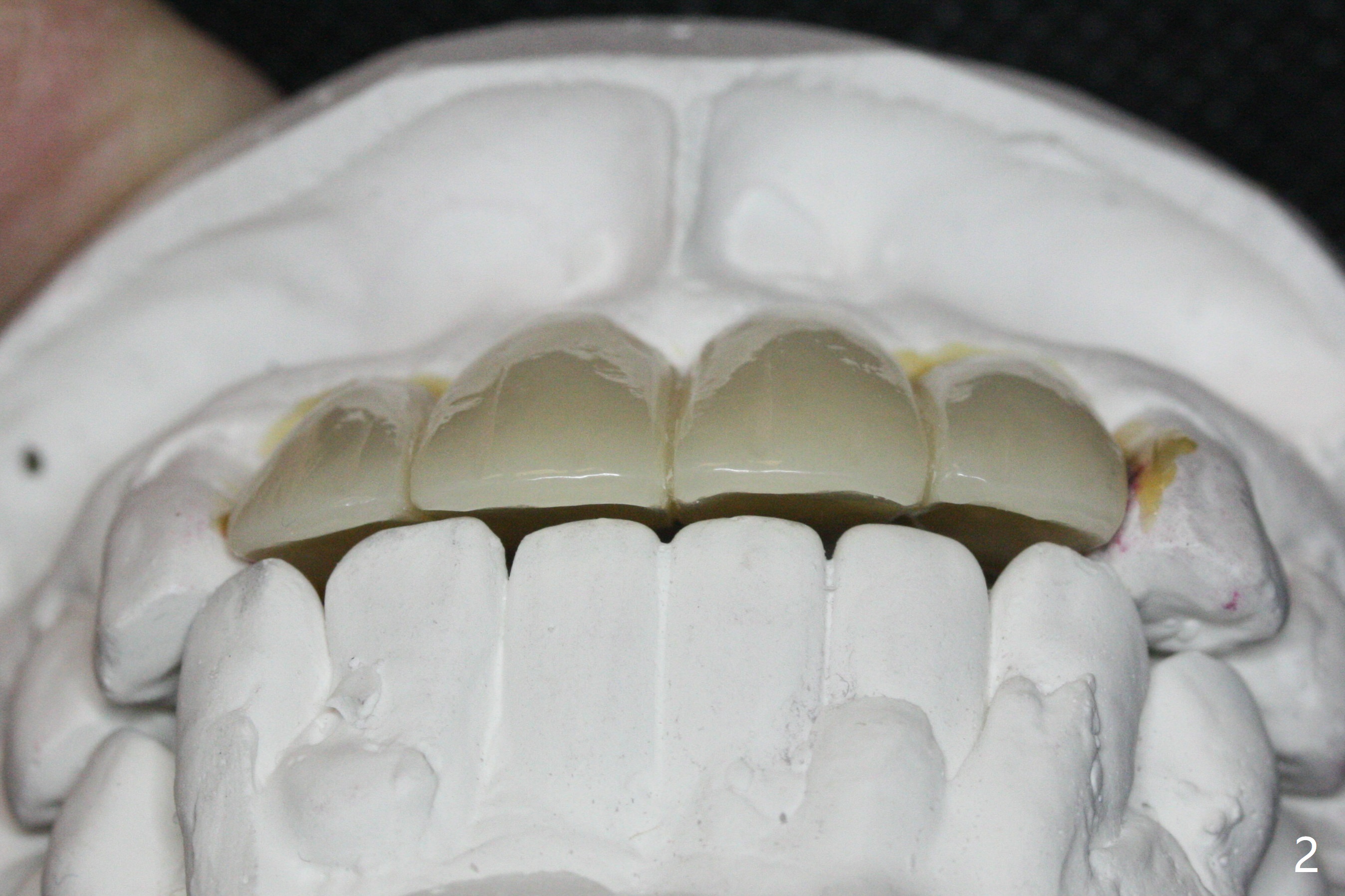
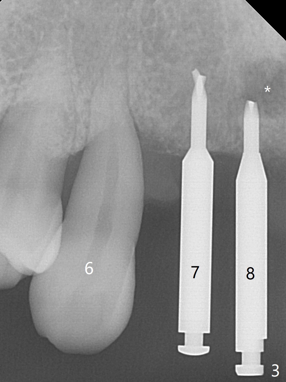
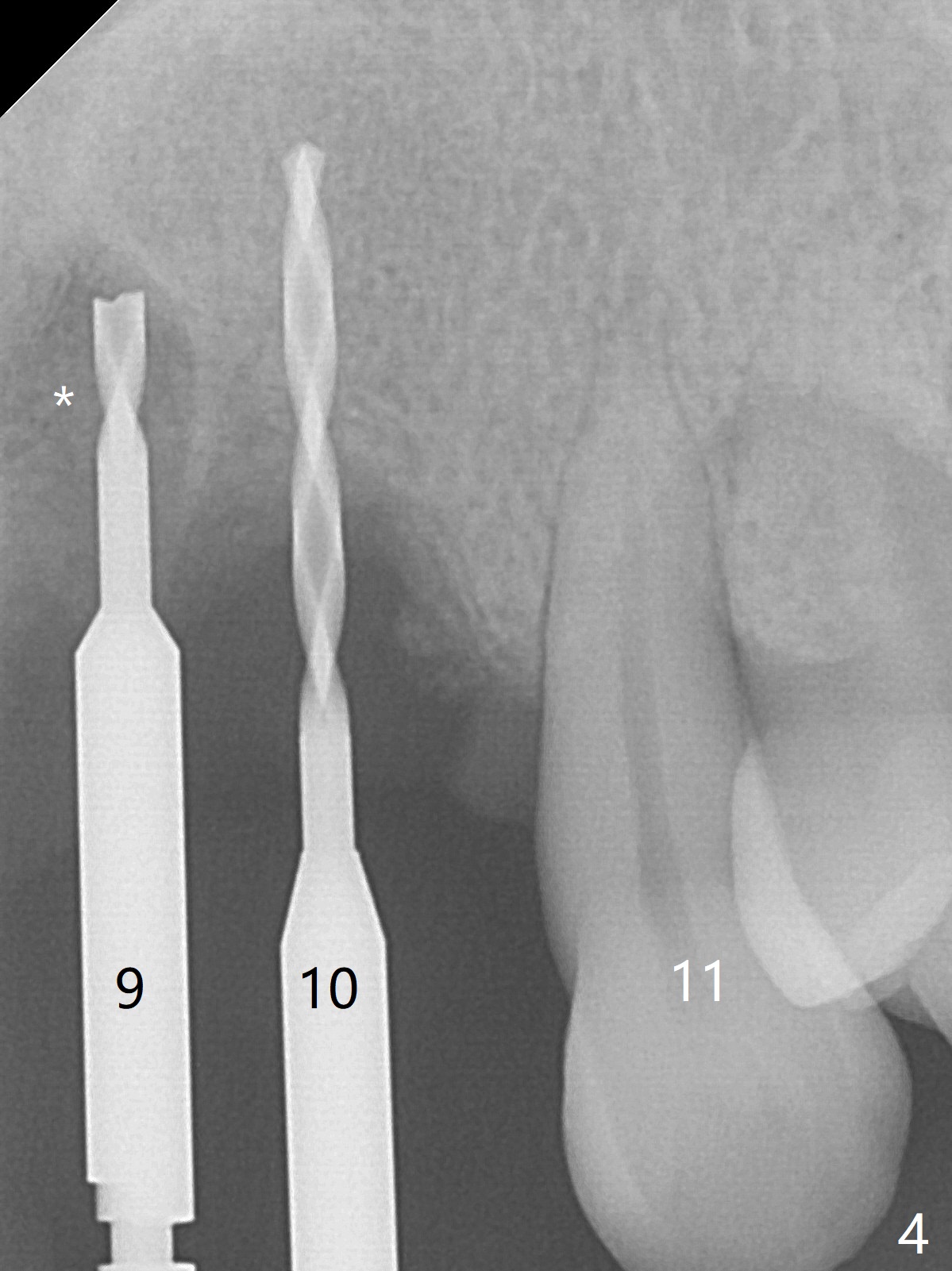
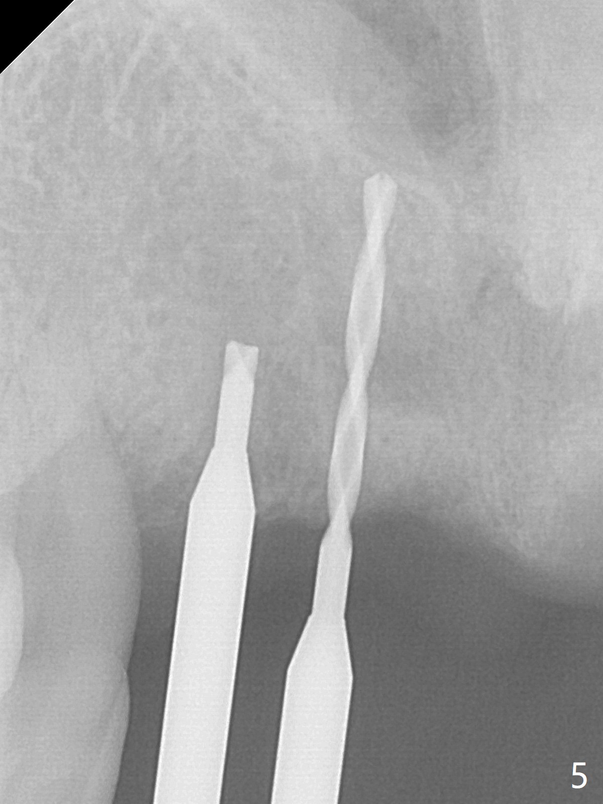
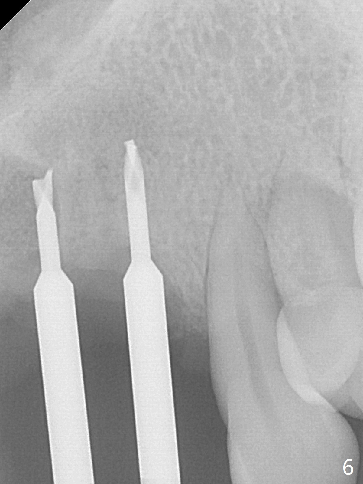
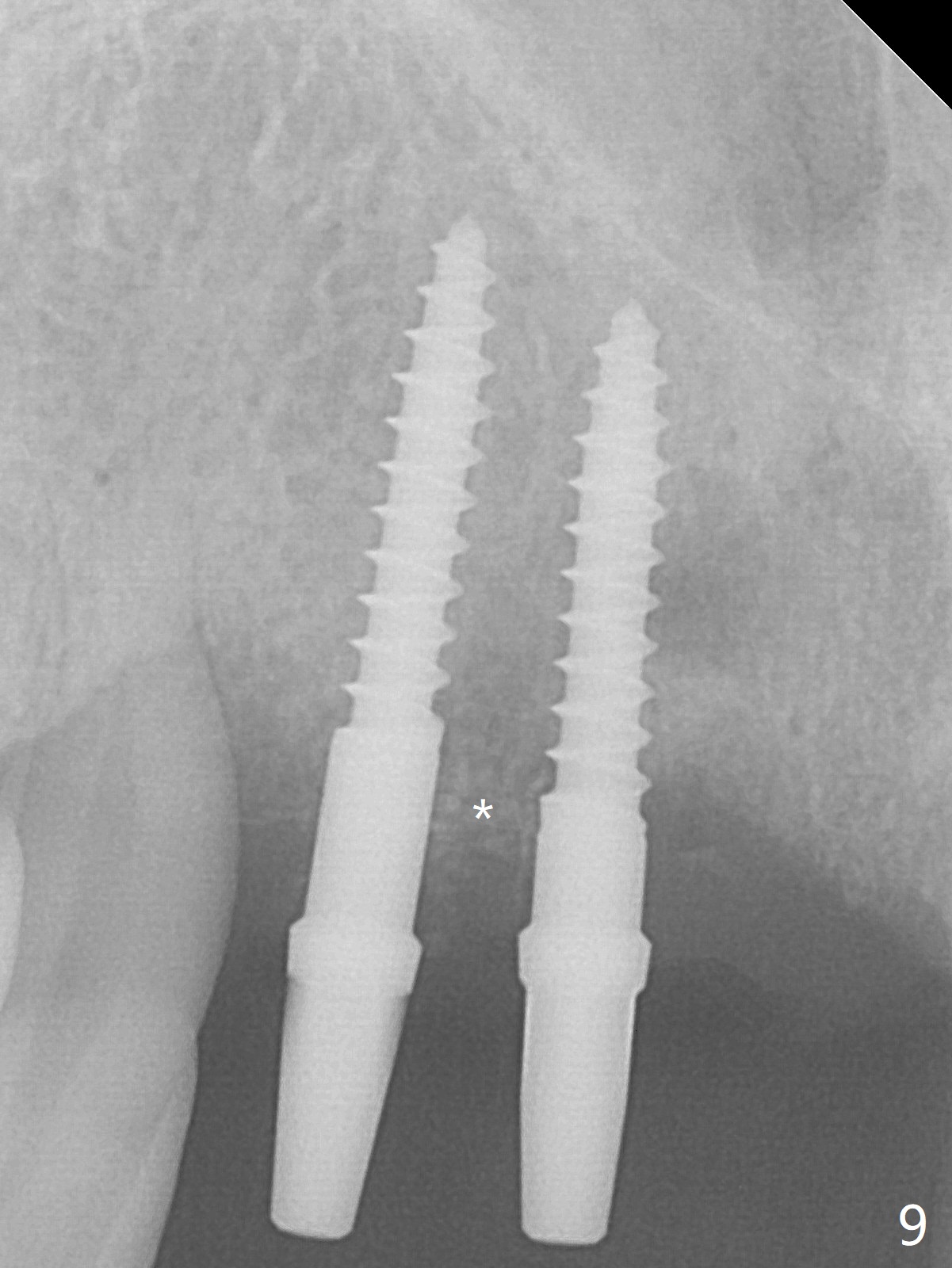
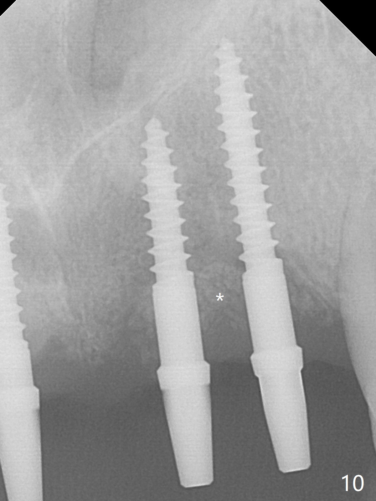
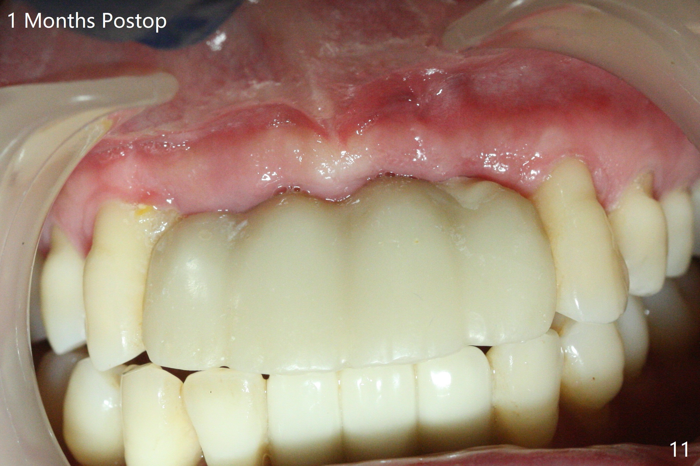
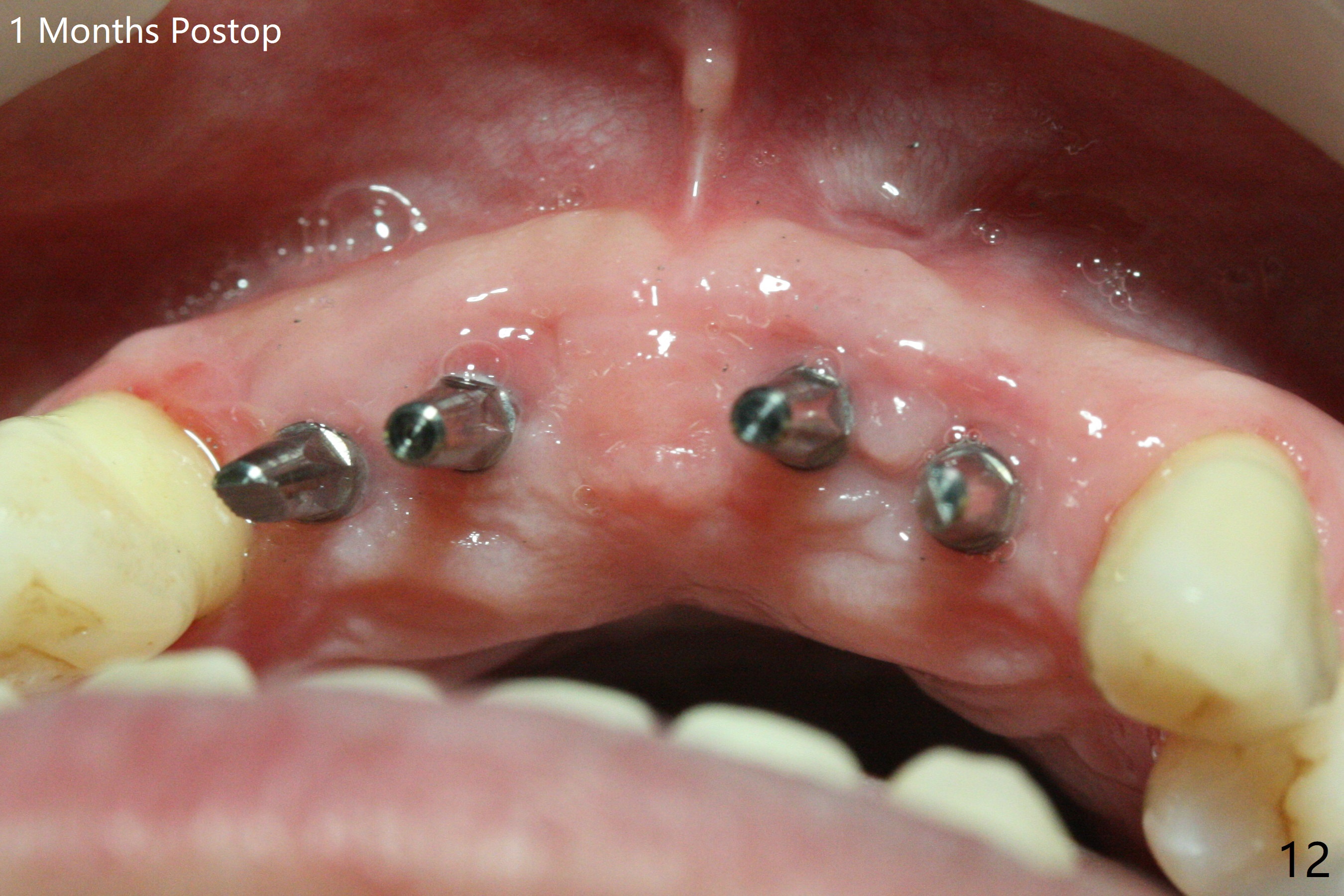
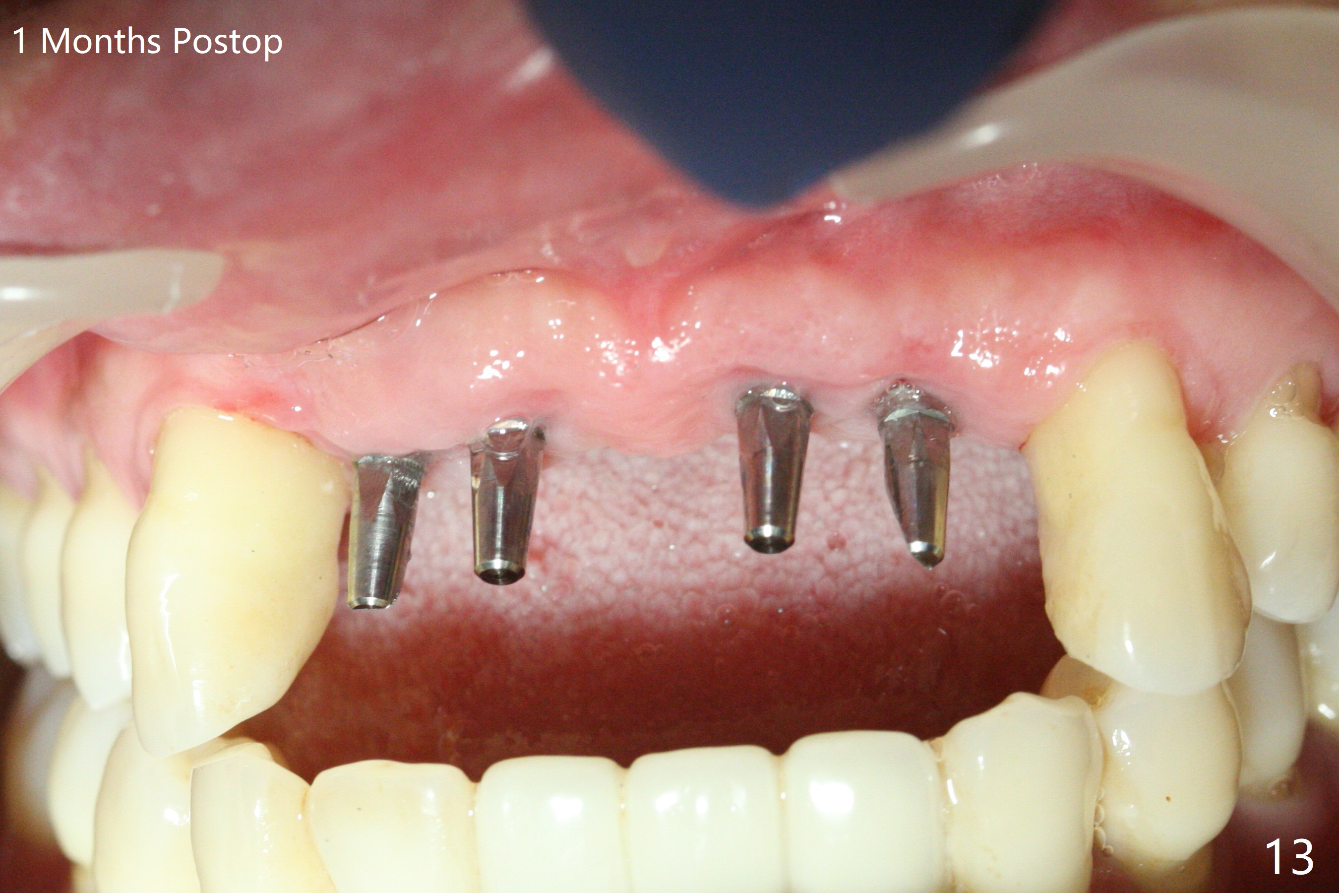
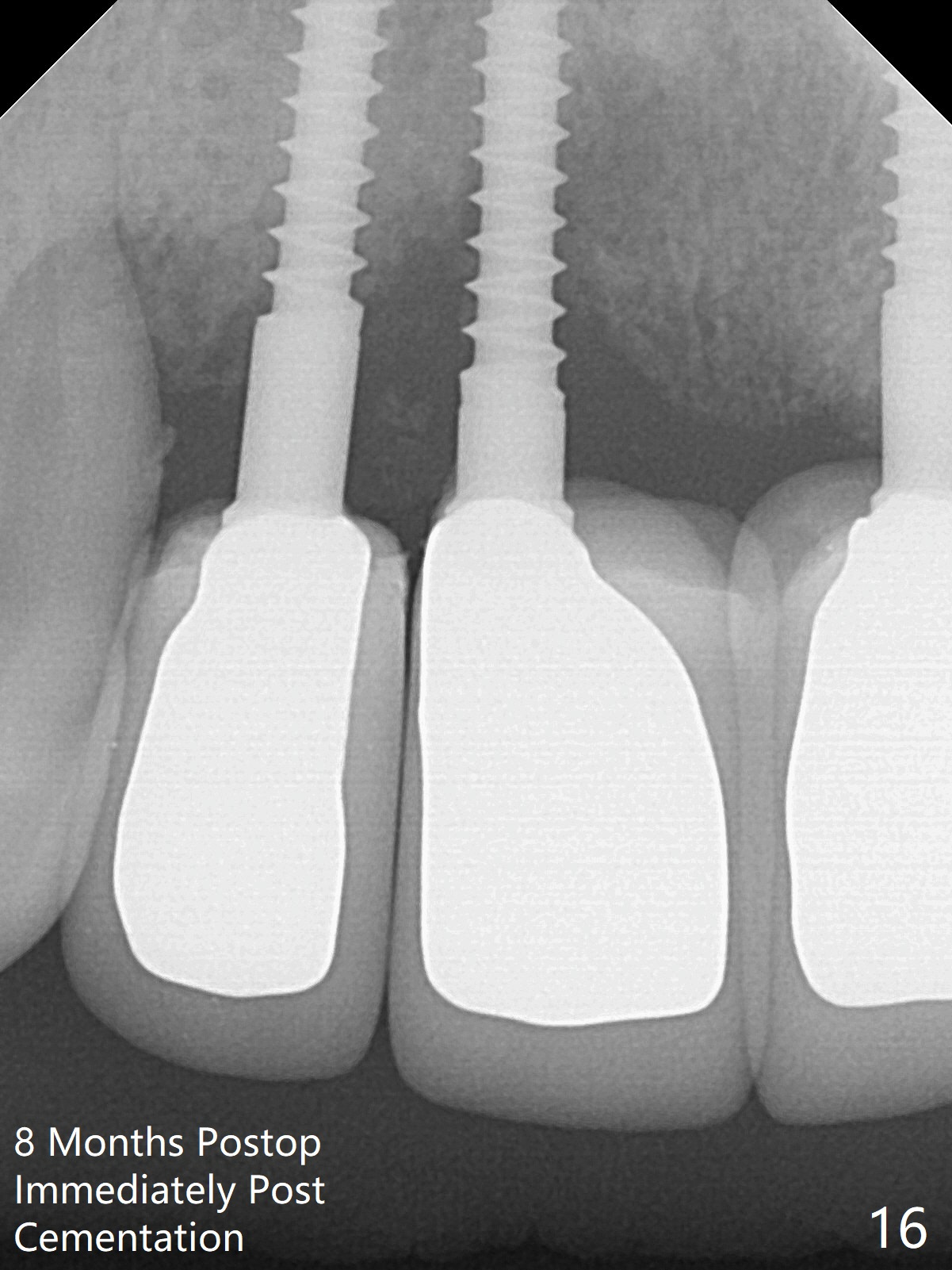
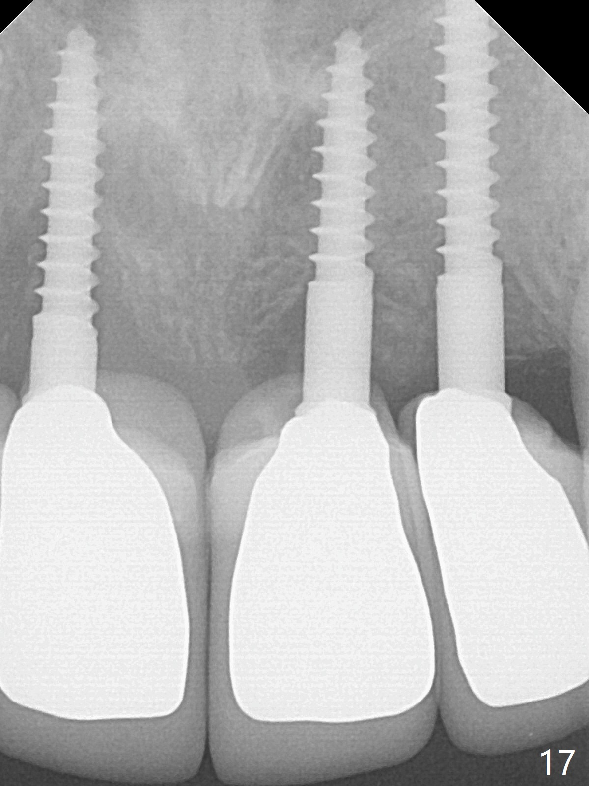
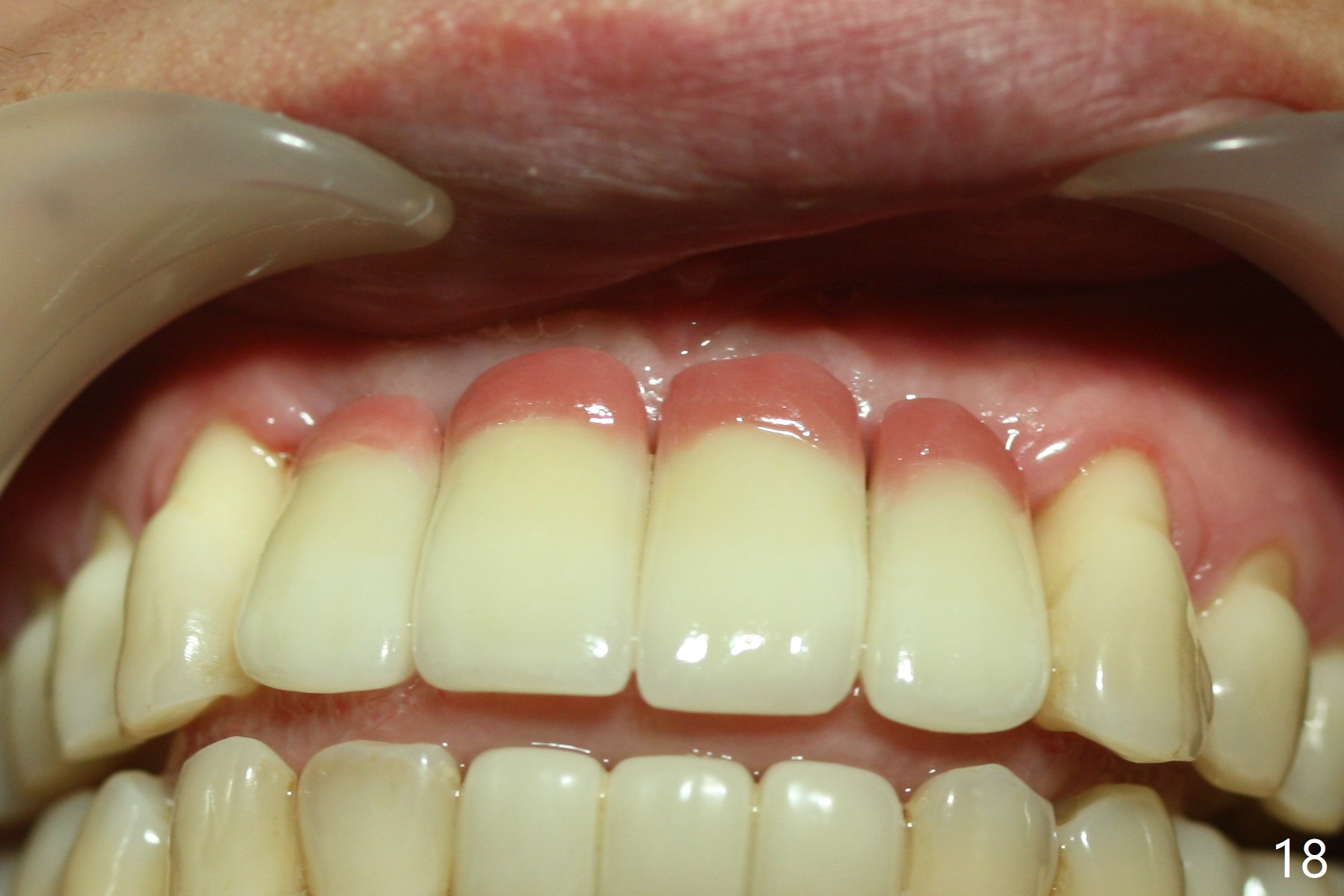
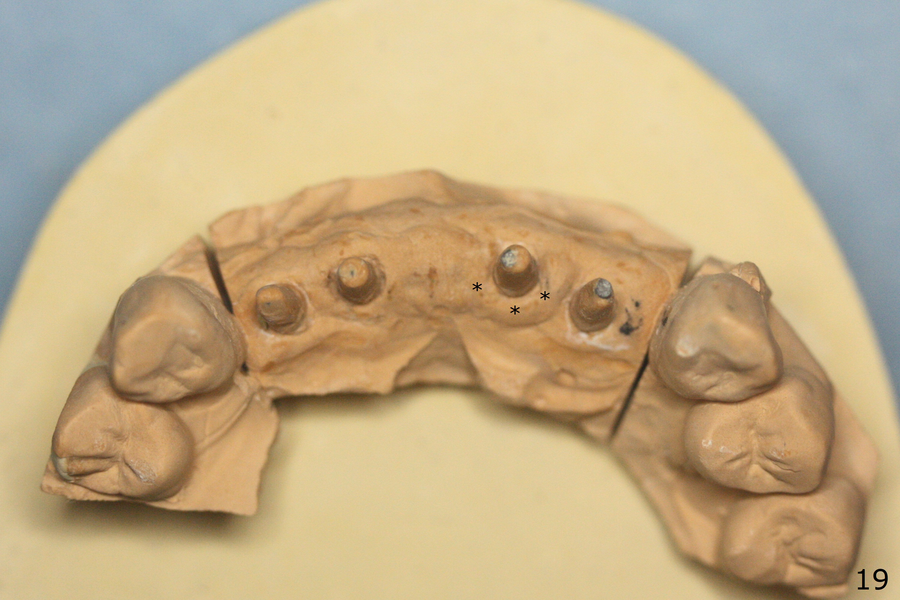
Although the ridge of the maxillary anterior ridge looks moderate in width (Fig.1), the bone is ~4 mm buccopalatally. Suction down surgical stent (Fig.2') made from the lab-fabricated provisional (Fig.2) will be used to check the position and trajectory of osteotomies. PAs taken after initial osteotomies (1.2 mm drill) show those at the central incisors tend to be mesial (close to the Incisive Canal *), while those at the lateral incisor sites distal (Fig.3,4). After adjustment, the position and trajectory of the osteotomies are acceptable (Fig.5,6). To reduce the chance of perforating the Incisive Canal (Fig.3,4 *), 2.5 mm 1-piece implants are inserted with >40 Ncm (Fig.7,8). After deep placement of the implants, Vanilla graft is placed at the crest (Fig.9,10 *). An immediate splinted provisional is fabricated from the suction down stent. The gingiva is healthy around the provisional (Fig.11) and the implants (Fig.12,13) 1 month postop. The provisional is adjusted monthly so that the interdental papillae can be elongated. No bone resorption is observed 6 months postop (Fig.14,15). Crowns are cemented 8 months postop (Fig.16-18). The keratinized gingiva appears to have formed the abutments 8 months postop immediately pre-cementation (Fig.19).
Return to
Upper
Incisor Immediate Implant,
Armaments,
Atrophic Ridge
Xin Wei, DDS, PhD, MS 1st edition 01/04/2018, last revision 04/20/2020