|
|
|
|
|
|
|
|
|
|
|
|
|
|
|
|
|
|
|
|
|
|
|
|
|
|
|
|||
Post-Implant Bone Graft
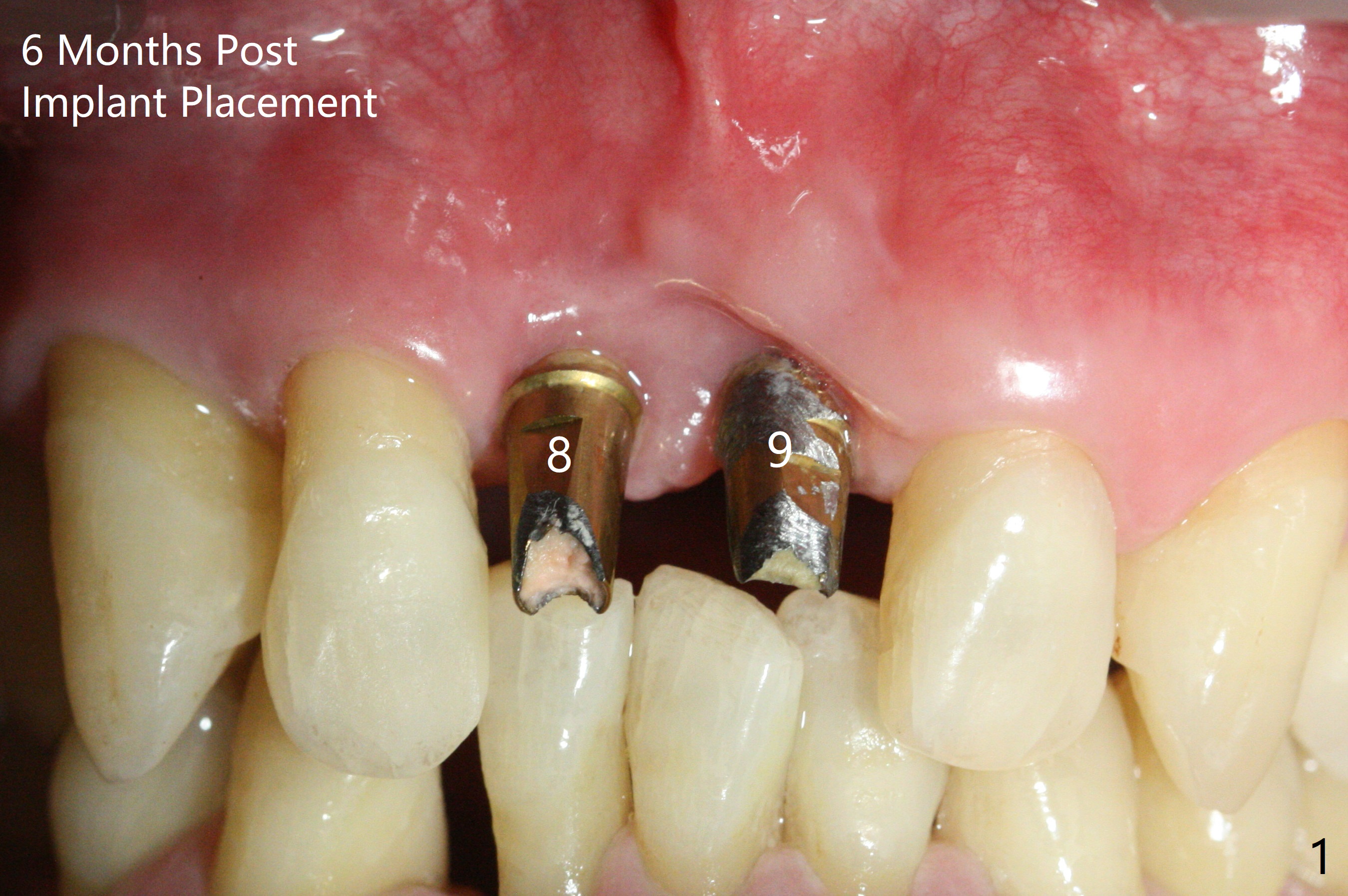
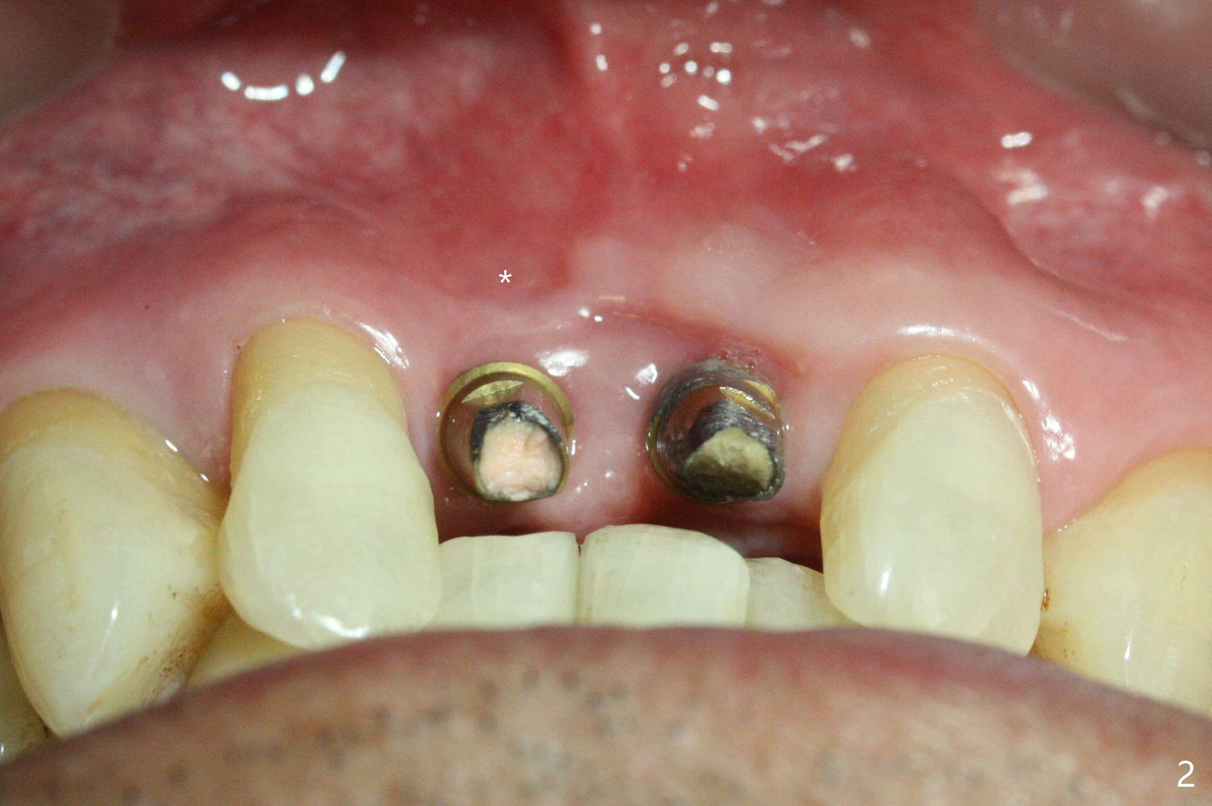
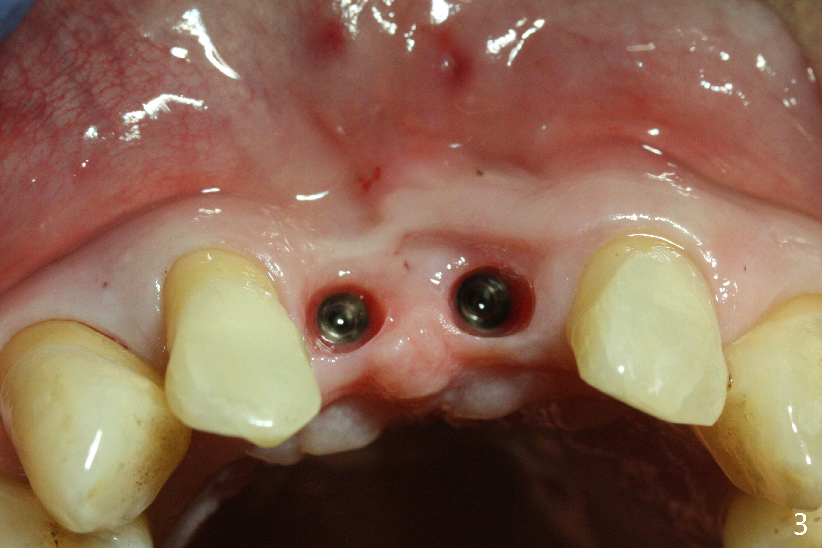
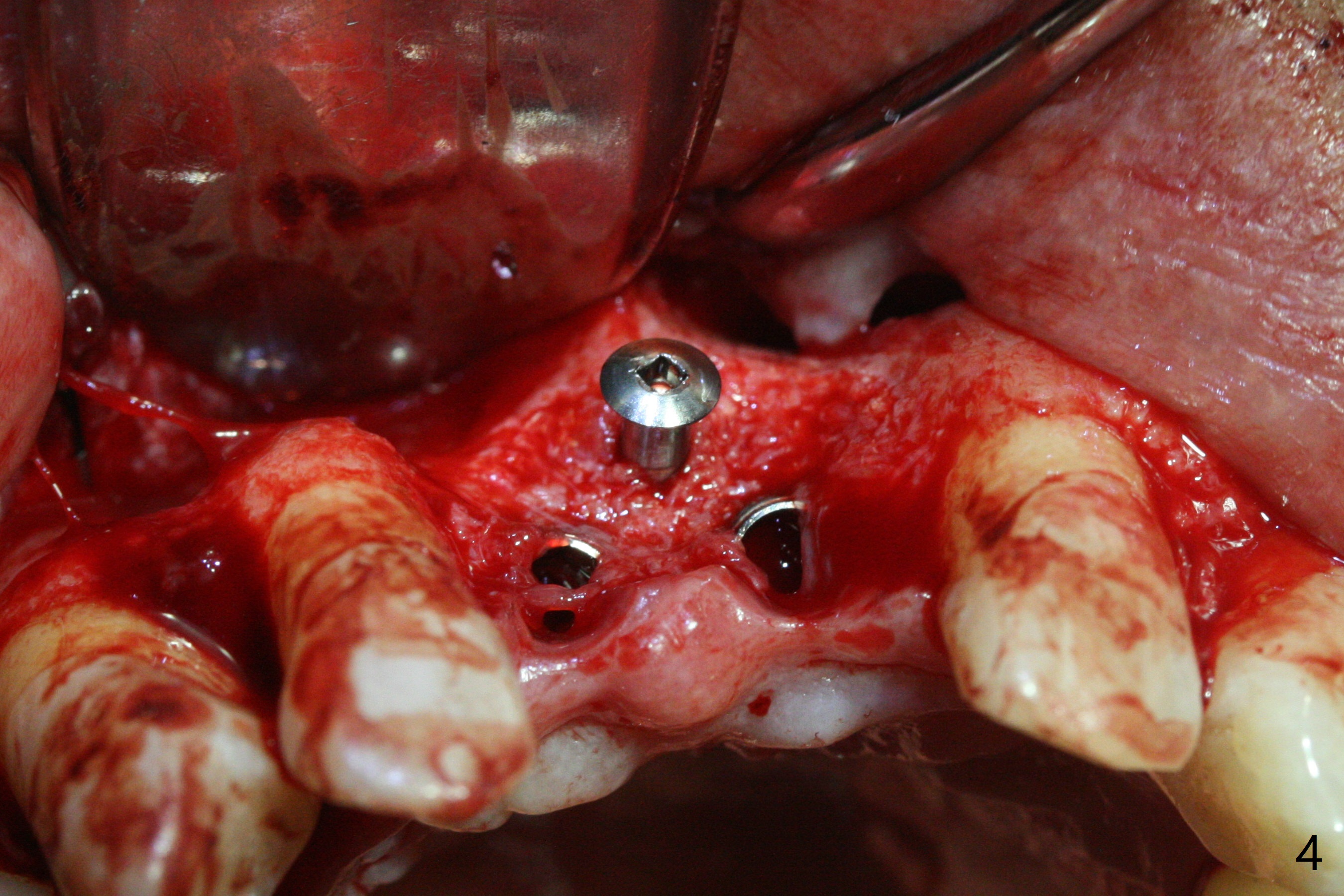
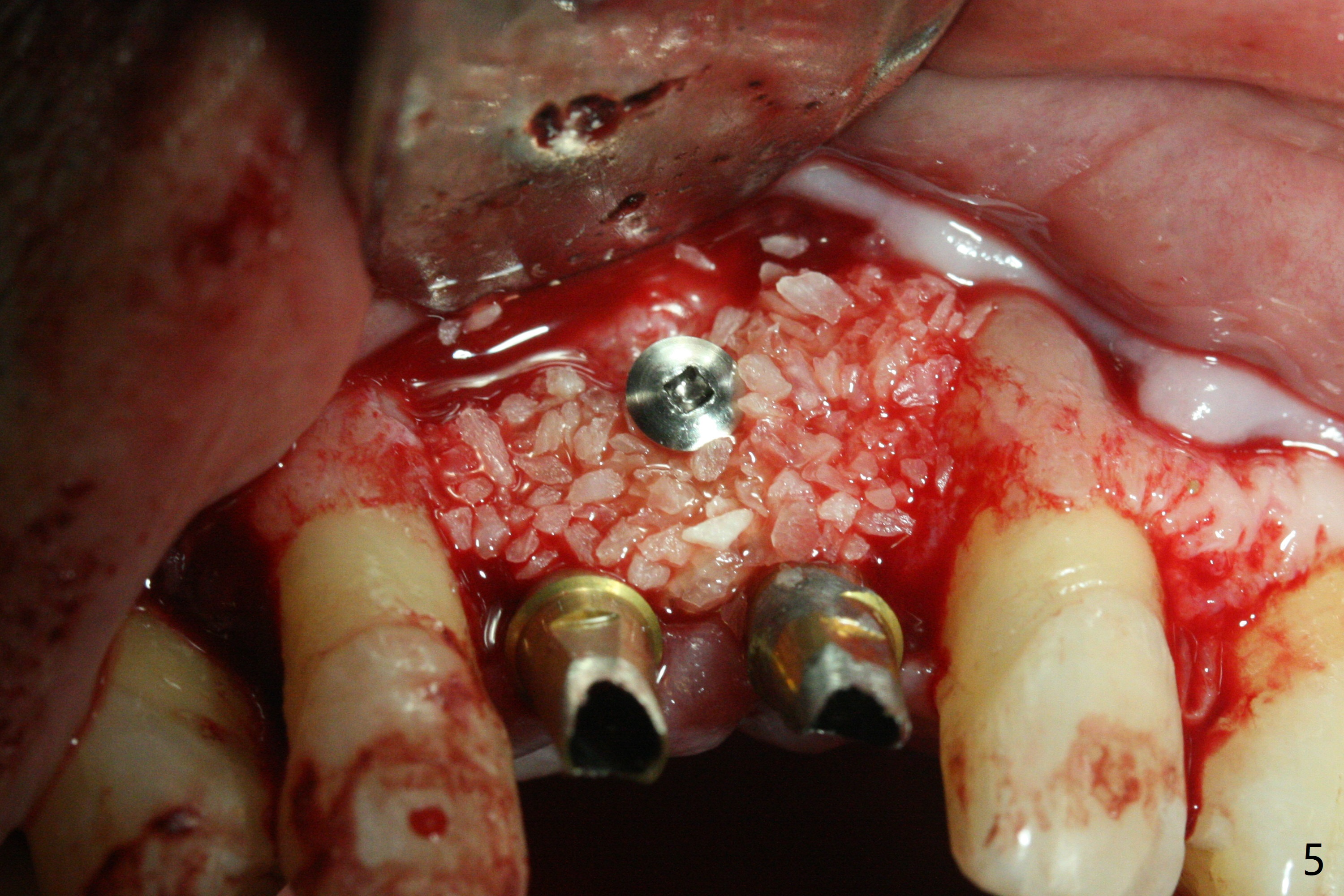
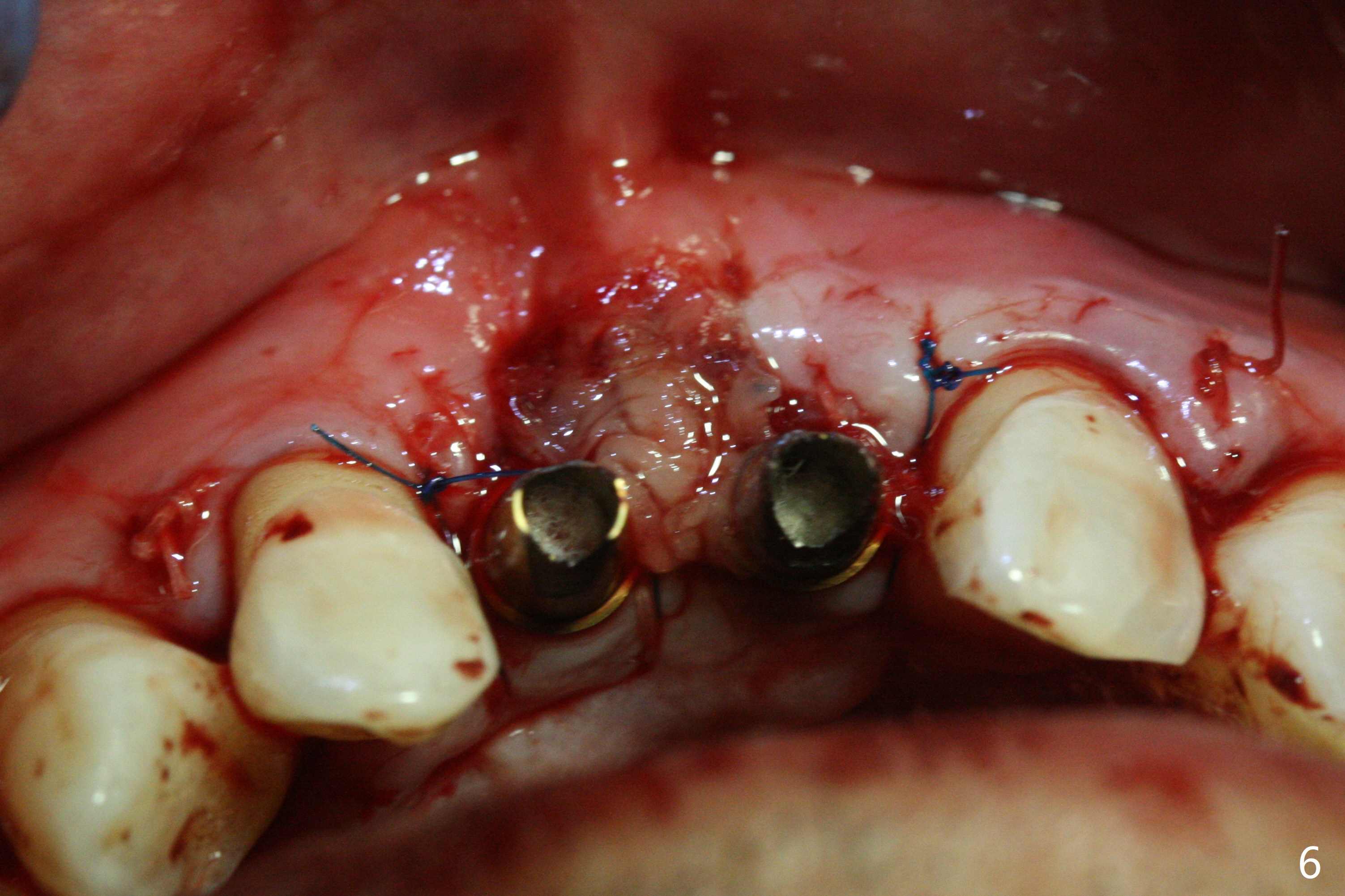
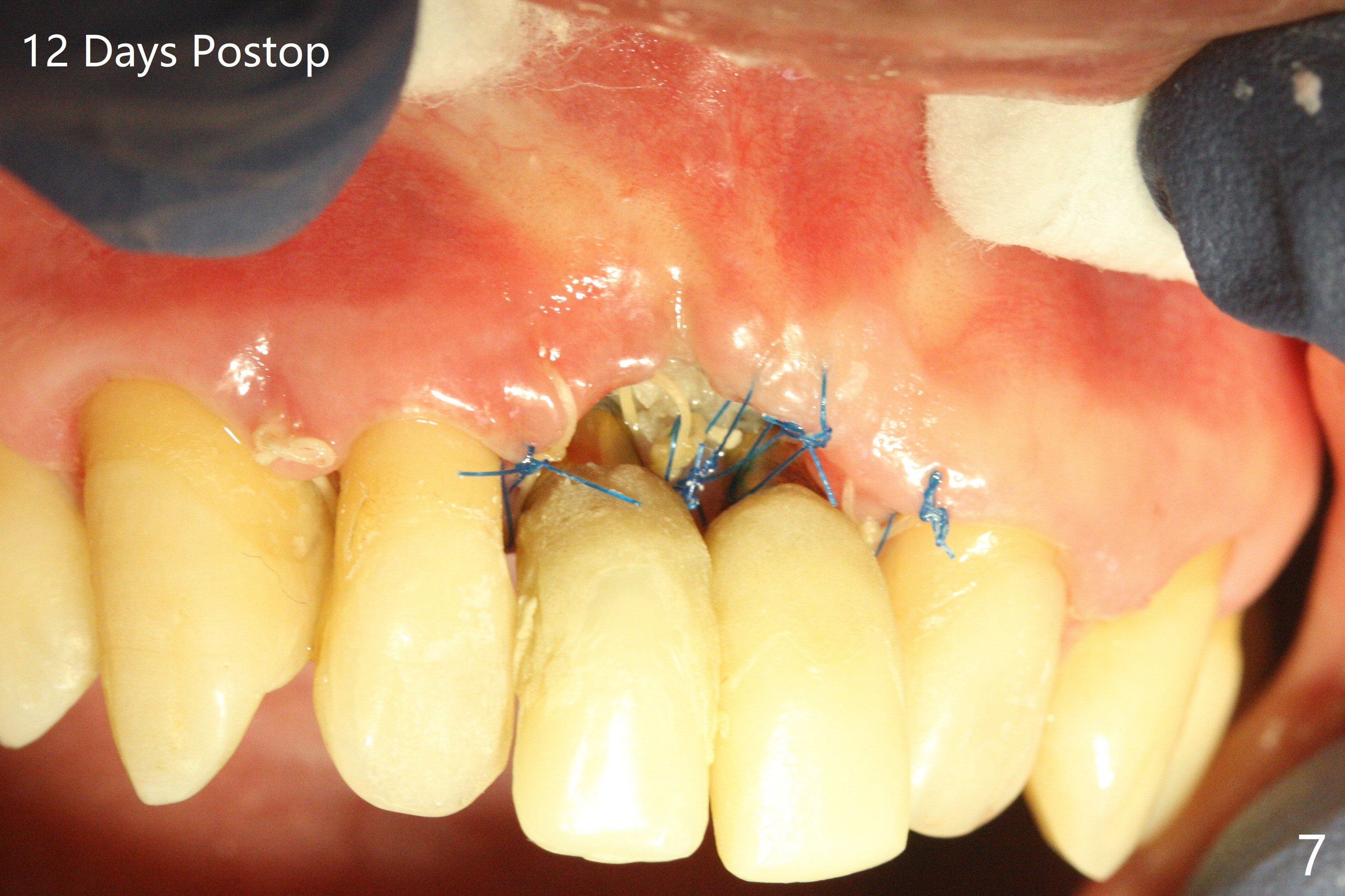
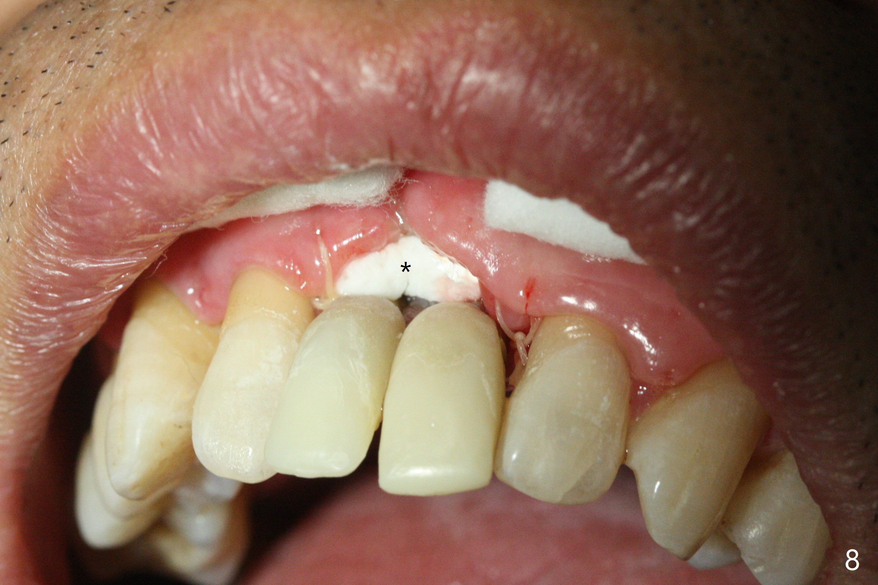
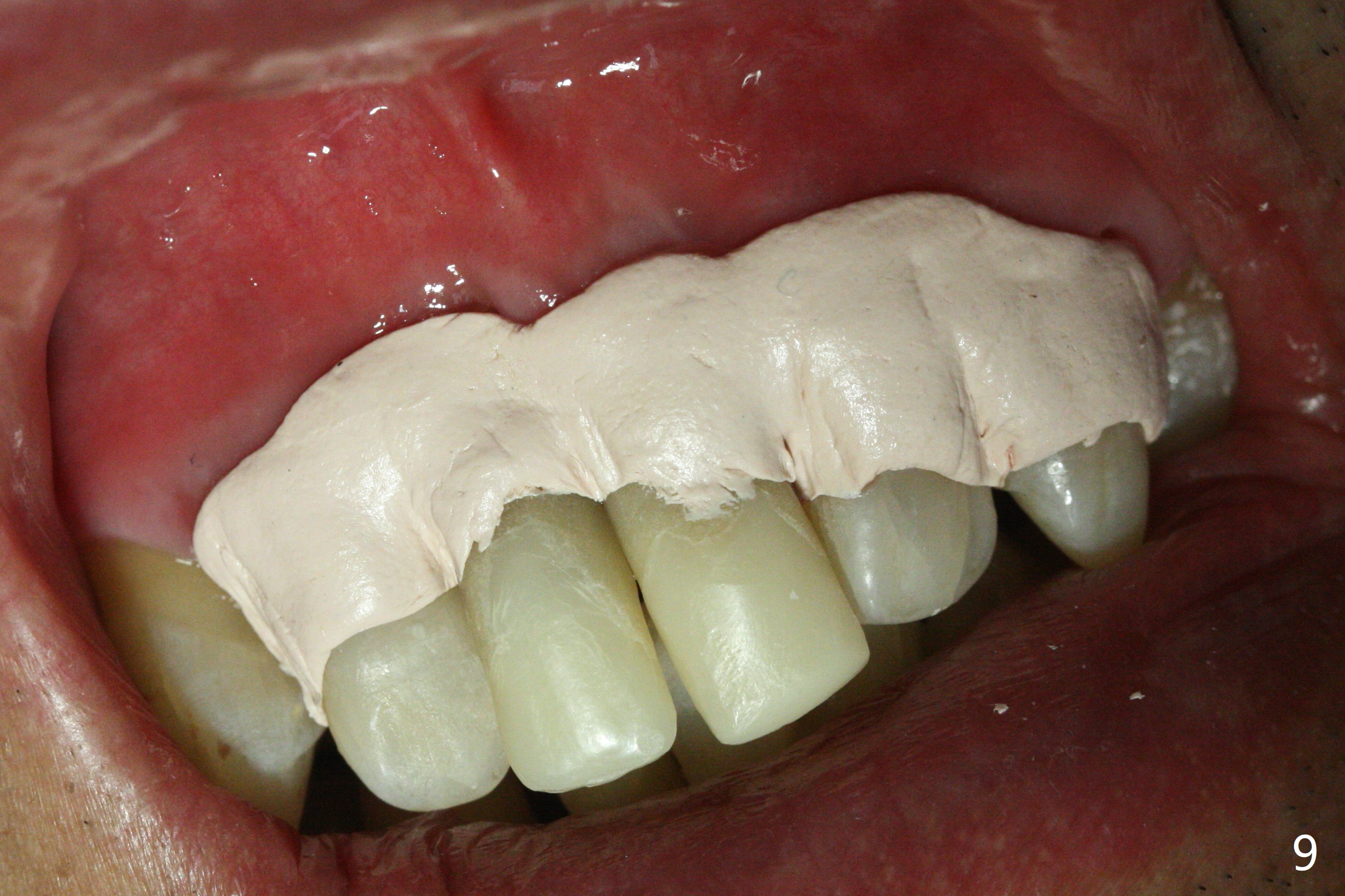
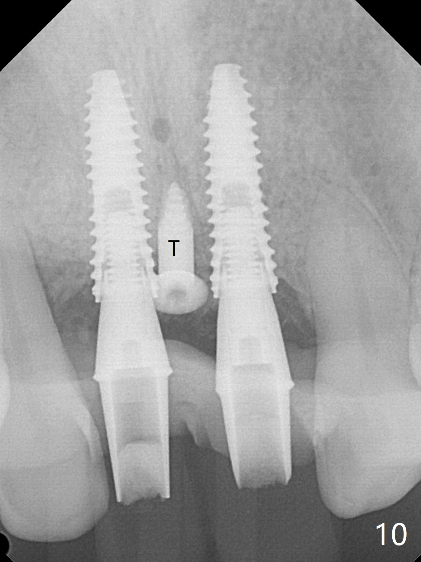
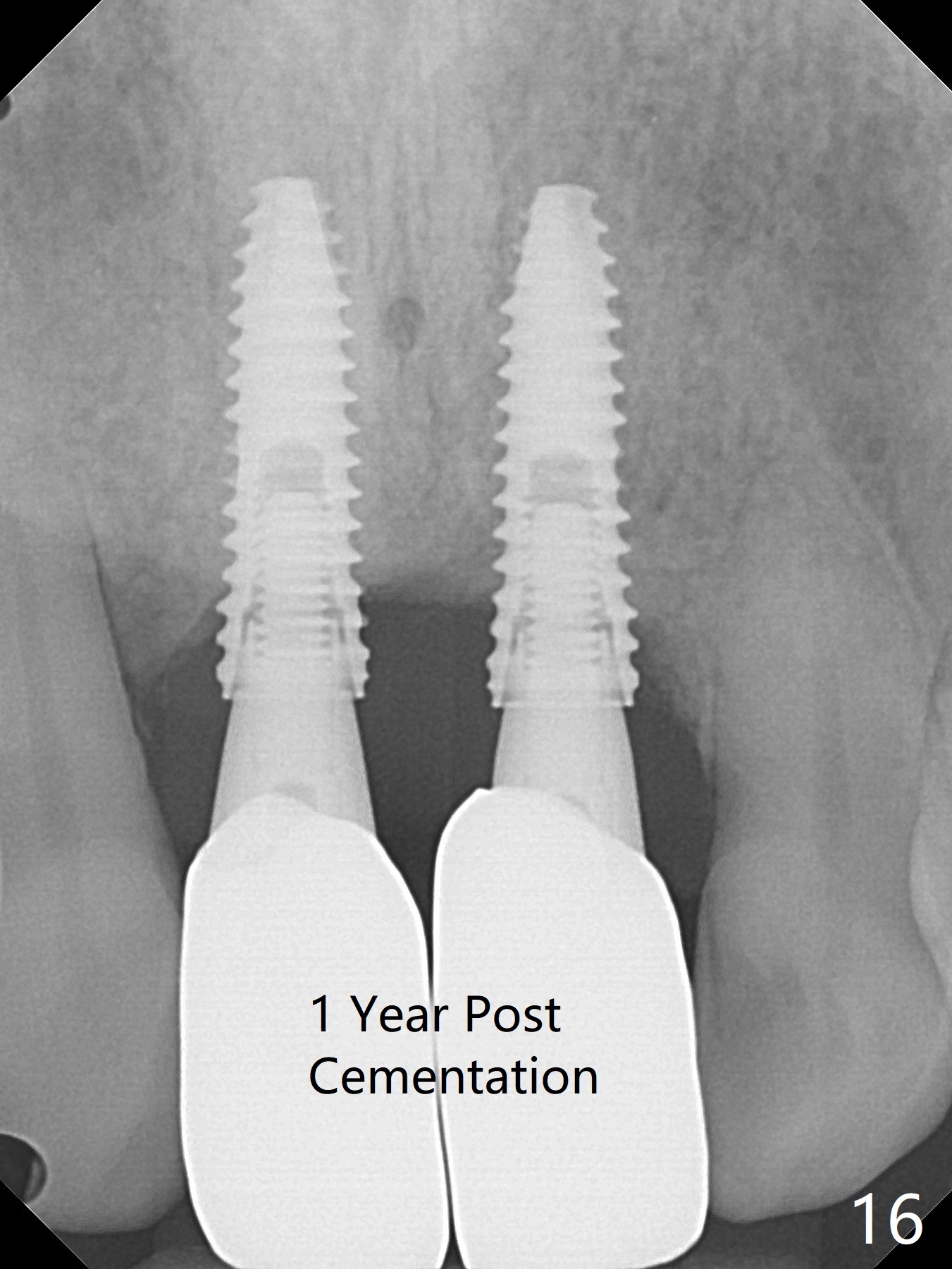
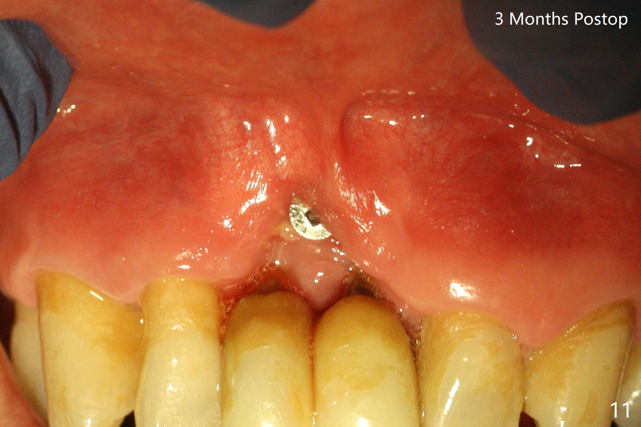
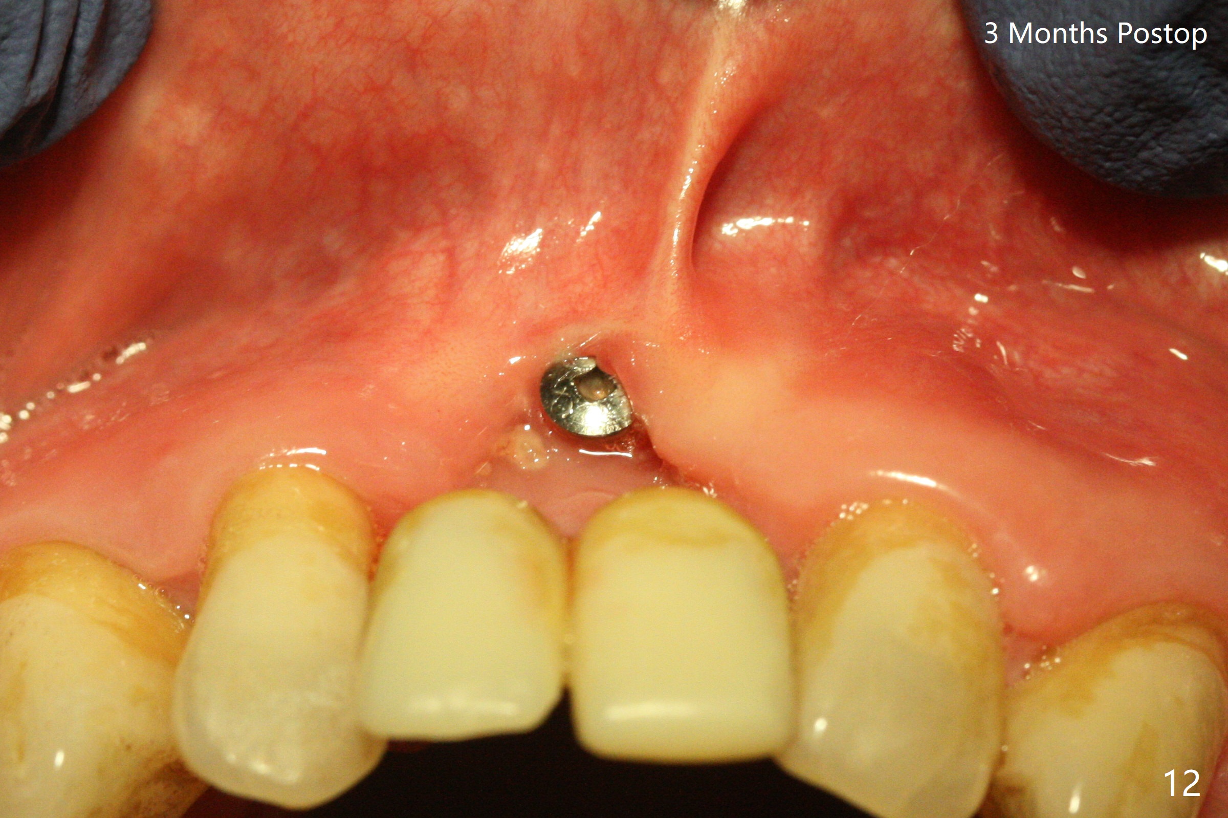
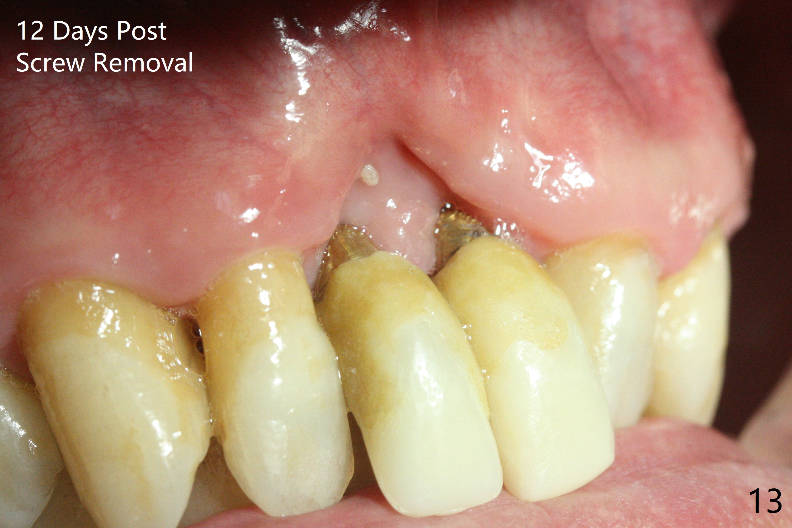
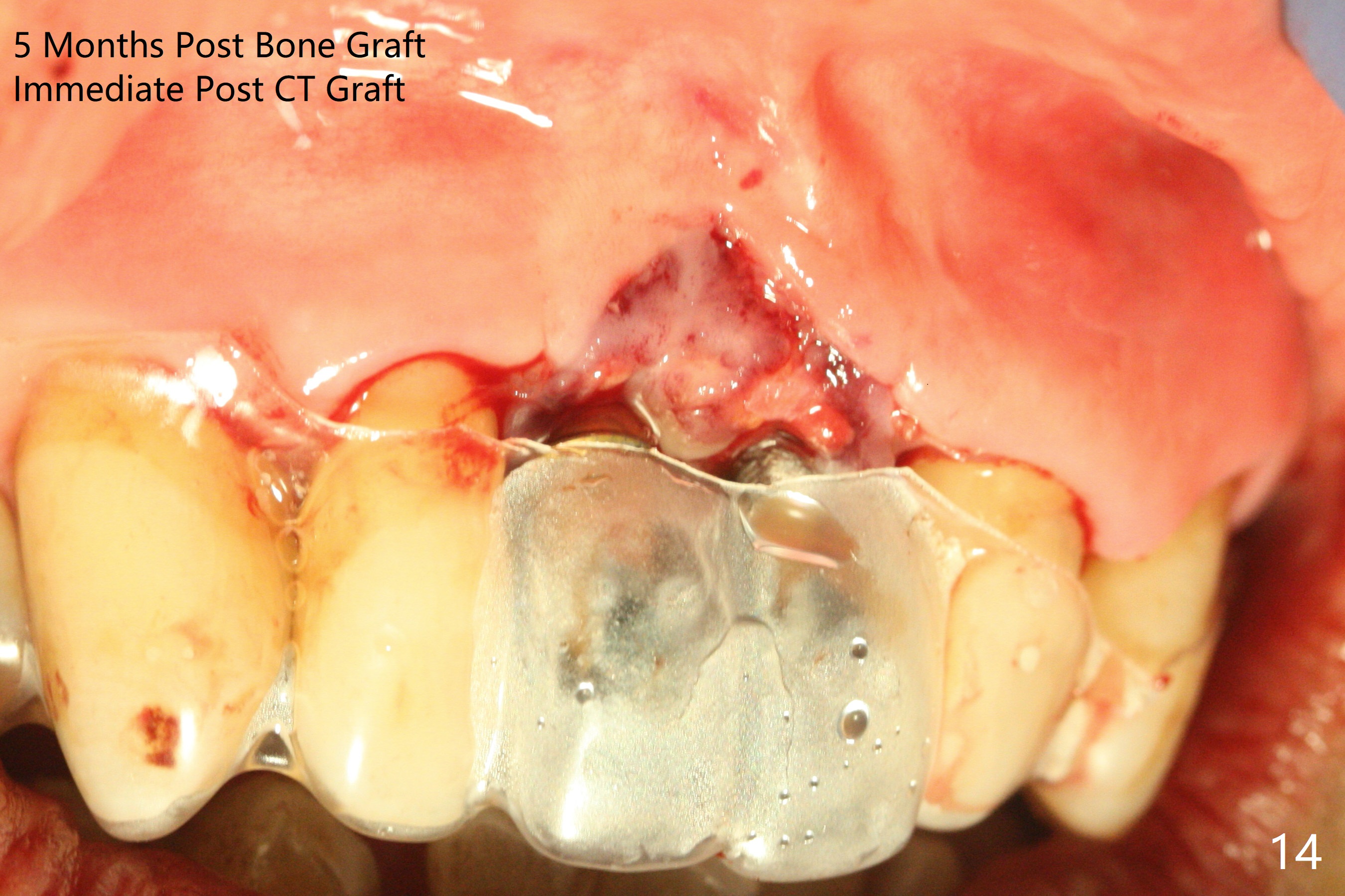
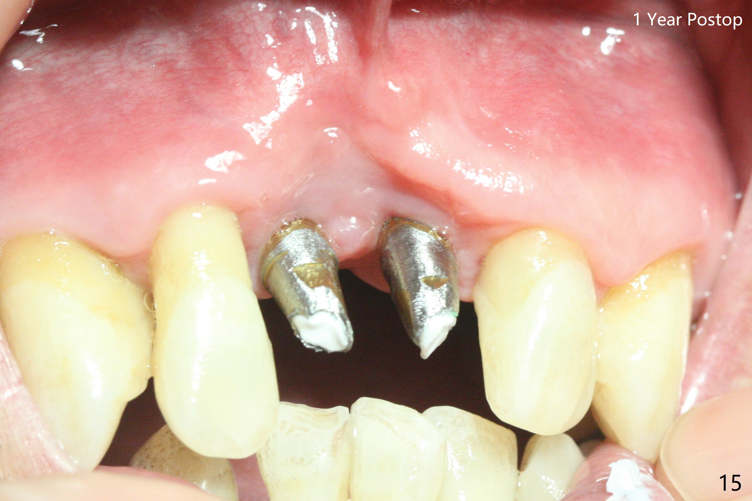
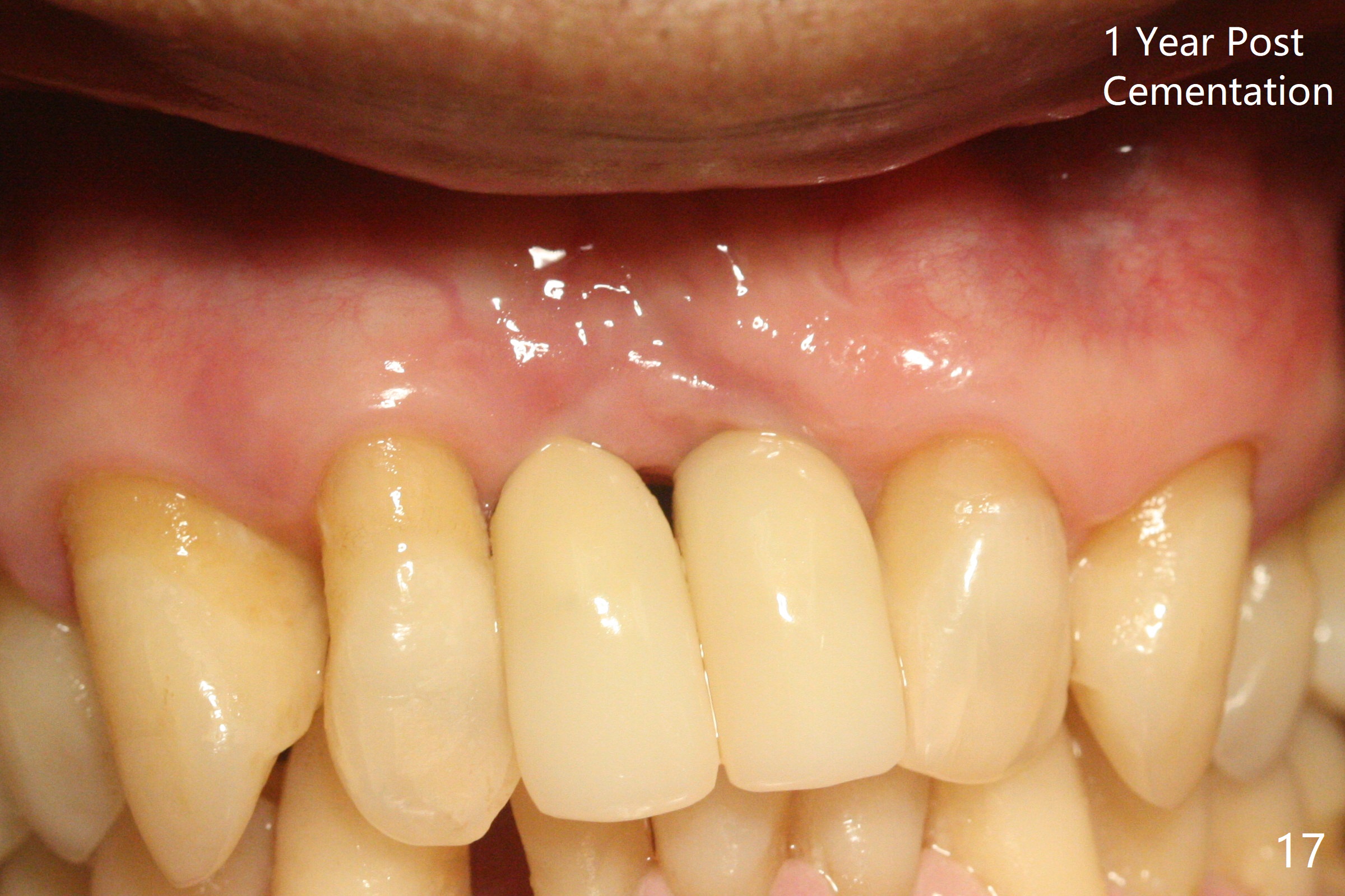
The gingiva remains recessive at #8 and 9 six months post immediate implant placement with bone graft (Fig.1). The buccal plate at #8 is particularly concave before (Fig.2 *) and after (Fig.3) abutment removal. The bony defect repair is assisted by placing a 4 mm tenting screw between the 2 implants (Fig.4) and placing allograft mixed with PRF (as putty) around the screw (Fig.5 (after replacement of the abutments)). The buccal contour improves because of the tenting screw and the bone graft placement (Fig.6 (as well as PRF and 6-month membranes)). The wound dehisces 12 days postop and immediately before leaving country for months (Fig.7). The sutures are removed, Osteogen plug is inserted (Fig.8) and periodontal dressing is applied (Fig.9). PA is taken to show the tenting screw (Fig.10 T). The latter is exposed 3 months postop (Fig.11,12). It appears that gingival graft is a must (Fig.13). Make a palatal stent, remove the temp with abutments and create a bleeding surface before harvesting a large piece of tissue. Connective tissue graft is done 5 months post bone graft (Fig.14). In fact there is no implant thread exposure. In fact the connective tissue graft does not survive. The abutments are re-prepared for pink porcelain (Fig.15). The bone loss is stable 1 year post cementation in spite of incomplete abutment seating (Fig.16). The soft tissue is nearly normal (Fig.17).
Return to
Upper Incisor Immediate Implant,
Trajectory
Xin Wei, DDS, PhD, MS 1st edition
04/25/2019, last revision
01/12/2021