|
|
|
|
|
|
|
|
|
|
|
|
|
|
|
|
|
|
|
|
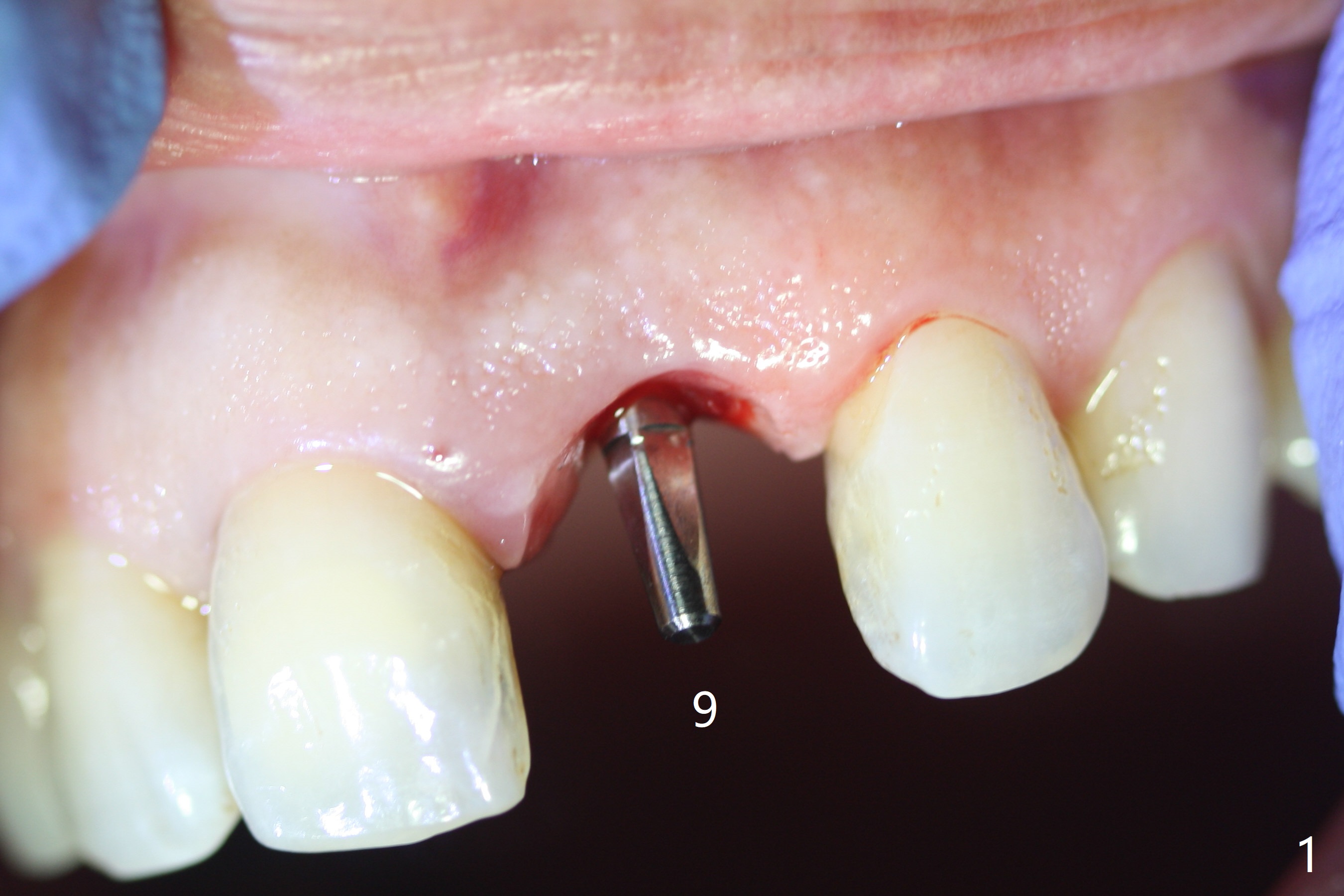
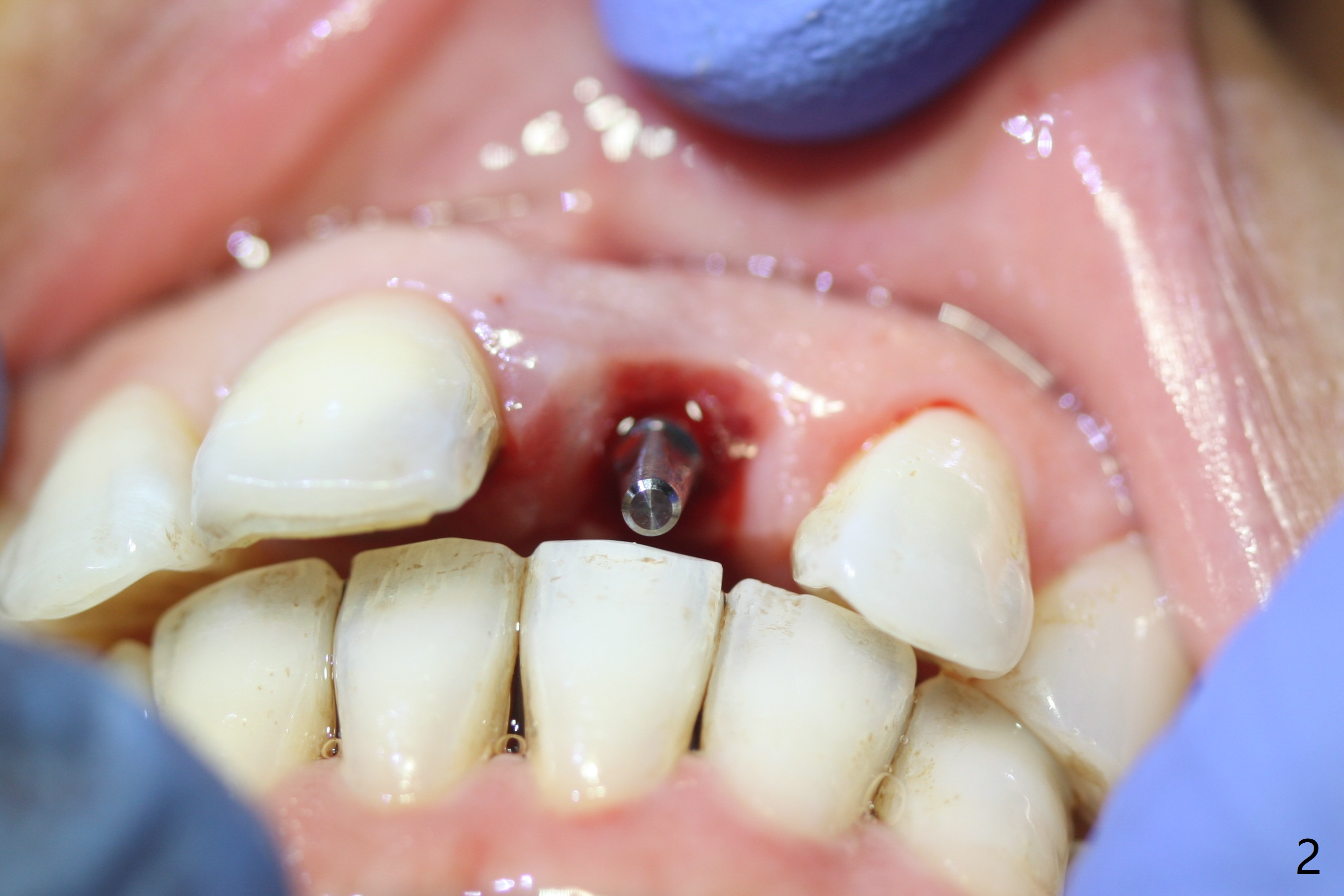
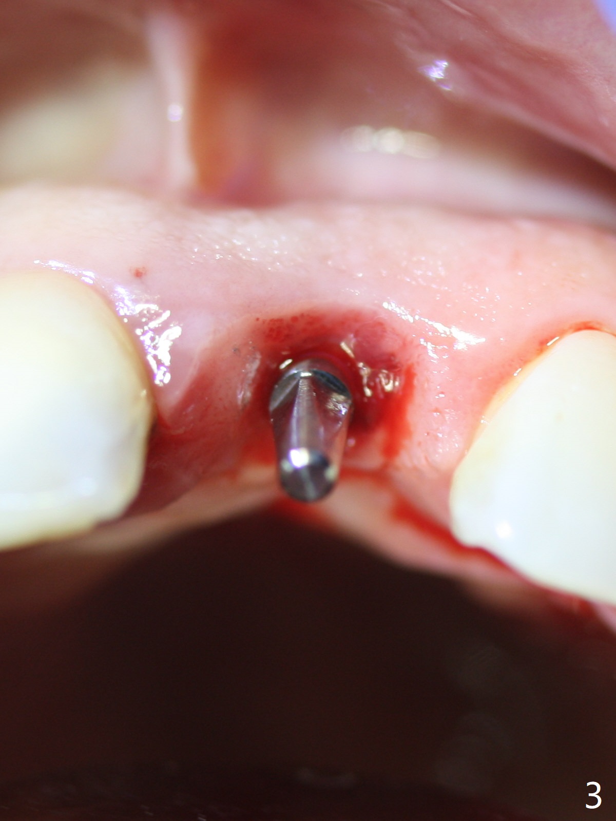
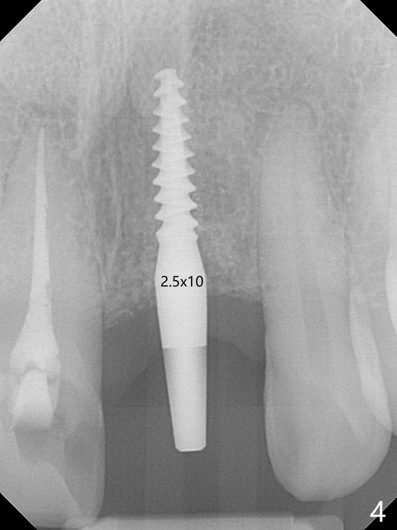
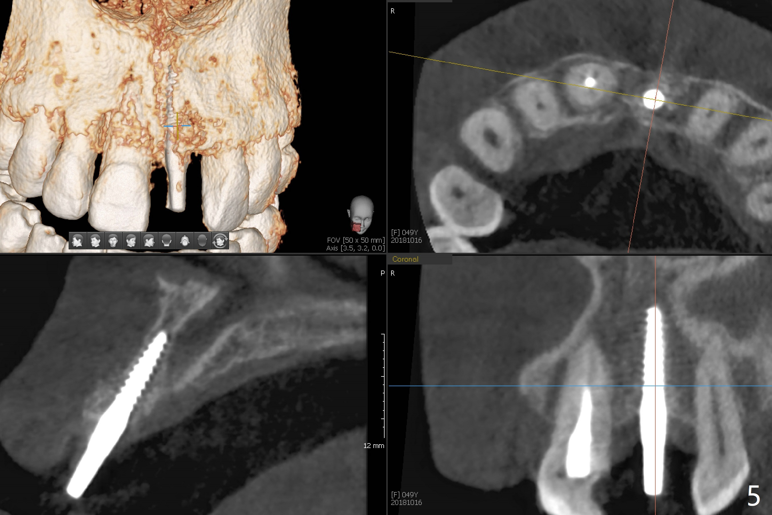
Guide for Narrow Bone
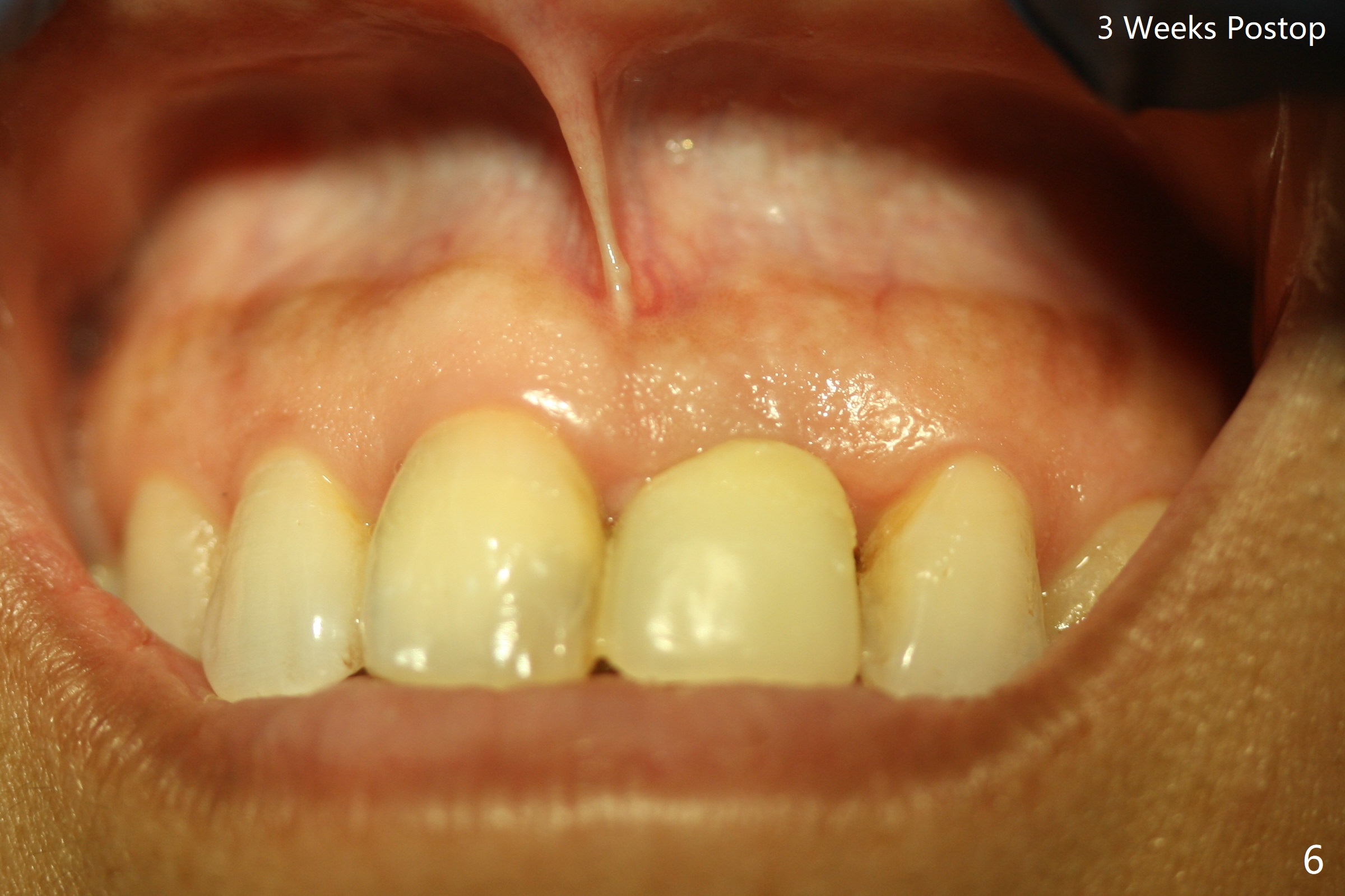
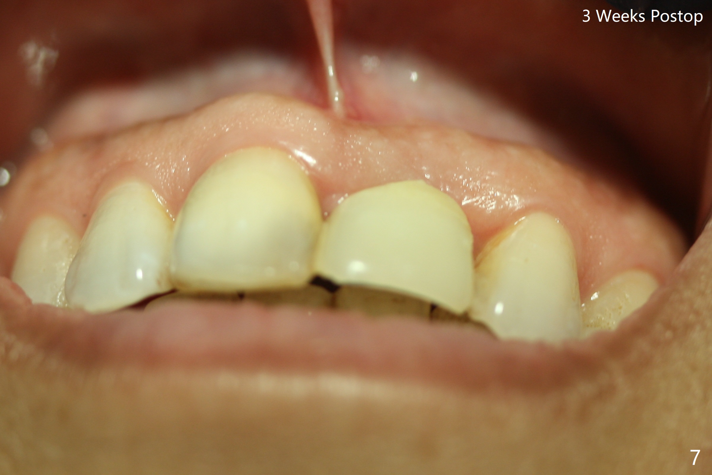
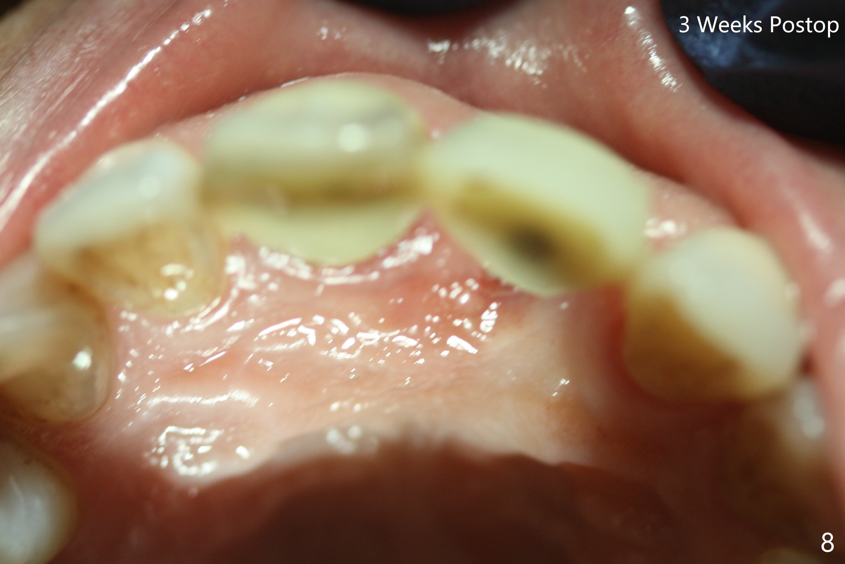
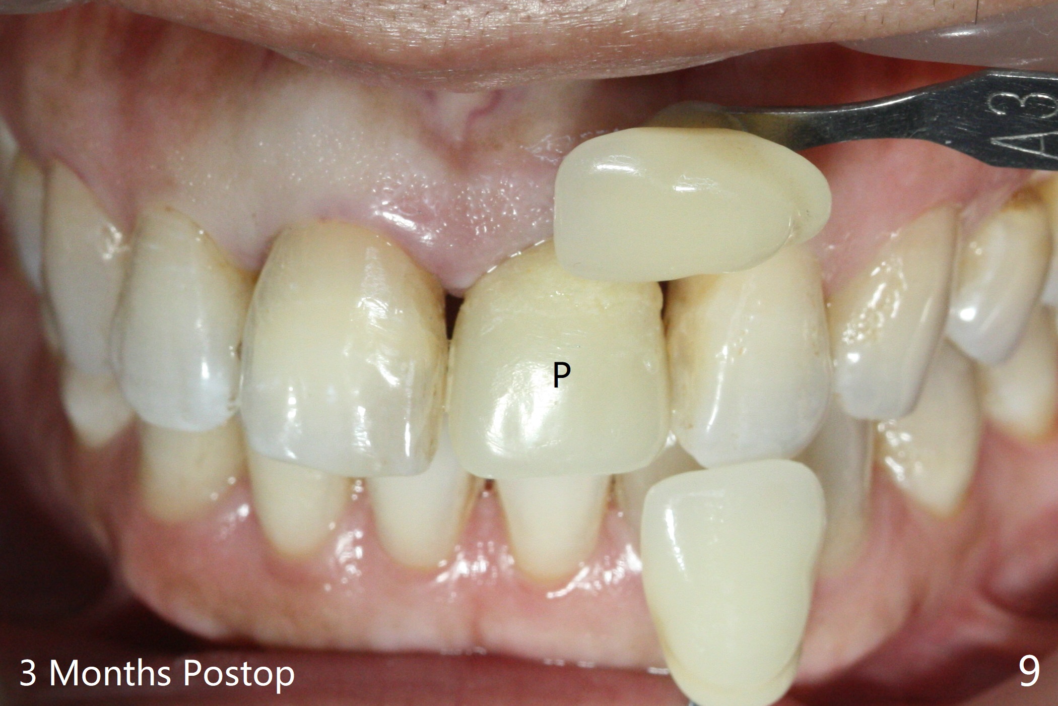
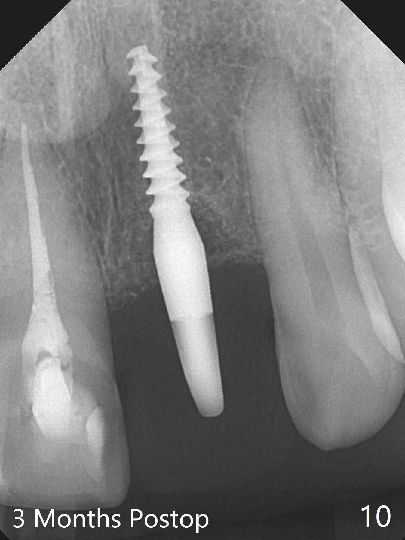
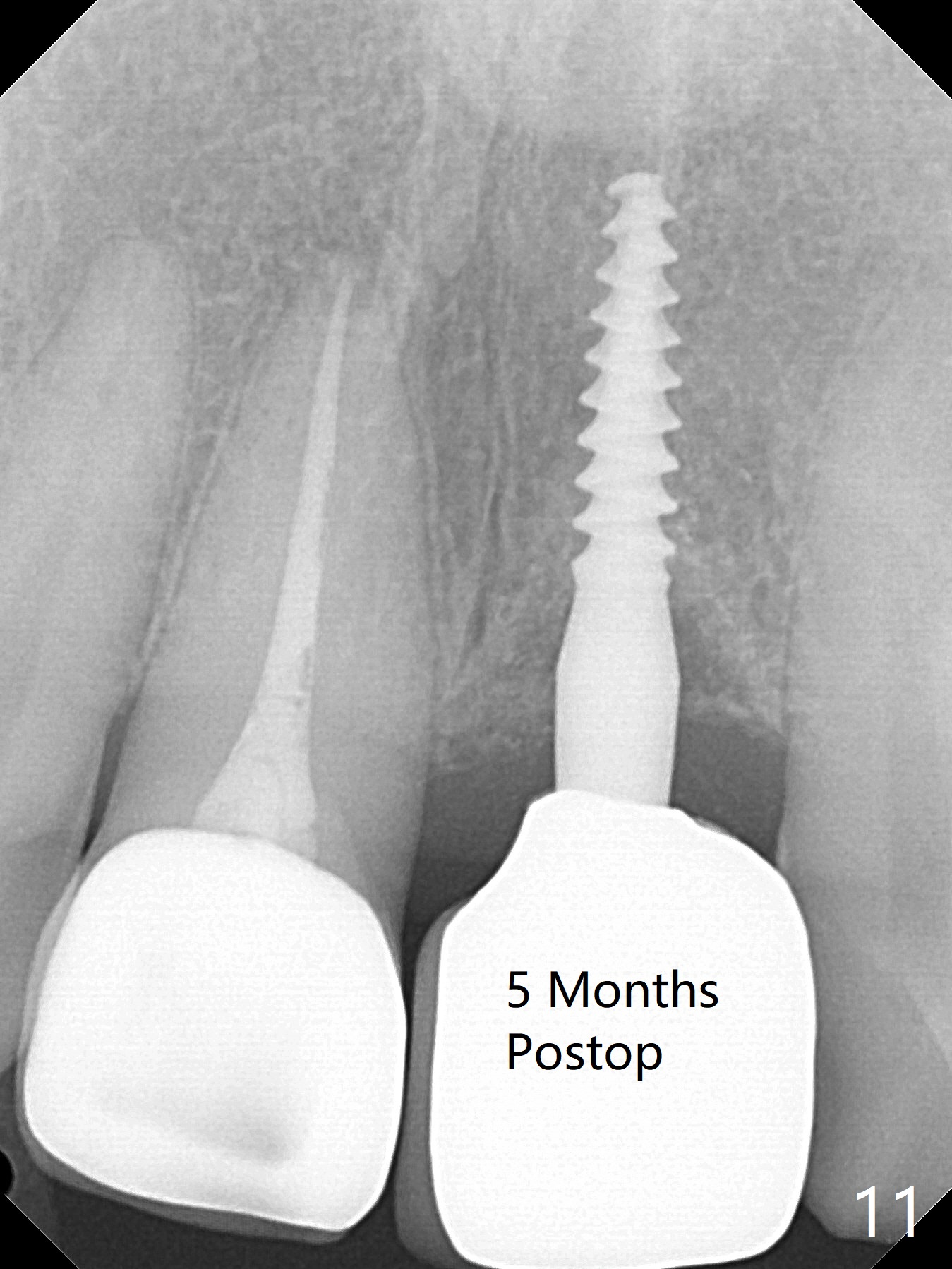
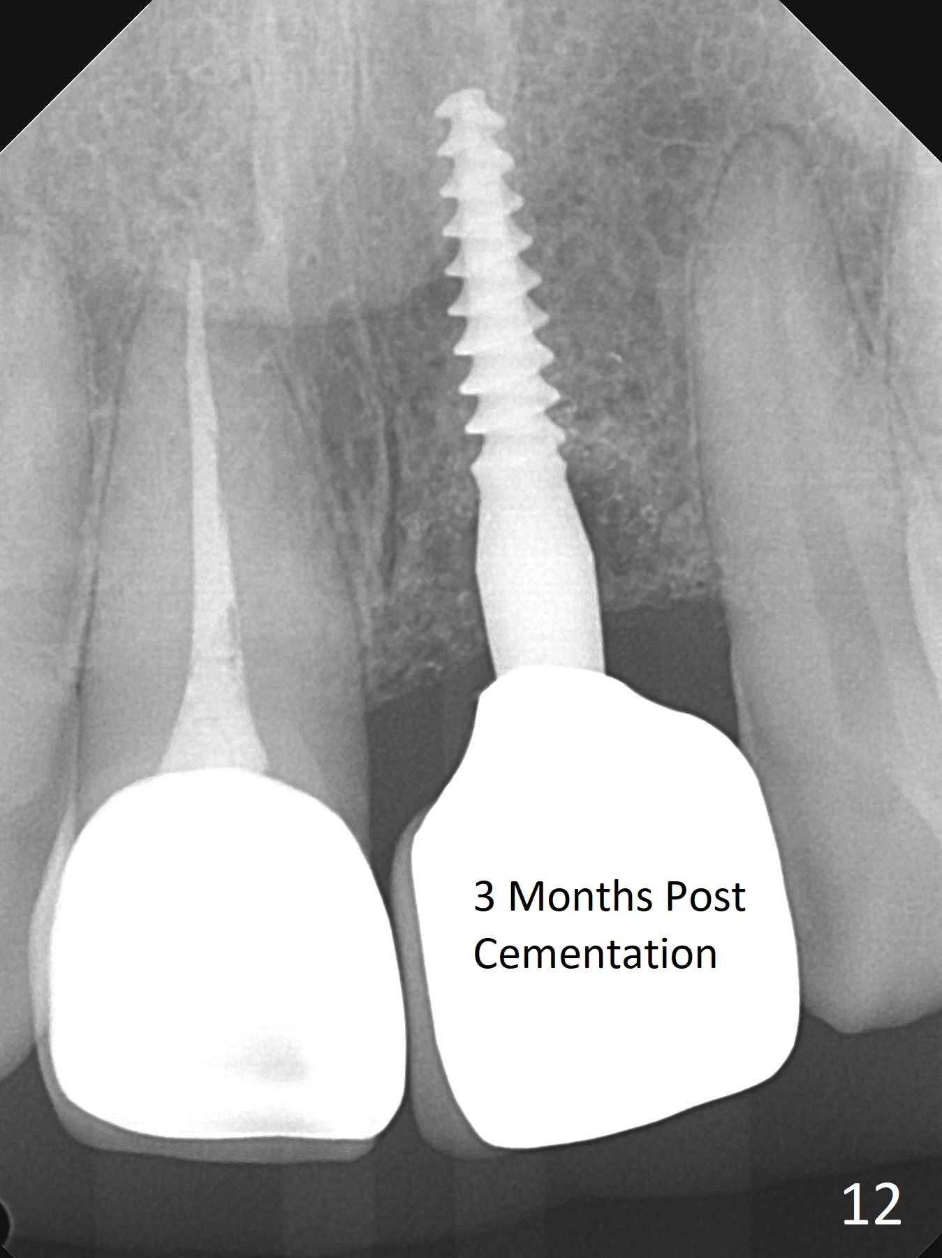
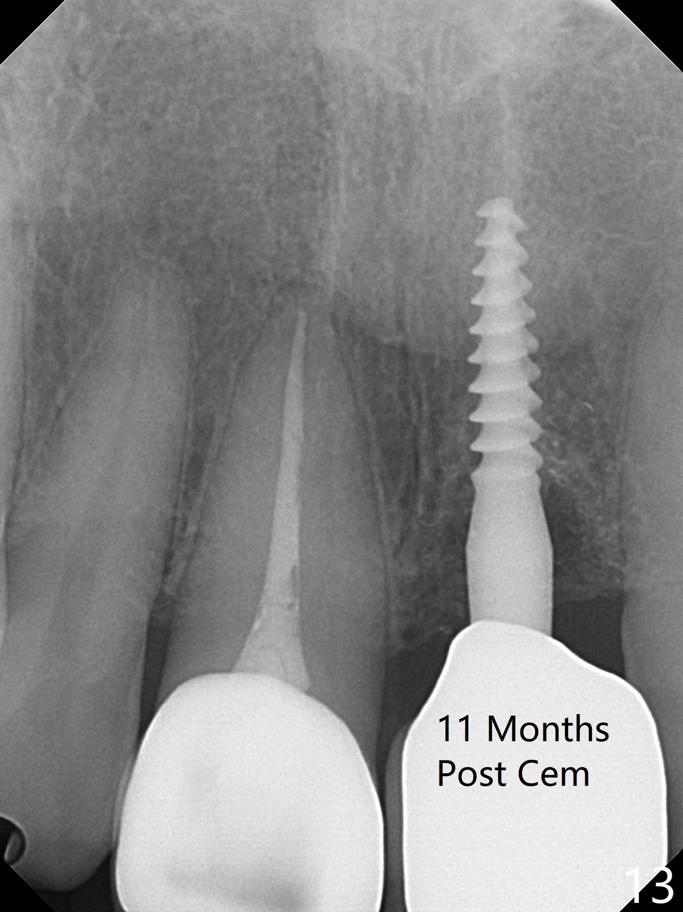
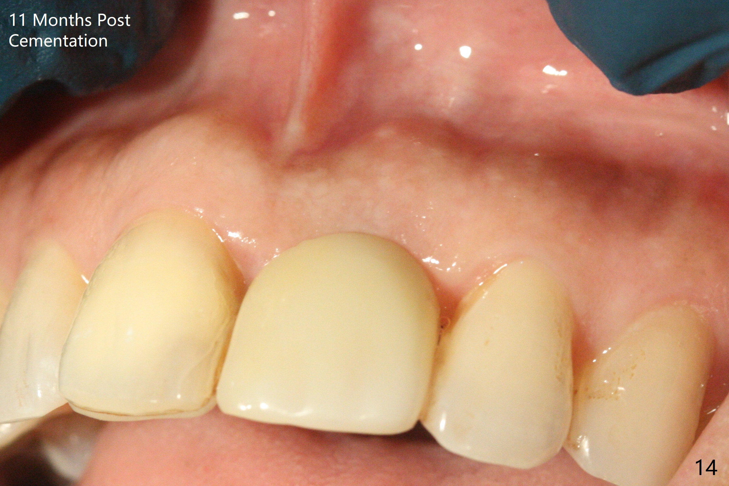
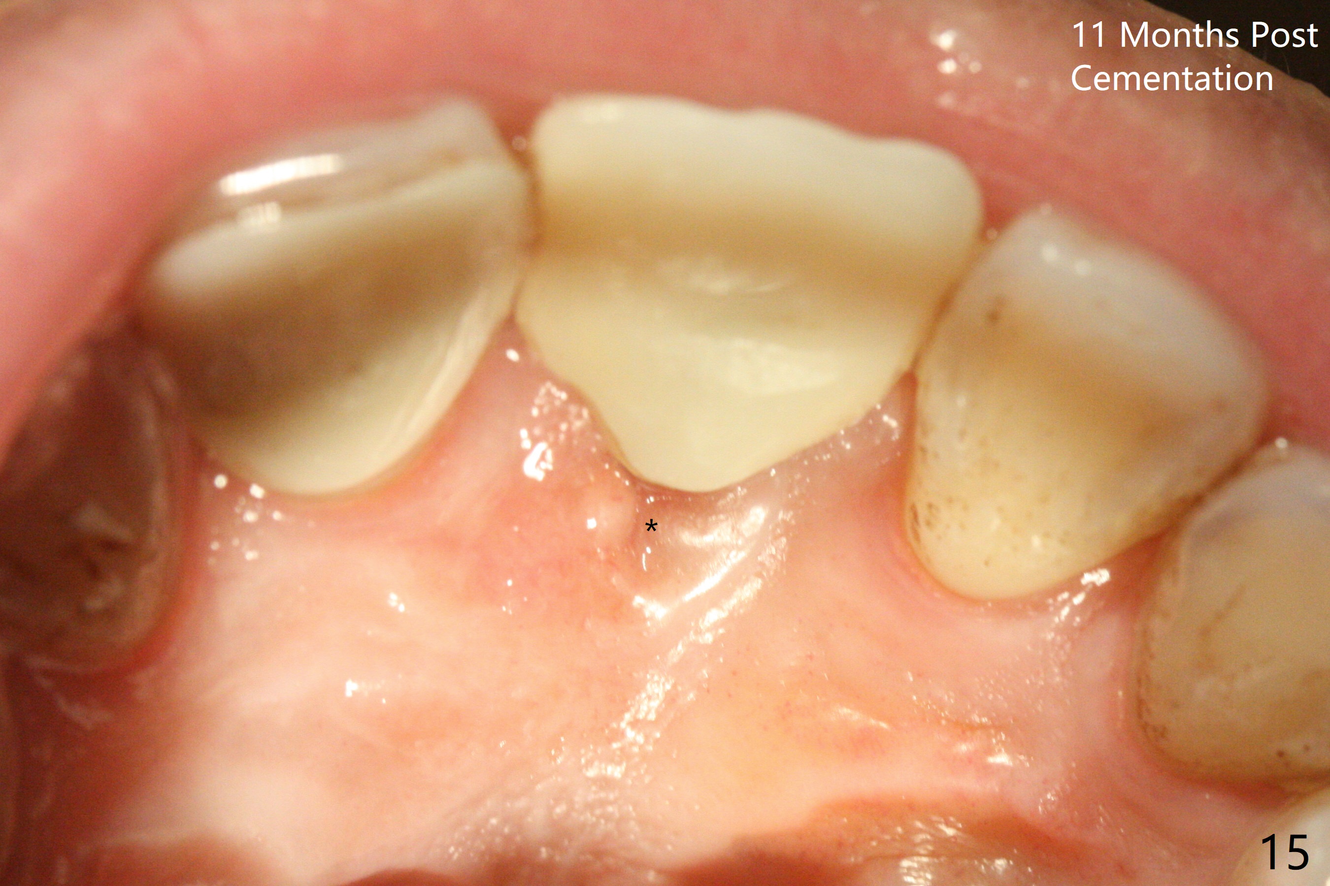
Except the depth, a 2.5x10 mm mini implant is placed with precision (in trajectory) at #9 (Fig.1-5). Confidence using surgical guide for the narrow ridge is enhanced due to placement of two digits against the buccal and palatal plates for tactile sensation. The depth issue is related to overprep with 2.2 mm drills. The torque is <15 Ncm. The immediate provisional is bonded to the neighboring teeth for retention. It appears that smaller drills should be made for guided surgery (such as 1.5 and 2.0 mm). The immediate provisional looks acceptable buccal and occlusal 3 weeks postop (Fig.6,7), although the palatal gingiva is erythematous (Fig.8, which is common after use of drill for access (tissue laceration), OHI offered). Three months postop (Fig.9), the palatal gingiva looks healthy (data not shown), while there is no bone loss around the implant (Fig.10). It remains the same 5 months postop (immediately post cementation, Fig.11) and 3,11 months post cementation (Fig.12,13). The labial gingiva is healthy (Fig.14), while the palatal one is less erythematous and edematous (Fig.15) than earlier (Fig.8).
Return to
Upper
Incisor Immediate Implant,
Trajectory
Guided Surgery
Atrophic Ridge
CT/前牙植牙
1-Piece
Xin Wei, DDS, PhD, MS 1st edition 10/16/2018, last revision 12/27/2020