.jpg)
|
|
|
|
|||
|
|
|
|
|
|
|
|
|
|||||
Socket
Shield Due to Root Fracture M
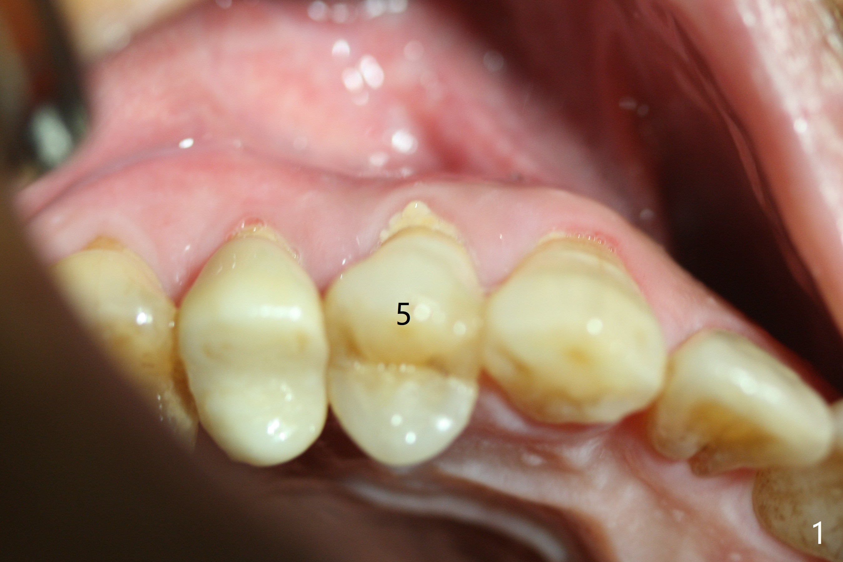
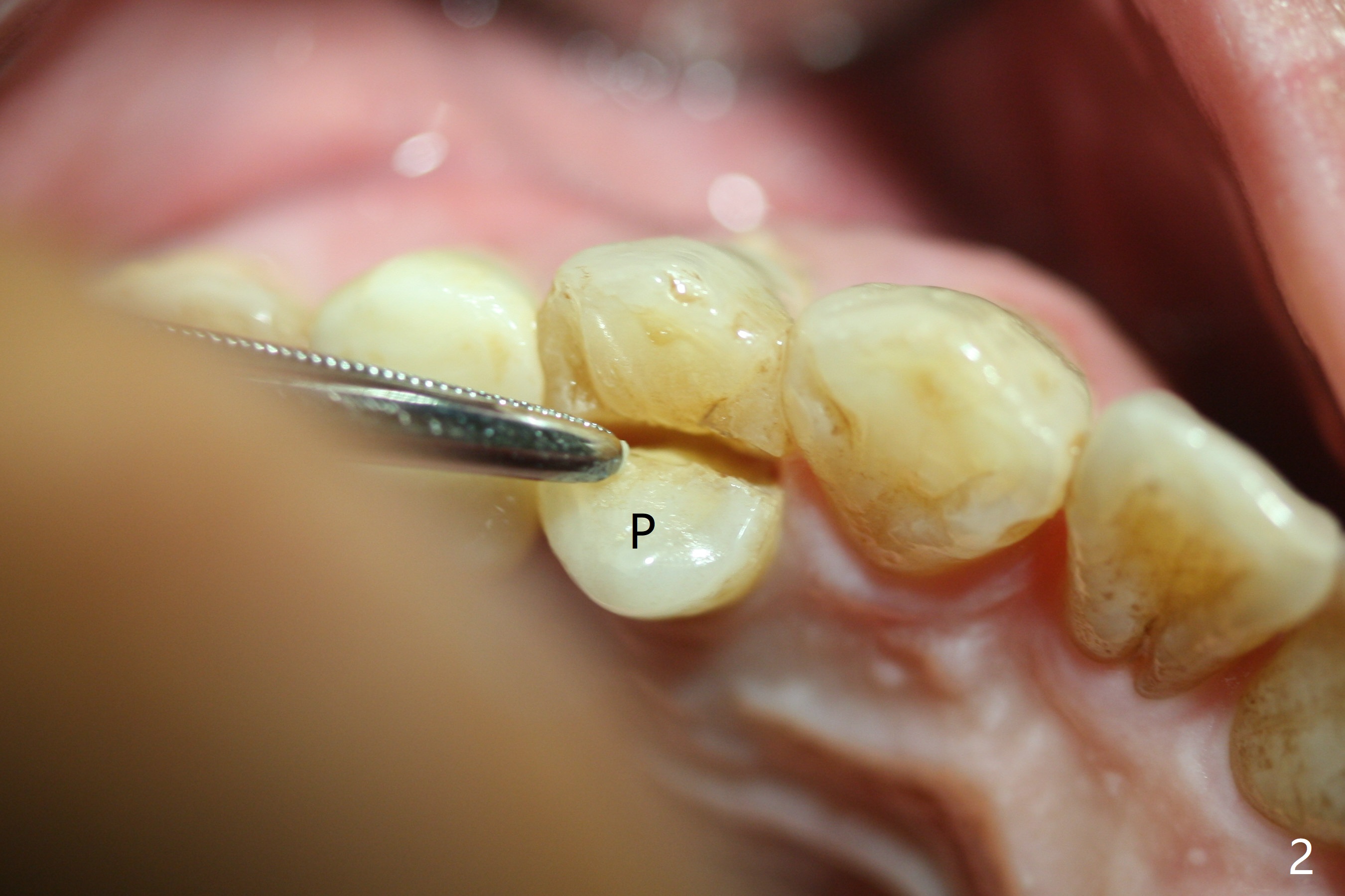
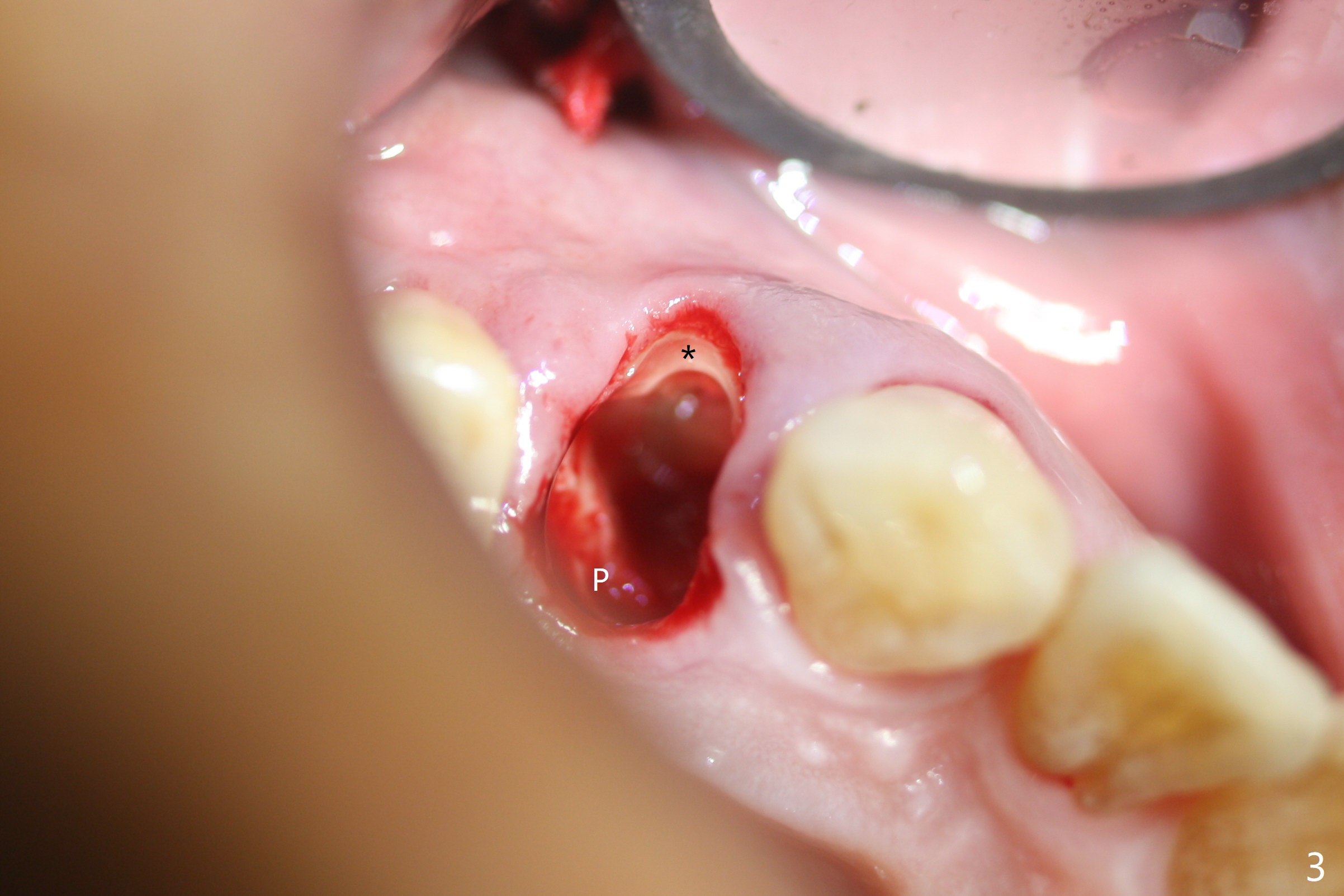
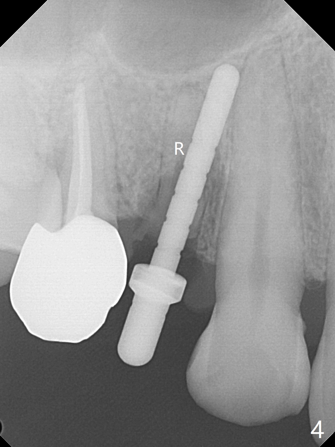
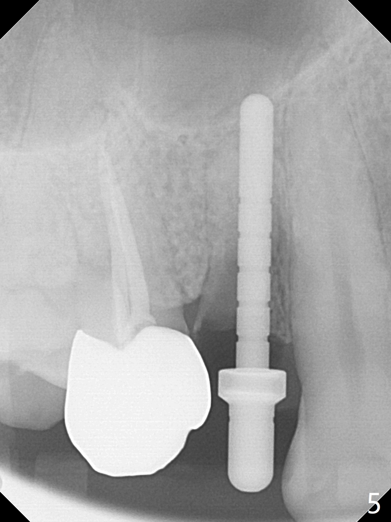
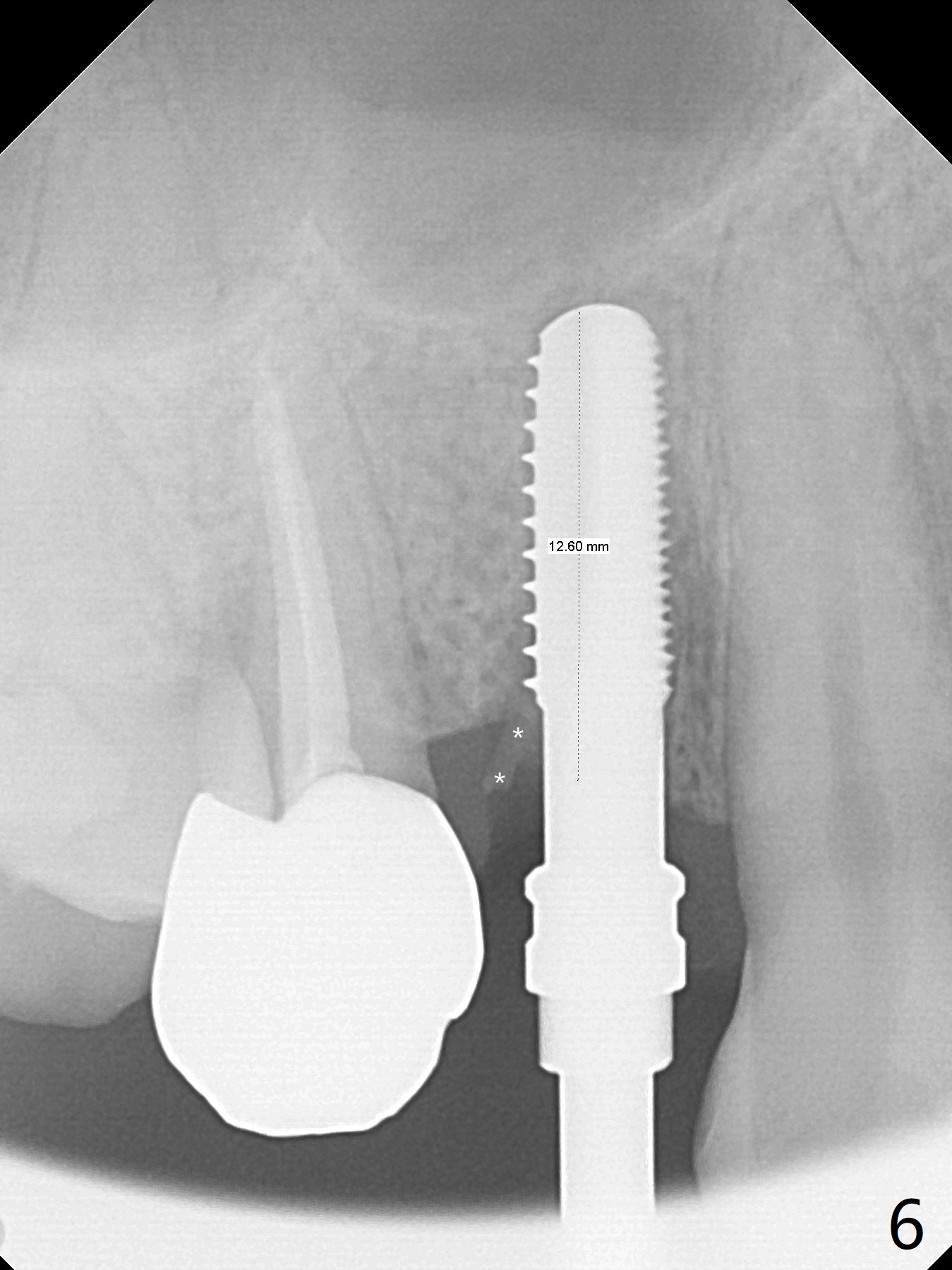
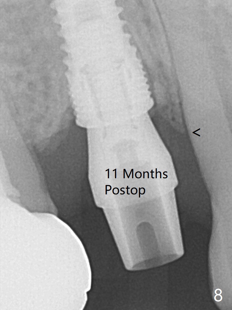
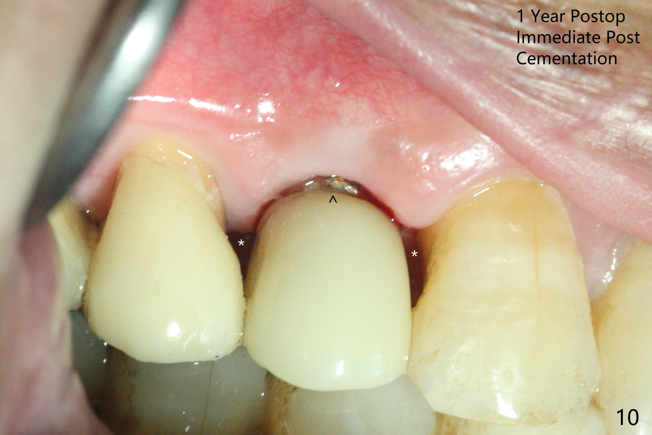
The buccal plate is normal at the tooth #5 (Fig.1) with the loose palatal fragment (Fig.2 P). Extraction leads to root fracture. Sectioning removes the palatal portion of the root and keeps the buccal semilunar piece (Fig.3 *); the mesiopalatal plate is resorbed (P). Initial osteotomy is off (Fig.4 (R: remaining root)). Redirection improves the trajectory (Fig.5). With the 2nd redirection (Fig.6 (4.5 mm tap)), a 4.5x12 mm implant is placed with 50 Ncm and sinus lift (Fig.7 black *); bone graft is placed with emphasis on the palatal defect (white *). As usual, an immediate provisional is fabricated. In fact the abutment may be not completely seated because of contact with the mesial crest. Prepare anesthetic and 5.5 mm profile drill. Take parallel BW or PA. Take occlusal photos to show no buccal or mesiopalatal atrophy. After 5.5 mm profile drill 11 months postop, the abutment has no contact with the mesial crest (Fig.8 <). Since the proximal contact between #3 and 4 is light with food impaction, the provisional at #5 is fabricated with tight distal contact. When the patient returns for final crown cementation (Fig.9), the food impaction is minimal between #3 and 4. The distal black triangle (Fig.10 *) and exposure of the abutment margin (^) are partially related to provisional fabrication and should dissolve over time considering socket shield.
Return to
Upper Premolar Immediate Implant,
Trajectory,
Socket Shield
Xin Wei, DDS, PhD, MS 1st edition
02/19/2019, last revision
03/08/2020