|
|
|
|
|
|
|
|
|
|
|
|
|
|
|
|
|
|
|
|
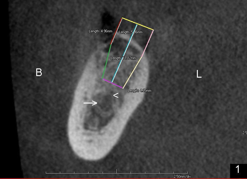
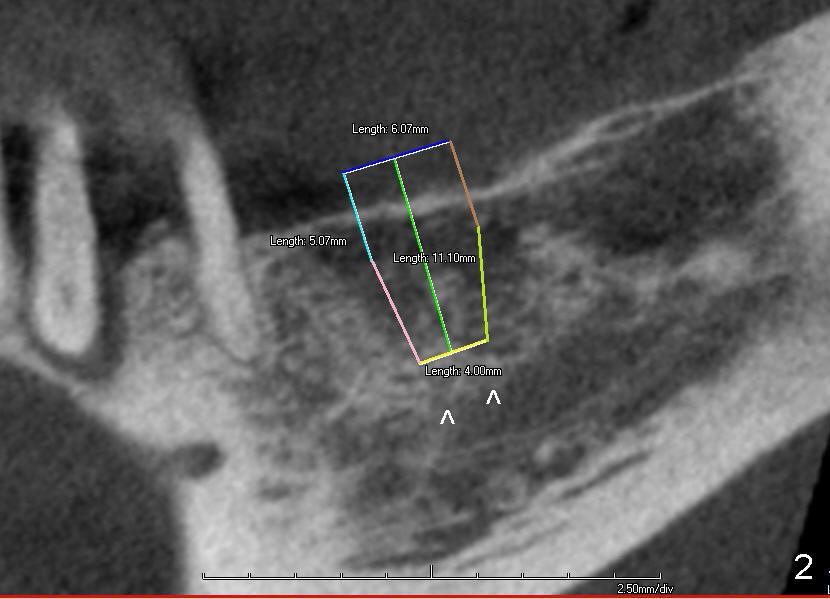
Implant placement at the Site of #30 as Future Anchorage
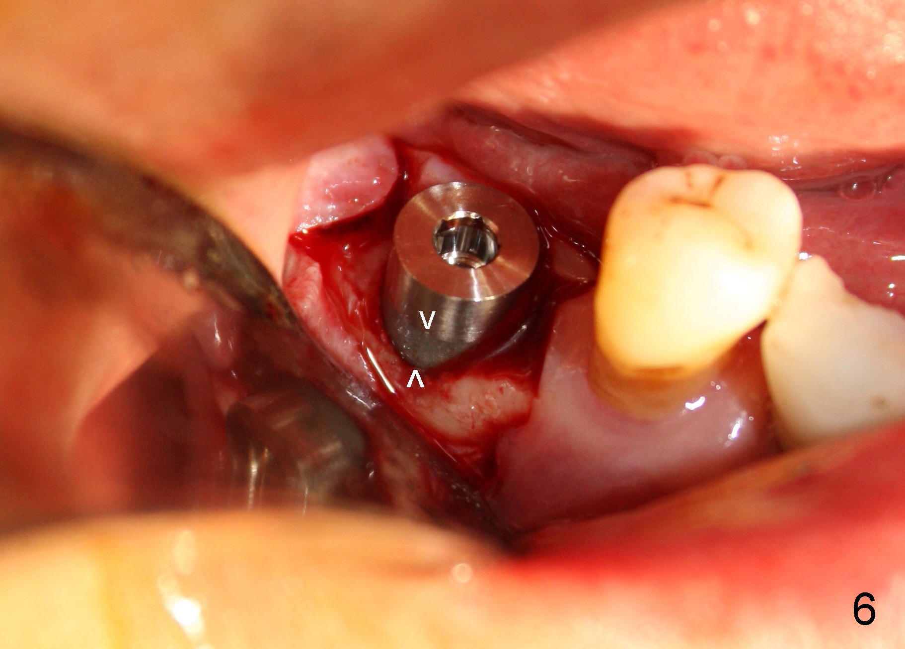
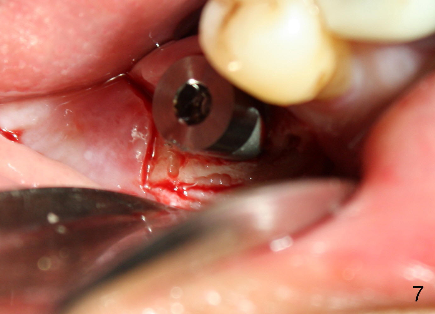
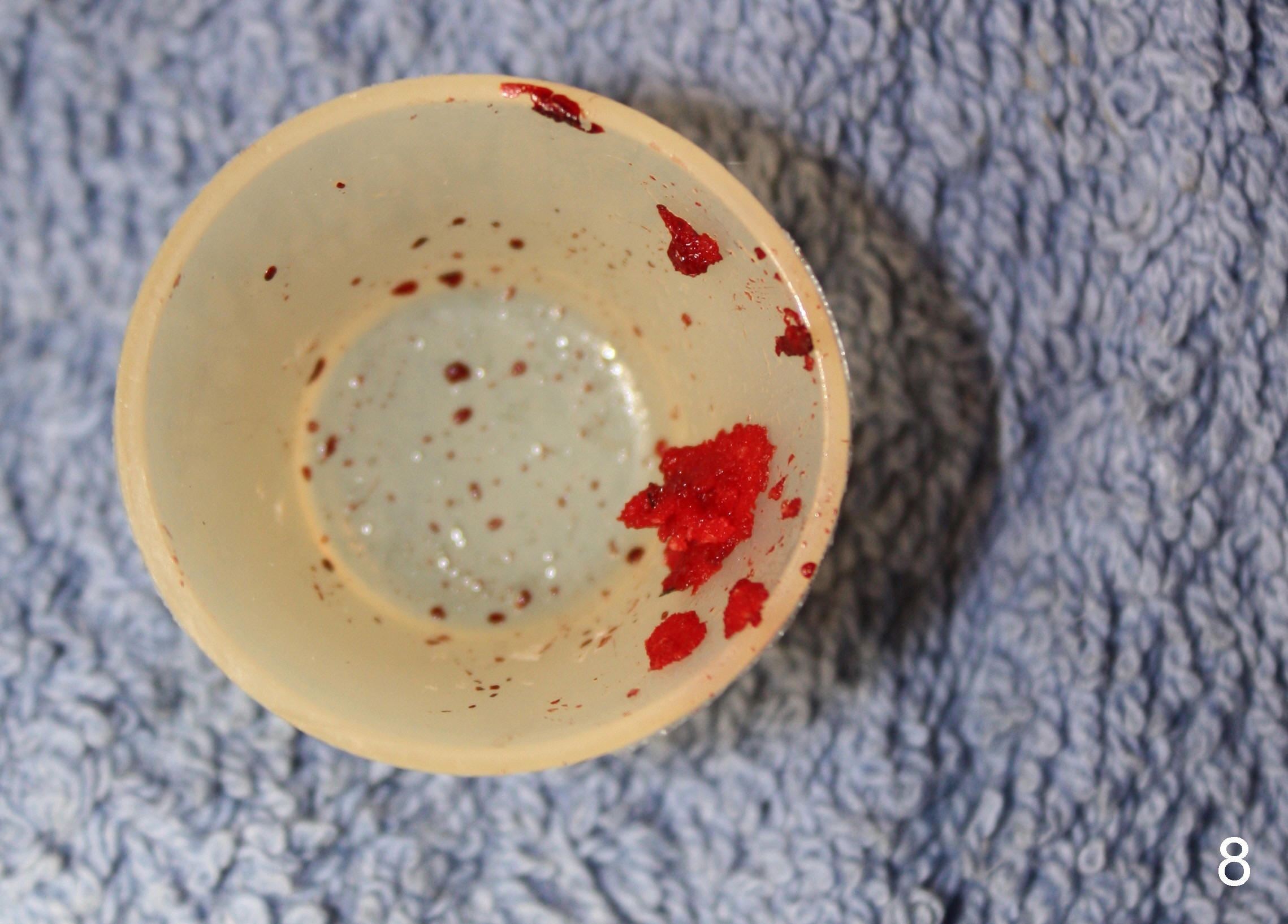
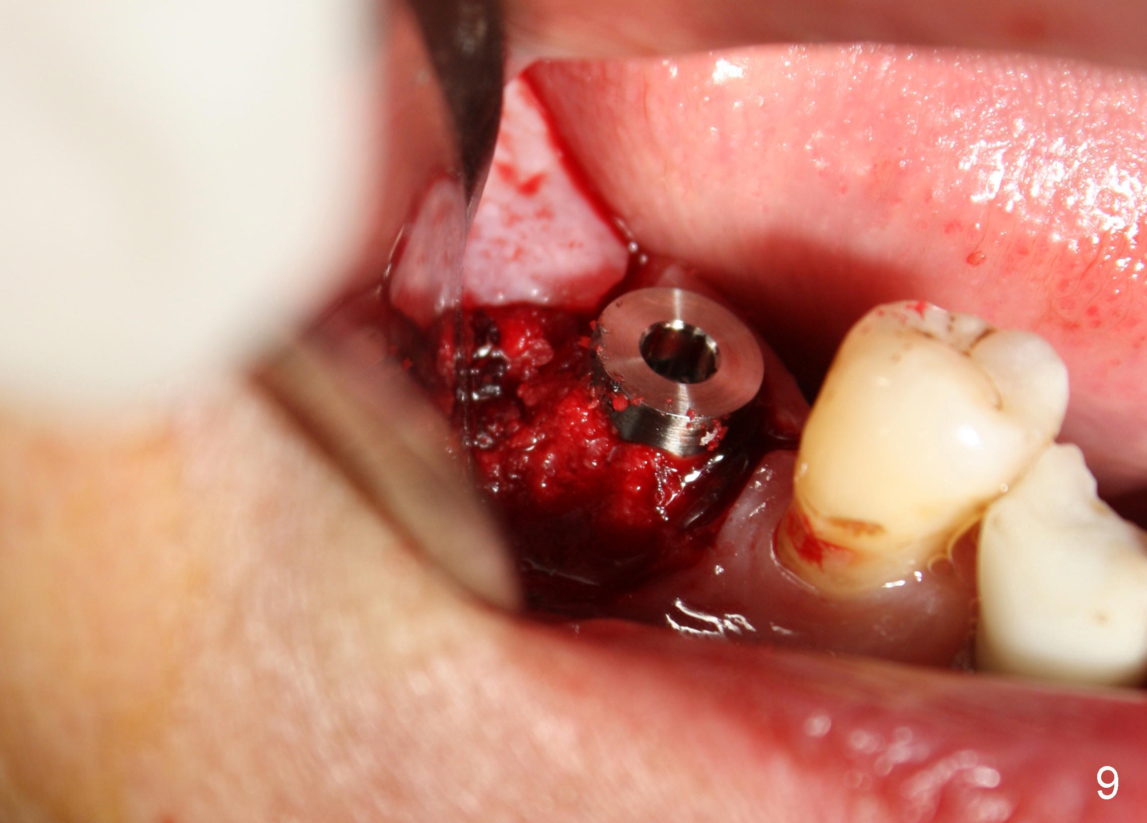
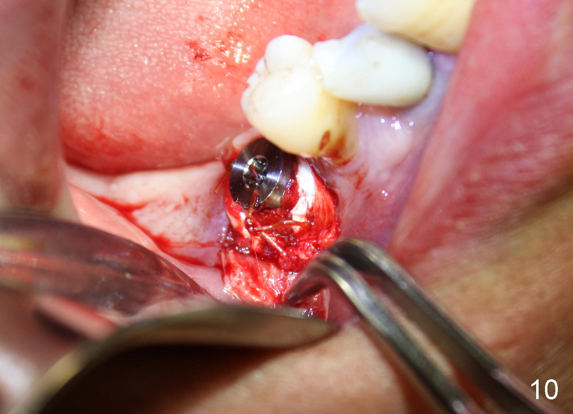
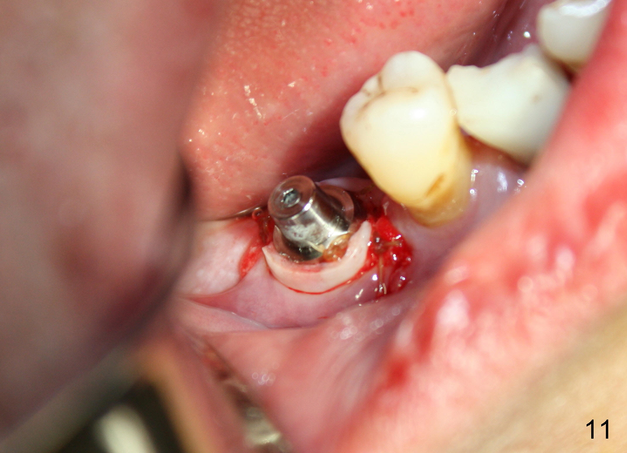
CBCT studies show that a 6x11 mm implant can be safely placed at the site of the tooth #30 (Fig.1 (coronal), 2 (sagittal section). Osteotomy is initiated by 2 mm pilot drill at the depth of 8 mm from the crest (the prospective implant is 3 mm above the crest); X-ray is taken with a parallel pin (Fig.3 P). It appears that there is 12 mm of bone from the crest to the upper border of the inferior alveolar nerve canal. The depth of osteotomy is accordingly adjusted to 11 mm below the crest; osteotomy finishes with insertion of 6x14 mm tap (Fig.4); the patient feels pressure while the tap is being inserted. Following further infiltration with Lidocaine, the depth of the osteotomy is intended to increase in order to bury the implant deeper, because the coronal portion of the buccal plate starts to perforate. The patient feels pain. Finally a 6x14 mm implant is placed ~ 1 mm above the inferior alveolar canal (Fig.5). As mentioned earlier, the rough surface of the implant is exposed buccally (Fig.6 between arrowheads). The nearby buccal plate is decorticated (Fig.7). The autogenous bone harvested during osteotomy (Fig.8) is going to be placed over the exposed portion of the rough surface of the implant (Fig.9); the graft is covered by collagen dressing (Fig.10). The buccal and lingual flaps are approximated with sutures mesial and distal to the implant (Fig.11). To increase the retention of perio dressing, a 4x3 mm abutment is placed.
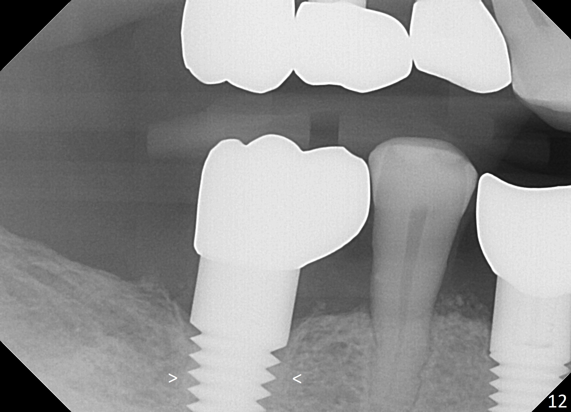
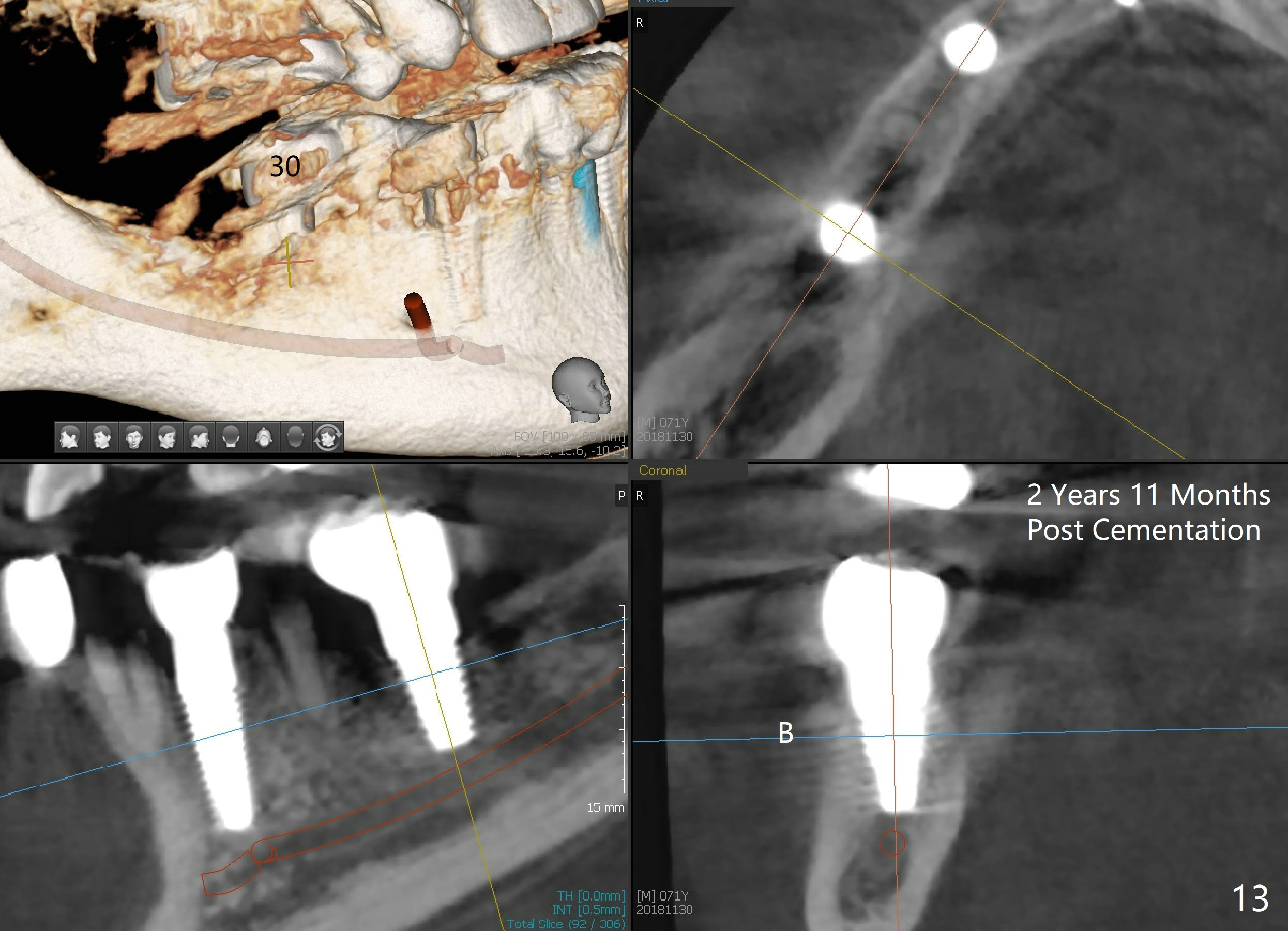
Placement of implant at the site of #30 is a part of full mouth reconstruction aided by orthodontic treatment (1). As Fig.3 demonstrates, the tooth #29 is supraerupted (arrow). Once the implant at the site of #30 is osteointegrated, it is used as one of anchorages (the other is #28 implant crown) to intrude #29. In fact, the patient is uncooperative in orthodontic treatment. There is cortical bone-like formation 3 years postop, 1 year 10 months post cementation (Fig.12 <). There is no bone loss 2 year 11 months post cementation (Fig.13 CT).
Return to Implant & Ortho,
Full Mouth Reconstruction
3 20/21
24/26
28
Xin Wei, DDS, PhD, MS 1st edition 04/01/2014, last revision 12/16/2018