
|
|
|||
|
|
|
|
|
|
|
|
|
|
|
|
|
|
|
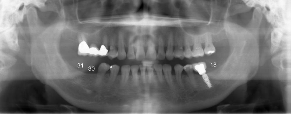
How to Choose Healing Abutments?
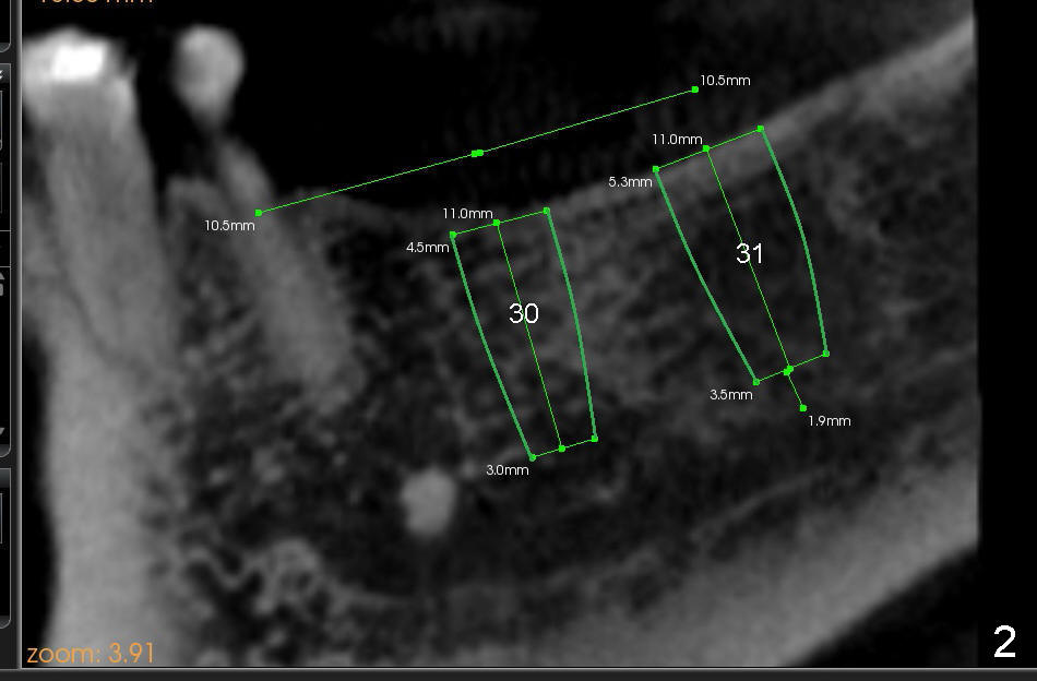
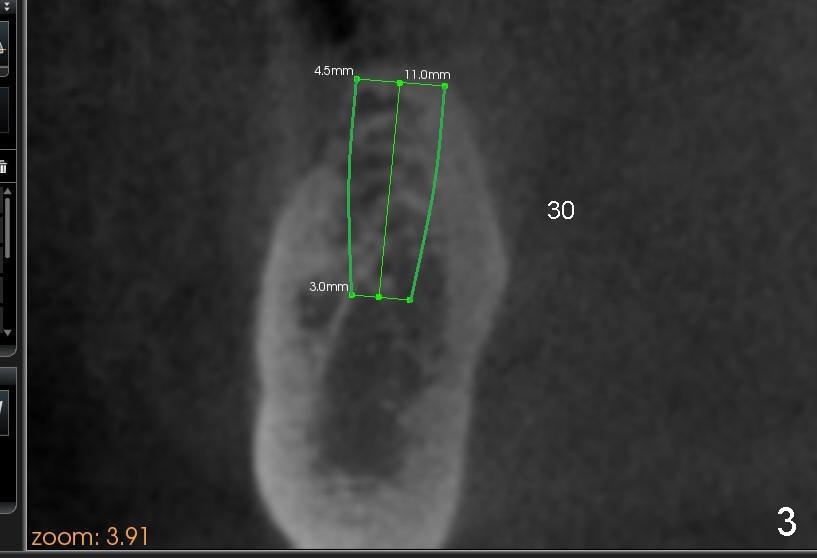
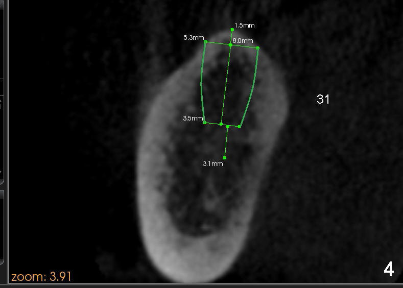
A 56-year-old lady requests implant placement at the sites of #18,30 and 31 (Fig.1, the last two first). CBCT shows preliminary design for implants at #30 and 31 (Fig.2 (sagittal section), 3,4 (coronal)).
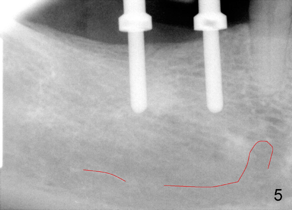
PA is taken after insertion of parallel pins at the depth of 10 and 8 mm at the sites of #30 and 31, respectively (Fig. 5, red line outlines the upper border of the inferior alveolar canal). It appears to be safe to increase the lengths of the implants by 2 mm.
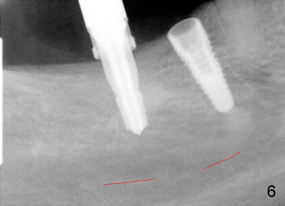
PA is retaken after placement of a submerged (SM) implant (4.5x12 mm) at #30 and insertion of 4.5x10 mm drill at #31(Fig.6).
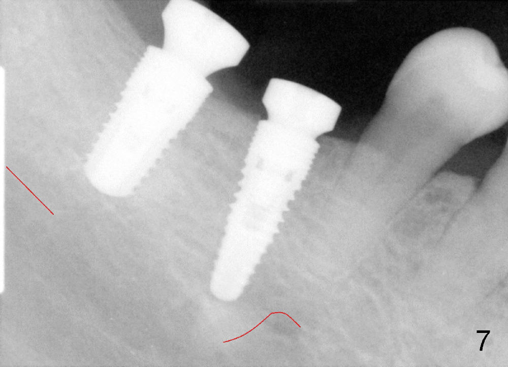
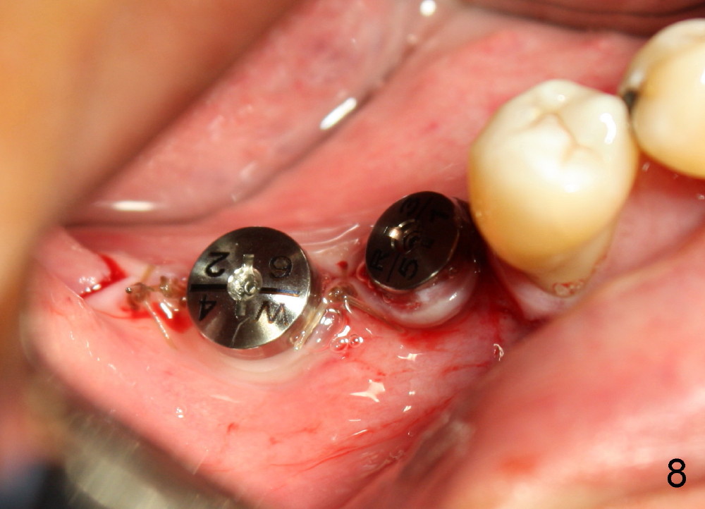
Finally 5.3x10 mm SM implant is placed at #31 (Fig.7). Healing abutments (Ɵ5.2x3(1) and 6.2x4(2) mm) are placed at #30 and 31, respectively (Fig.7-9).
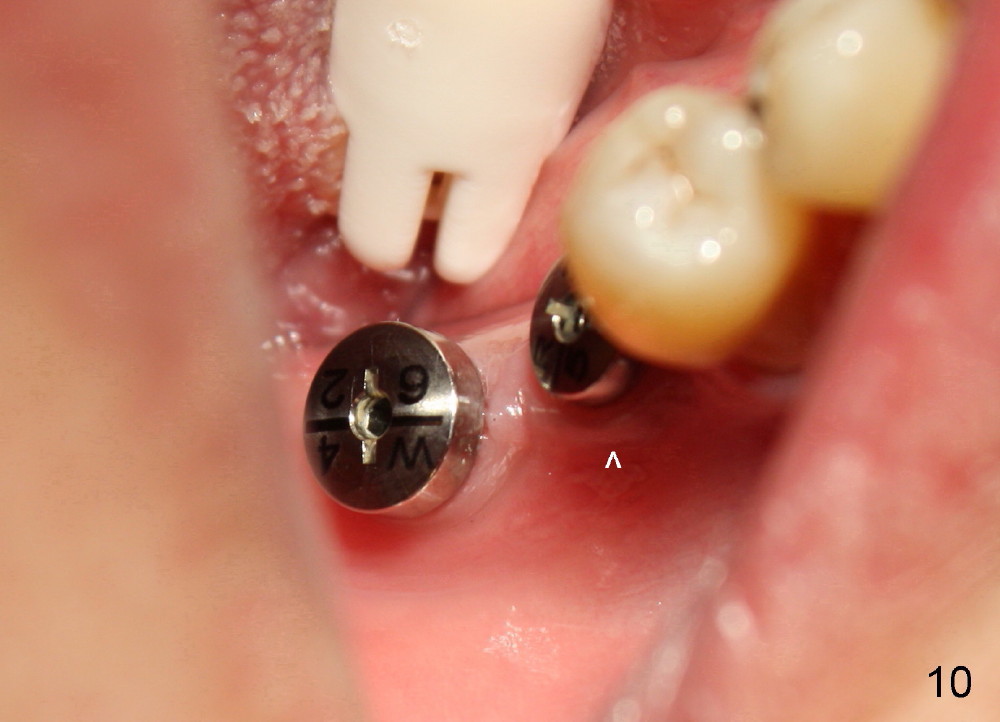
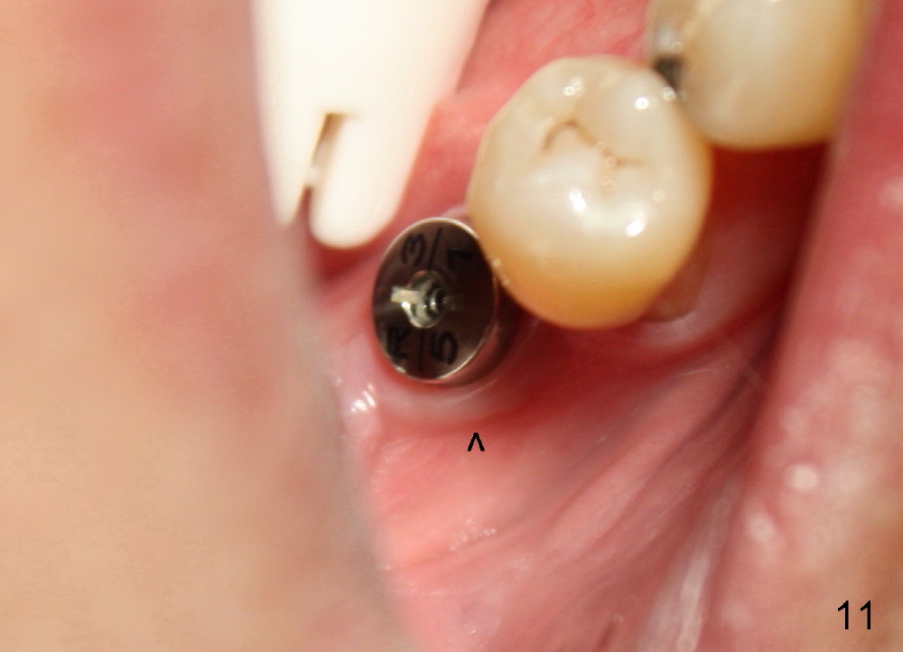
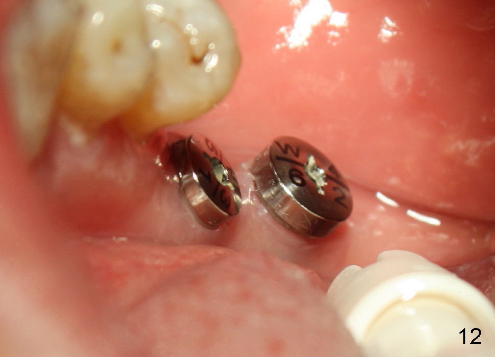
One week postop, the gingiva buccal to #30 healing abutment was reddish (at the area indicated by arrowheads in Fig.10,11). The patient complained facial swelling. Clindamycin was taken one extra week to help resolve postop infection. By 24 days postop, there is no infection around these two healing abutments (Fig.10-12). The patient is pleased and makes an appointment for #18 implant placement (Fig.1), which is more challenging.
Xin Wei, DDS, PhD, MS 1st edition 12/18/2013, last revision 01/26/2014