|
|
|
|
|
|
|
|
|
|
Small, Short Implant
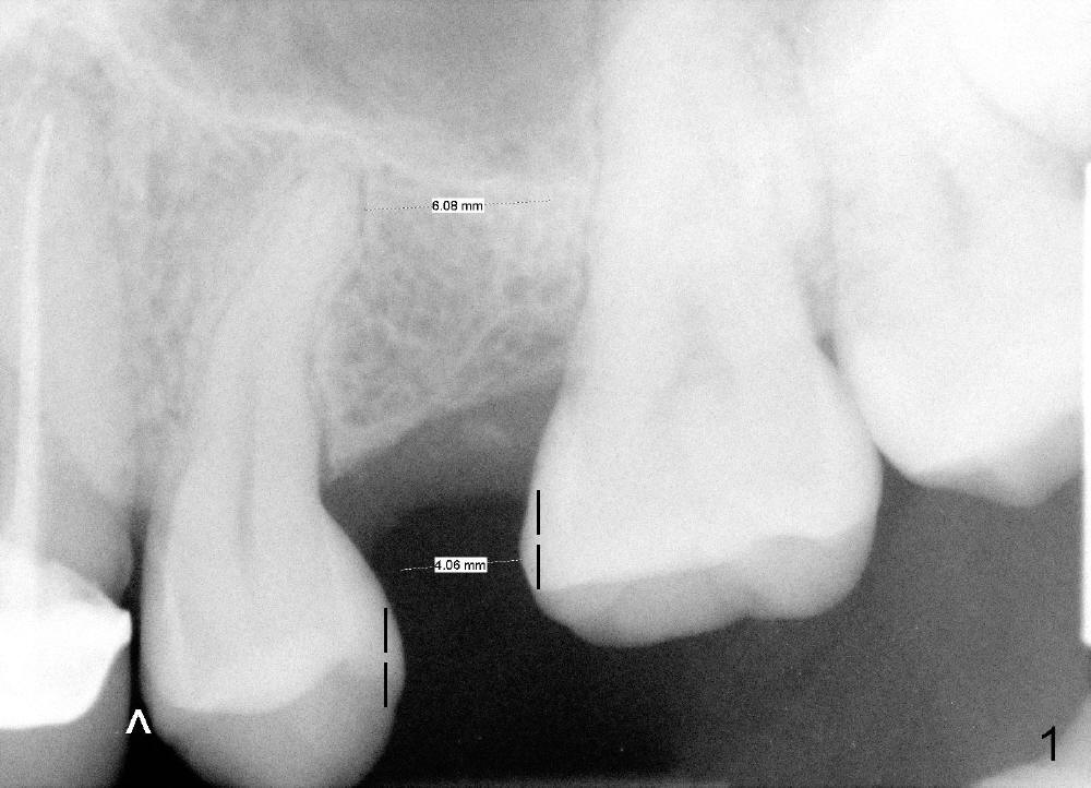
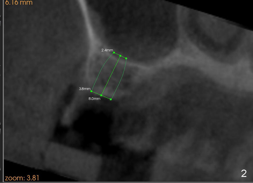
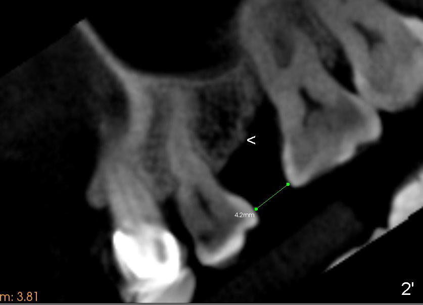
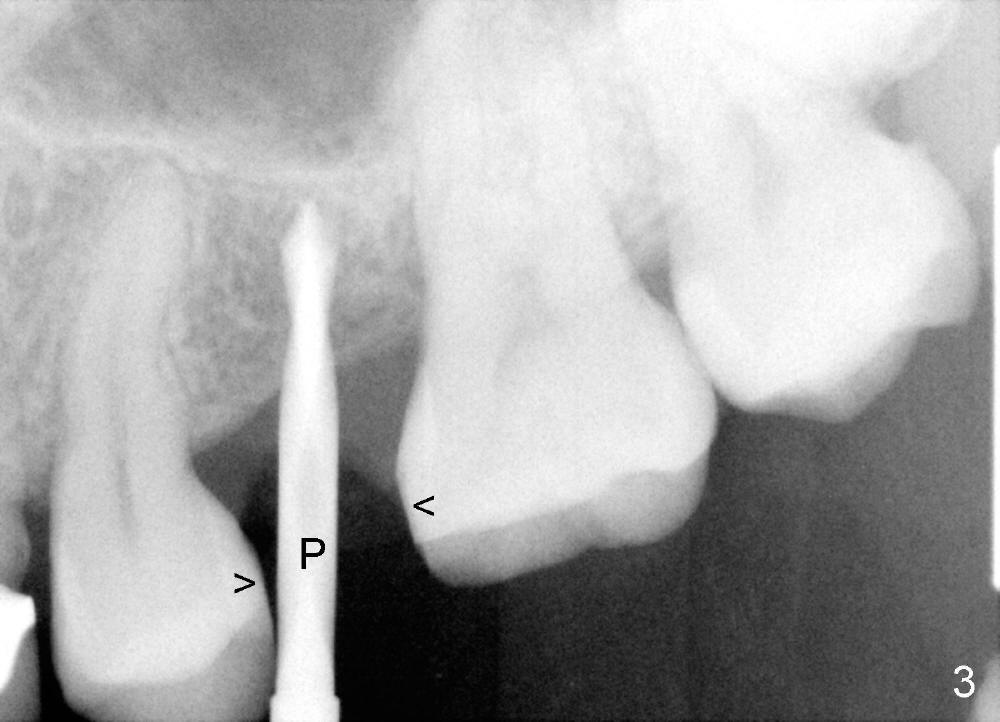
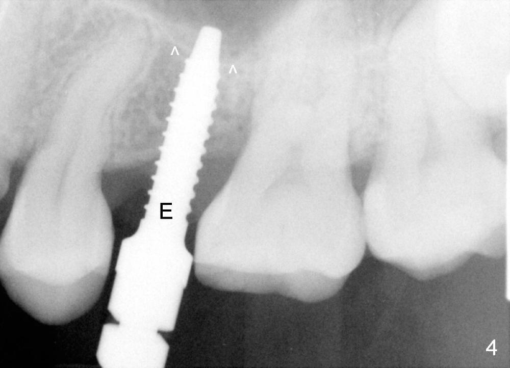
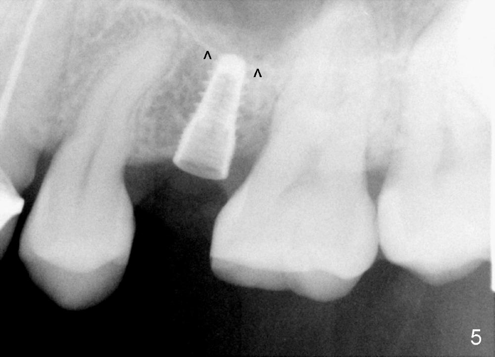
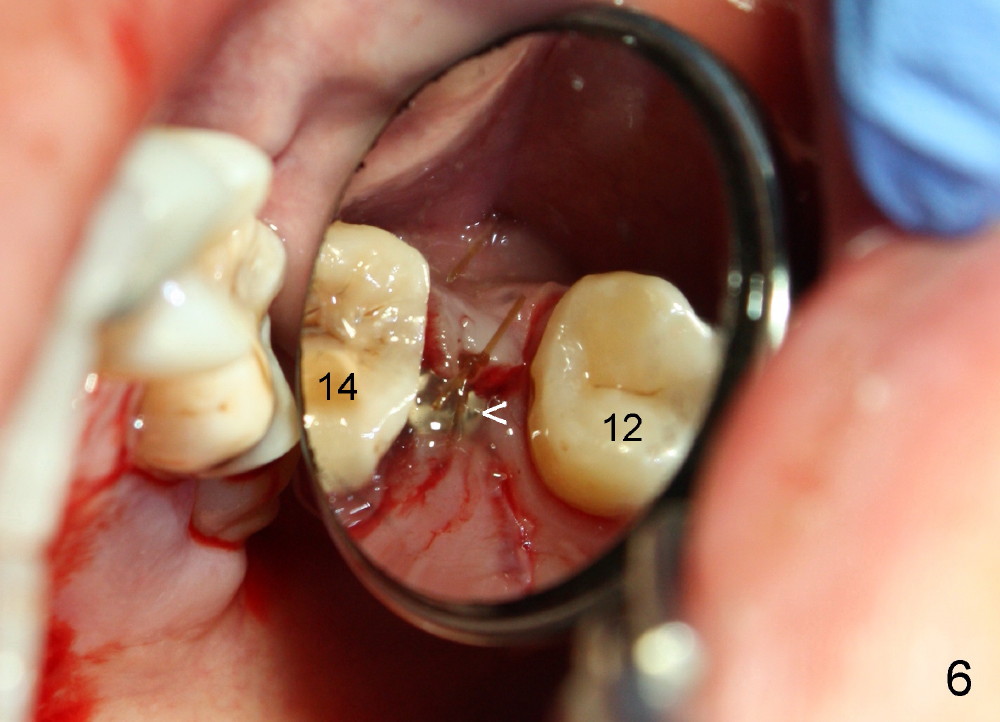
A 39-year-old lady is a dental phobic and finally agrees to restore long-termed missing upper left 2nd premolar (Fig.1). The adjoining teeth tilt toward the edentulous space. To place a 3.8x8 mm submerged implant (Fig.2, CBCT coronal section), enameloplasty is done (compare Fig.1 dashed lines with Fig.3 <). Since the density of the cancellous bone (<100 Hounsfield units (HU)) is much lower than that of the crest (300-400 HU) (Fig.2' <), osteotomy is initiated by 2 mm pilot drill (Fig.3 P) and finished by osteotomes (2 and 3 mm), bone expanders (2.6 mm (Fig.4) and 3.2 mm) and 3.8 mm tap drill. The implant is placed as planned with insertion torque around 35 Ncm (Fig.5). The implant is further lifted into the sinus by 1 mm without bone graft following the last X-ray. A healing abutment (4.1x3) is placed (Fig.6 <).
Limited orthodontic treatment is planned to close the diastema between the canine and the first premolar (Fig.1 ^) and to upright the molars, probably using osteointegrated implant as an anchorage.
In fact the implant exfoliated 18 days postop. It is probably due to over-preparation. The second placement should try to solve the problem of the over-preparation (2).
Return to Professionals, Implant Failure, Upper Bicuspid Immediate Implant
Xin Wei, DDS, PhD, MS 1st edition 03/13/2014, last revision 01/19/2018