|
|
||||
|
|
|
|
|
|
|
|
|
|
||
|
|
|
|
|
|
|
|
|
|||
How to Place D Implant in Upper First Bicuspid?
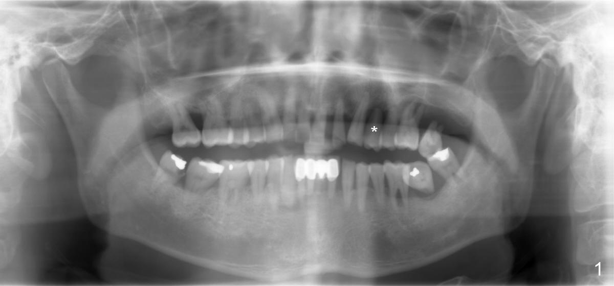
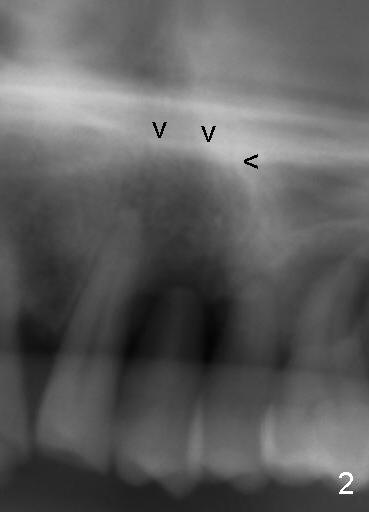
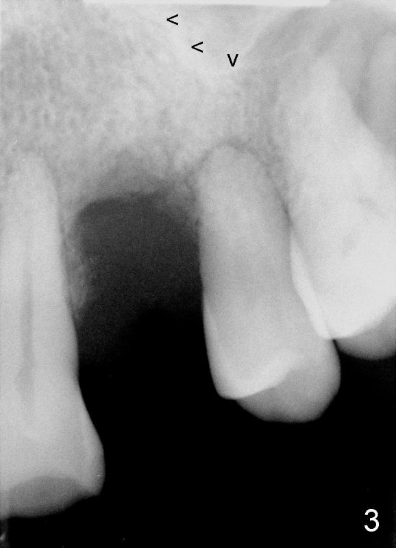
Dear Dr. Borgner: This Friday I may try D implant in a 50-year-old perio patient in #12 (Fig.1 *, Fig.2 is magnification of Fig.1 with sinus floor pointed by arrowheads), which was extracted ~ 1 month ago. PAs were taken yesterday (Fig.3). The socket must be partially empty. Can we start with RT2 or D1 spreader before using D2 one? #15 appears to be unnecessary. Tuesday, June 25, 2013 6:40 AM
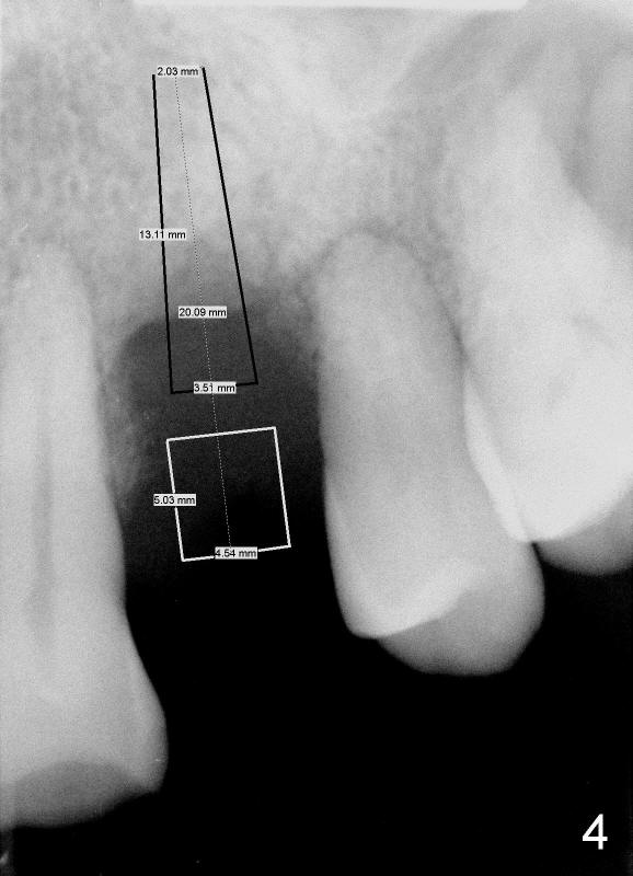
Dr Wei, This is exactly what we spoke about in the class. 1- You can go right to the D2 channel former turned 90 degrees (Fig.4: design of D2 implant turned 90 degree). 2- Engage all of the apical bone you can without entering the sinus. 3- Do not remove any bone with a bur in order to not remove any remaining interseptal bone ! 4- Place a D2 implant as directed previously. This should work perfectly if you follow these steps. Dr Borgner Thursday, June 27, 2013 2:32 PM
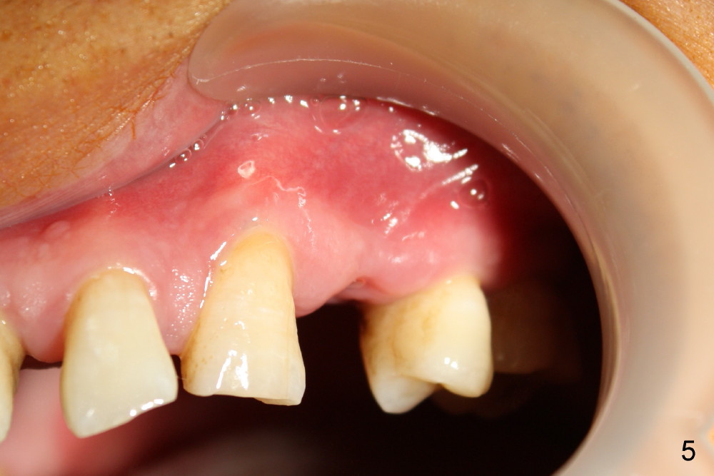
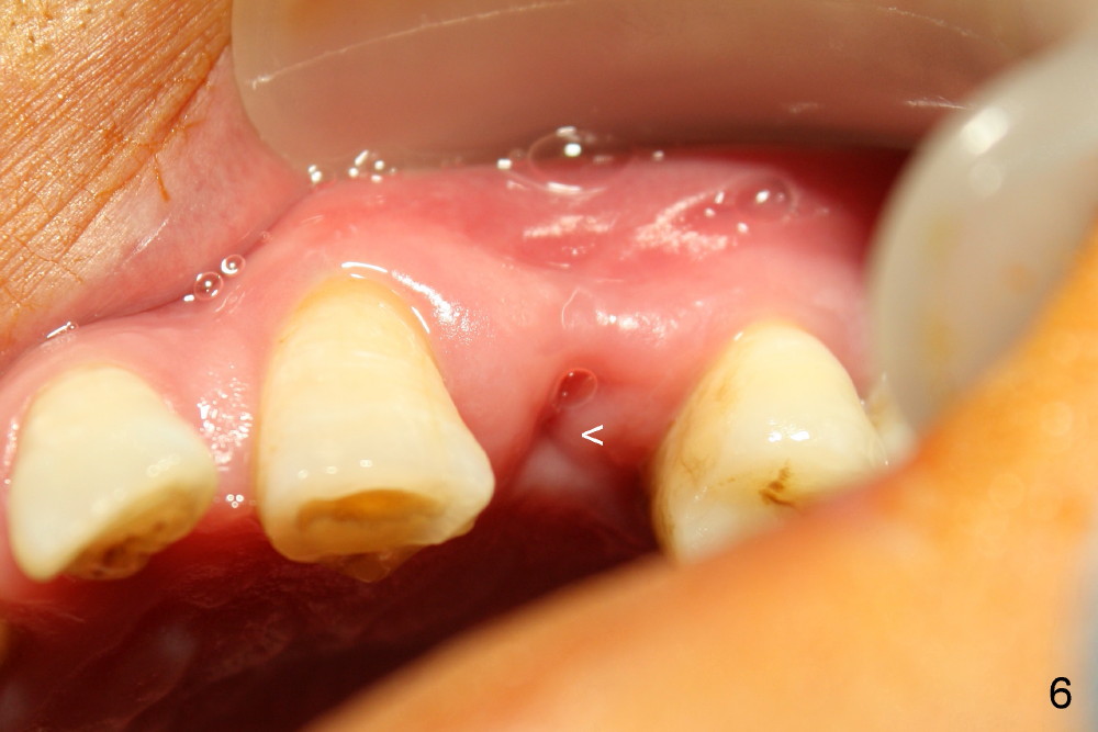
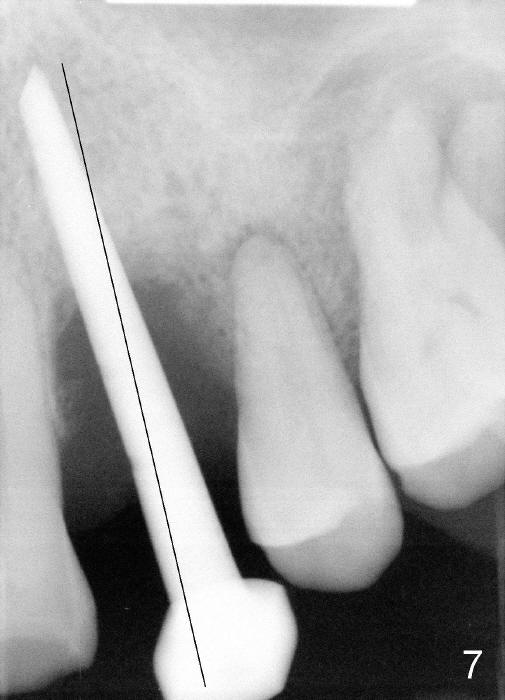
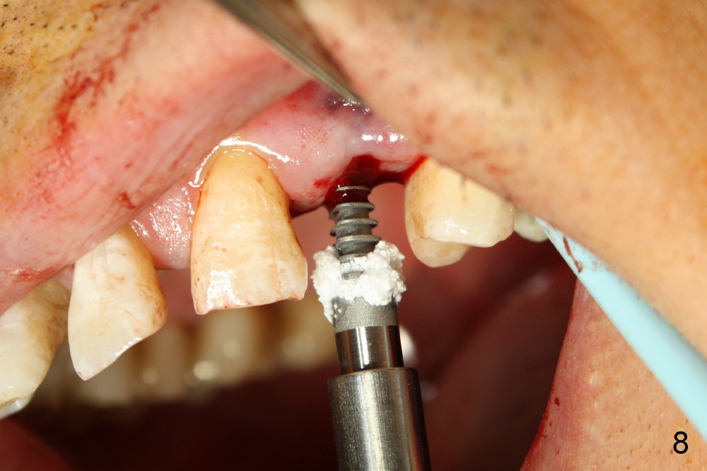
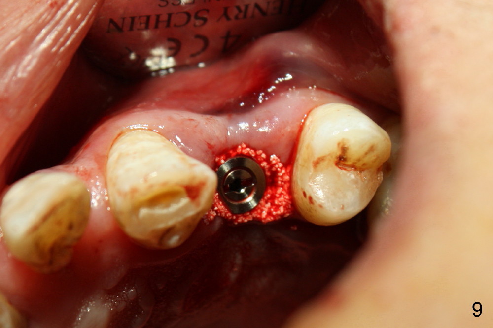
Dear Drs. Dunson and Borgner: Thank you for support. Fig.5 and 6 taken immediately prior to surgery show that the healing socket is elliptical (arrowhead). After D1 and D2 channel formers, D2 thin socket former is tapped in (Fig.7). The osteotomy needs to be redirected as shown by the black line. D2 implant is being inserted with Synthograft applied to the 1st two threads (Fig.8) and is in place (Fig.9,10). But the implant is not as stable as expected. There is a gap mesial to the implant (Fig.10 arrowhead), which is most likely created by re-directing the osteotomy with channel and socket formers.
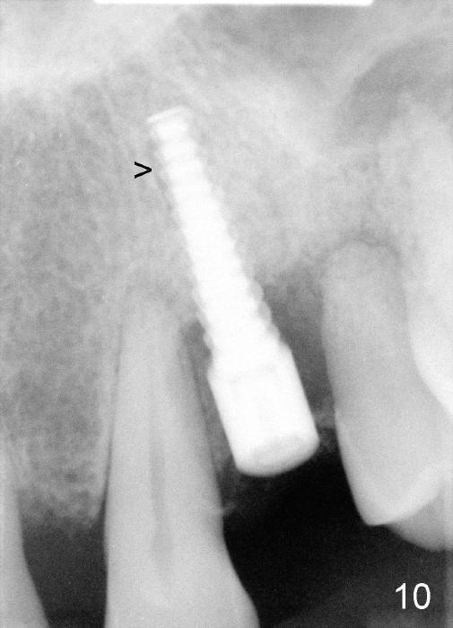
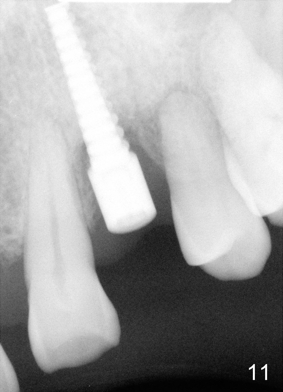
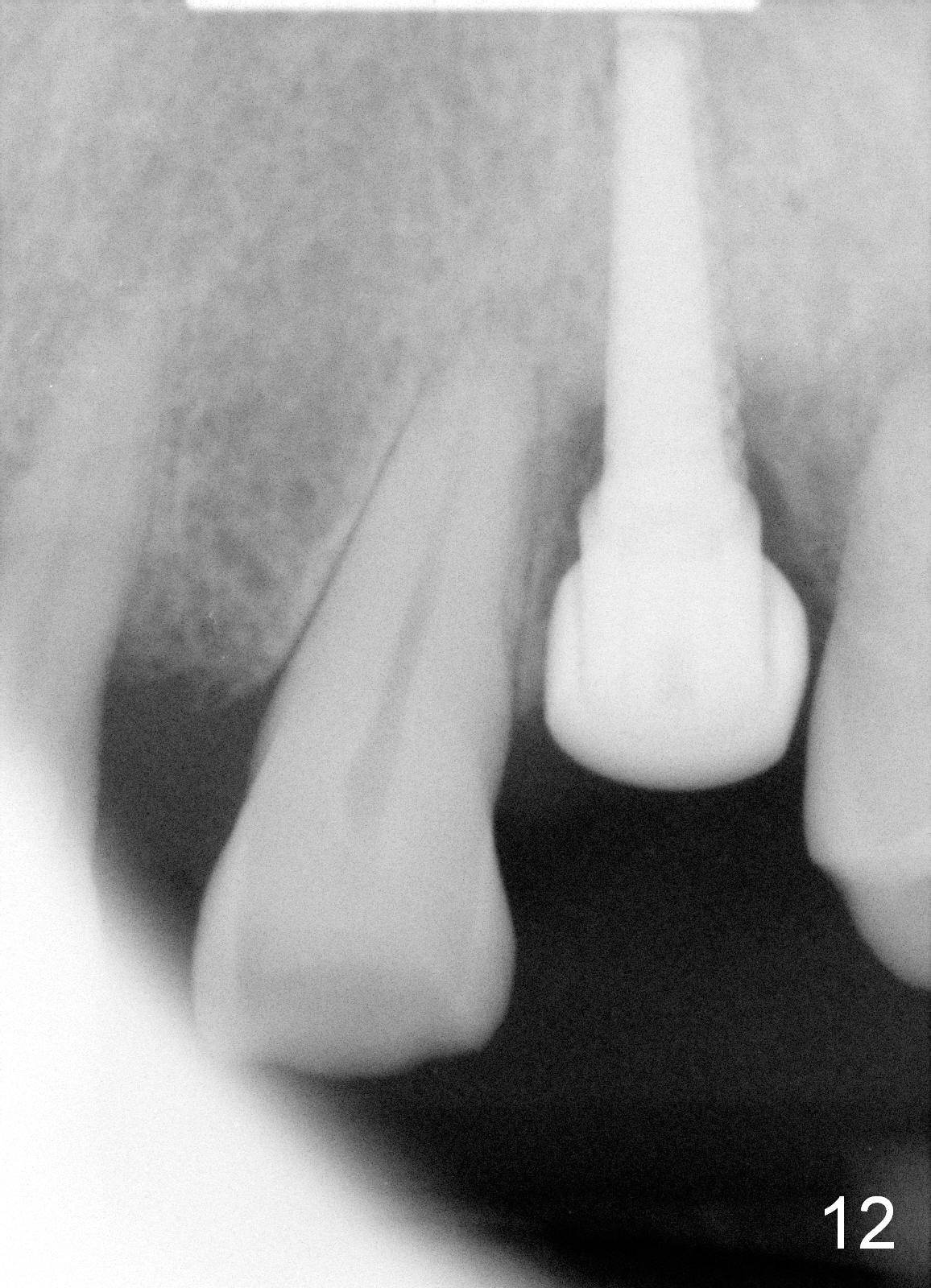
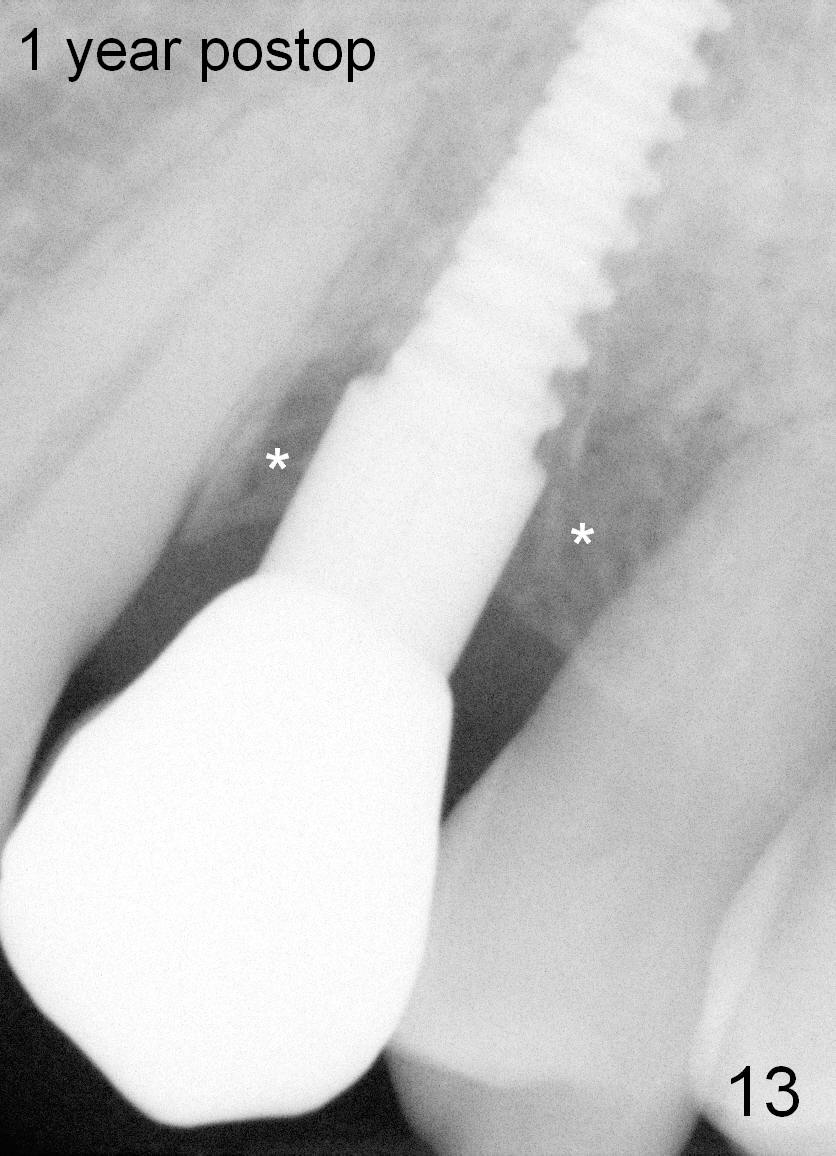
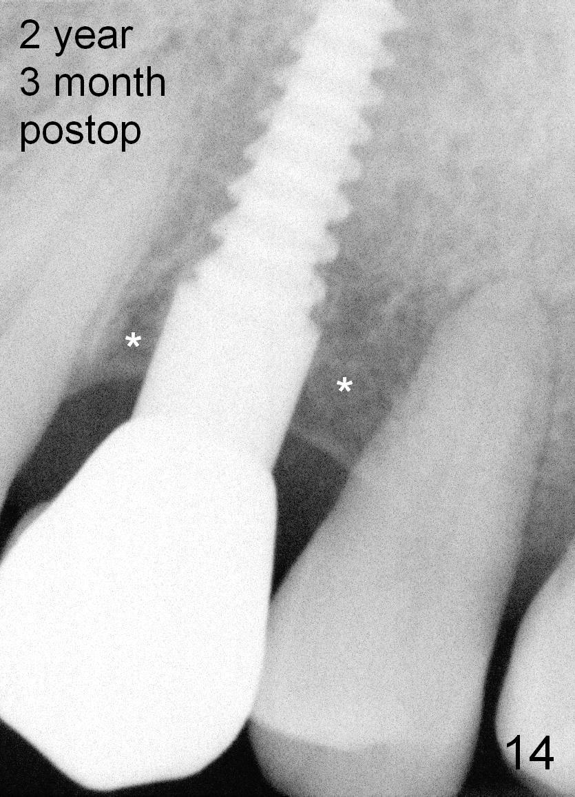
Crestal bone grows 2 months (Fig.11), 3 months (Fig.12), 1 year (Fig.13) and 2 years 3 months (Fig.14) postop. When the periodontally-affected tooth is removed and bone graft is placed around the most coronal thread of the implant, bone will regrow as time passes by.
Professionals, Upper Bicuspid Immediate Implant
Xin Wei, DDS, PhD, MS 1st edition 06/28/2013, last revision 10/24/2015