|
|
|
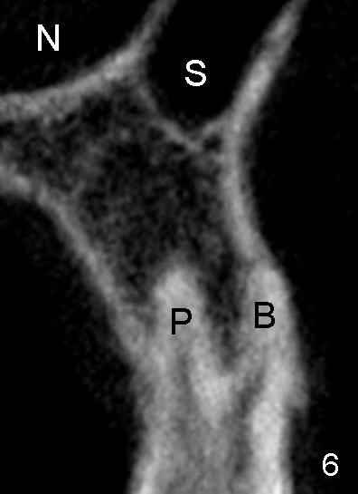
Fig.6 (preop coronal section of CBCT (not the same patient) shows the buccal (B) and palatal (P) roots of an upper 1st bicuspid in relation to the nasal cavity (N) and the maxillary sinus (S).
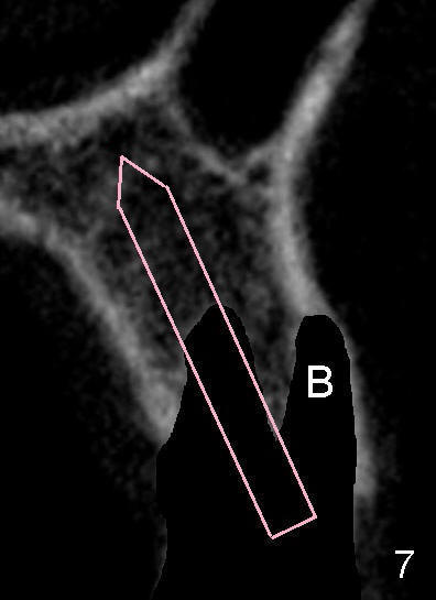
Fig.7: After extraction, an osteotomy is initiated in the palatal socket with a pilot drill, followed by a series of reamers (pink outline). The advantage of using the reamers is to collect autogenous bone from osteotomy, which is to be returned and placed in the buccal socket (B).
Return to Cosmetics with Upper Bicuspid Immediate Implant
Xin Wei, DDS, PhD, MS 1st edition 12/30/2012, last revision 02/23/2014