|
|
|
|
|
|
Is the tooth #19 Salvageable?
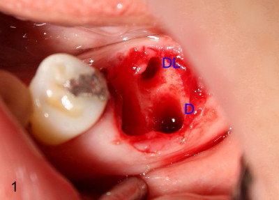
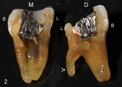
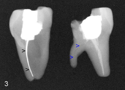
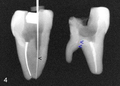
As discussed before, we plan to extract the affected tooth atraumatically by sectioning. When the distal portion of the tooth is taken out, there are two sockets distally (D, DL in Fig.1). There is an extra distal root (arrowhead in Fig.2). The mesial (M) and distal (D) portions of the tooth are laid out so that the buccal (B) is placed outside, lingual (L) inside of the photo. We are seeing the sectioned surfaces of the tooth. With the same arrangement, PA is taken (Fig.3). The silver cone (black arrowheads) is off the center of the mesial root, suggesting missing mesiolingual canal. The second missing canal, distolingual (blue arrowheads) is narrow and highly curvy. In fact the mesiolingual canal is also obliterated. It takes effort and time to go through it (Fig.4 black arrowhead). The distolingual canal is so obliterated that perforation is the end result (using rotary file) (blue arrowheads).
Xin Wei, DDS, PhD, MS 1st edition 03/02/2011, last revision 07/20/2011