
|
|
|
|
|
|
|
|
|
|
|
|
Abutments that can be changed during orthodontic treatment
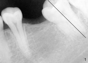
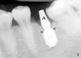
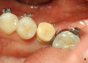
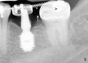
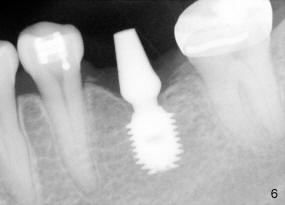
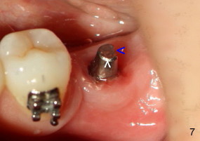
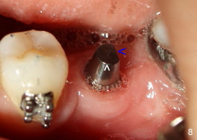
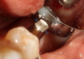
The tooth #19 has been lost for 8 years with tilting of #18 (black line in Fig.1). A 6x8 mm Bicon implant (3 mm post) was placed as distal as possible (I in Fig.2). Three months later, the implant was uncovered with evidence of osteointegration (arrowheads in Fig.3). A 4x6.5 mm 0 degree non-shouldered abutment with 3 mm post (A) was inserted into the implant well. The flat surface of the abutment needed to face distal in order to seat the abutment without interference. The implant/abutment complex was used as an anchorage to upright #18. The tooth #17 was extracted prior to orthodontic treatment (compare Fig.5,6 vs. 1-3). A bracket was bonded to Jet temporary crown of #19 and open coil spring was placed between #18 and 19. In the first two months, distalization of #18 is limited (between black and white lines in Fig.4), partially due to the fact that the bracket of #19 was debonded quite easily. A premolar band (B in Fig.5) was cemented to the temporary crown of #19. In another two months, the tooth #18 was pushed to desired position (arrowhead) under the tension of open coil spring (*). The mesial crest height of #18 was increased during uprighting (arrow, as compared to Fig.1-3). A new temporary crown was fabricated to fit the enlarged space of #19 with cementation of a molar band. The next problem is that the new molar temporary crown was easily dislodged from the 4x6.5 mm abutment even with a permanent cement. A larger abutment (5x6.5 mm) was used (Fig.6), with relining the temporary crown. The upper end of the larger abutment has sharp edge (Fig.8: white arrowhead, as compared to rounded edge of the smaller abutment (Fig.7). This may also contribute to better retention. In another 3 months, the temporary crown was dislodged again. An even larger abutment (6.5x6.5 mm with two flat surfaces (blue arrowheads in Fig.9) was adopted.
The unique connection between Bicon implant and abutment makes it easy to change abutments in accordance with changing clinical situation during orthodontic treatment.
Return to Fellowship Candidate Case Documentation Form
Xin Wei, DDS, PhD, MS 1st edition 05/02/2011, last revision 07/07/2014