
|
|
|
|
|
|
|
|
|
|
|
|
|
|
|
|
|
|
Why Are His Front Teeth Not Straight?
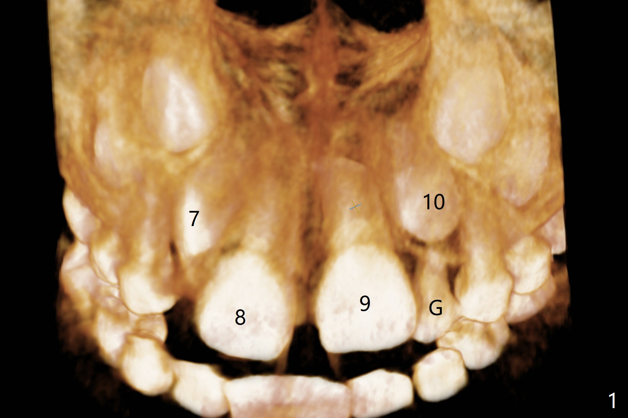
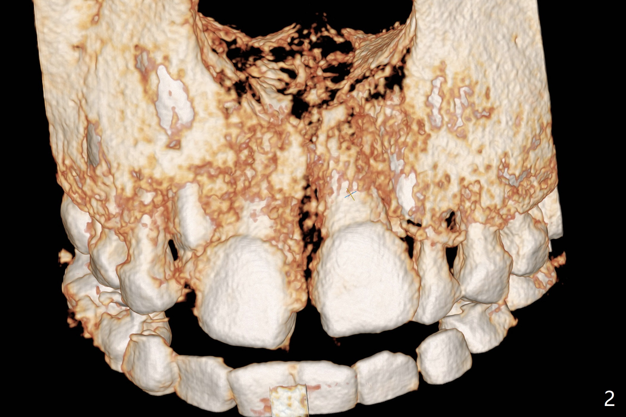
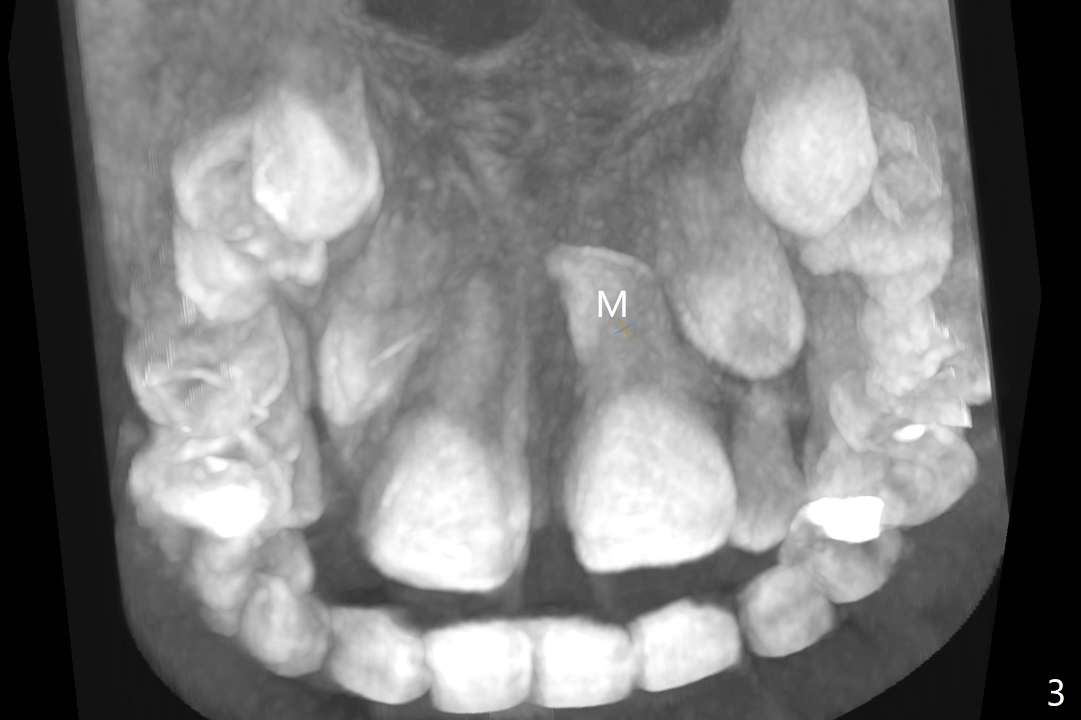
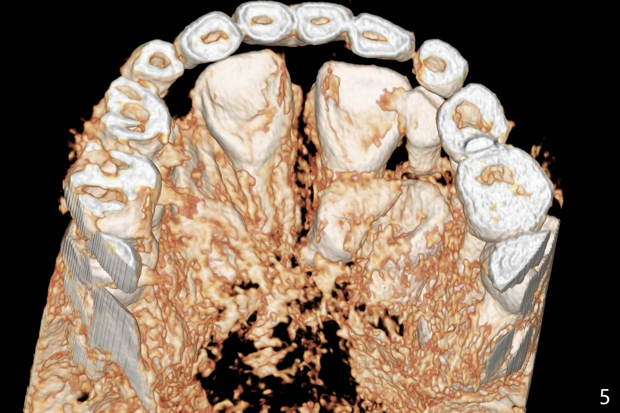
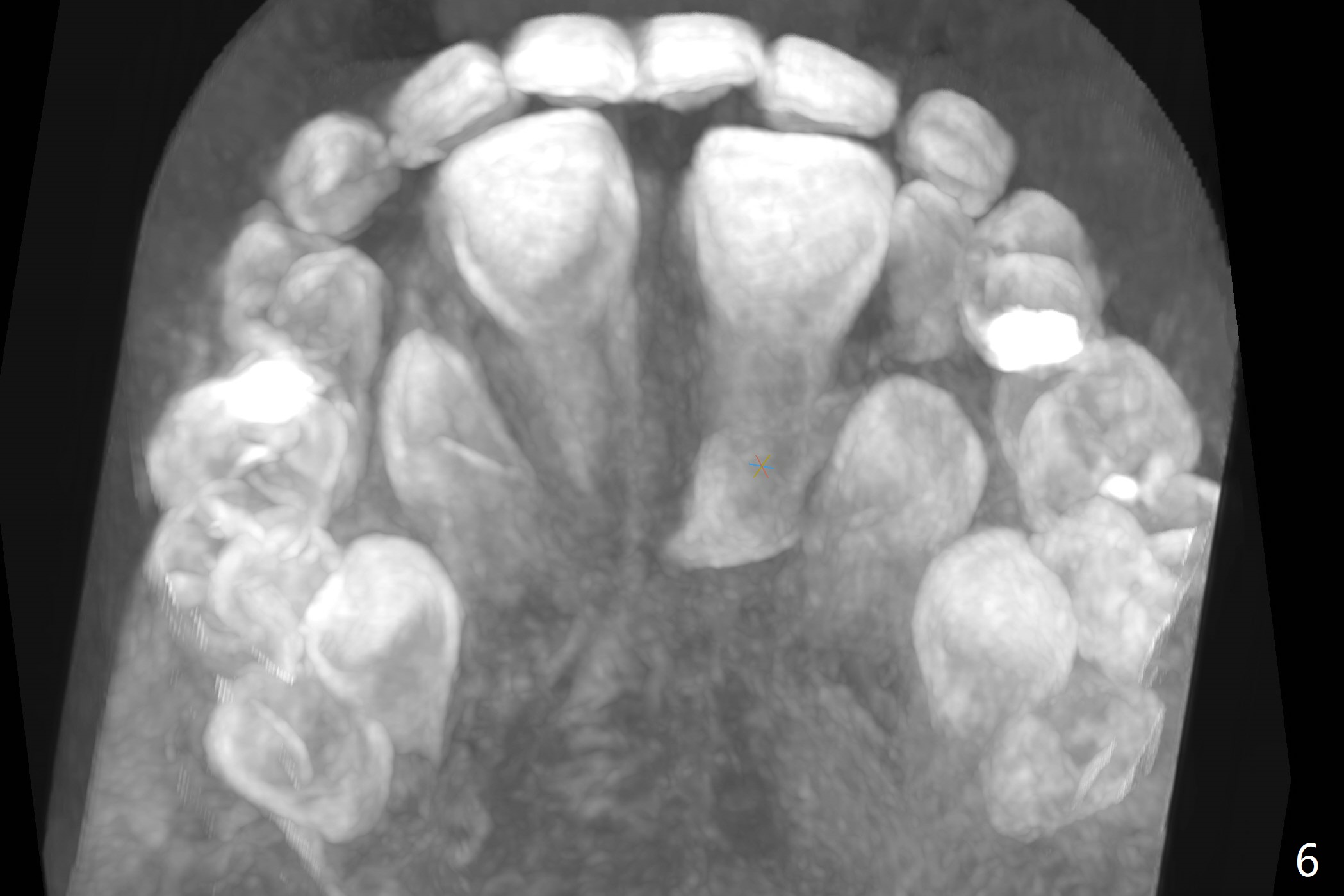
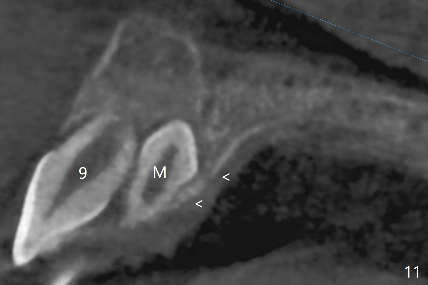
The father of a 7-year-old boy wonders why the upper central incisors, particularly #9, are off (Fig.1 3-D CT tooth coloring). Fig.2 (bone coloring) does not reveal much. Fig.3 (MIP, more transparent) reveals a supernumerary tooth, called mesiodens (M, an extra central incisor, abnormally shaped (cone), near the midline). Palatal view shows that the mesiodens points to the midline and distal (arrow). A palatal gingival sulcus incision will be made to expose the extra tooth (Fig.5). An elevator will be used as midline and distal as possible to avoid damage to the root of #9 and the tooth bud of #10 (Fig.6 MIP). Photos will be taken frontally and palatally preop and after incision and exposure of the mesiodens (palatal).
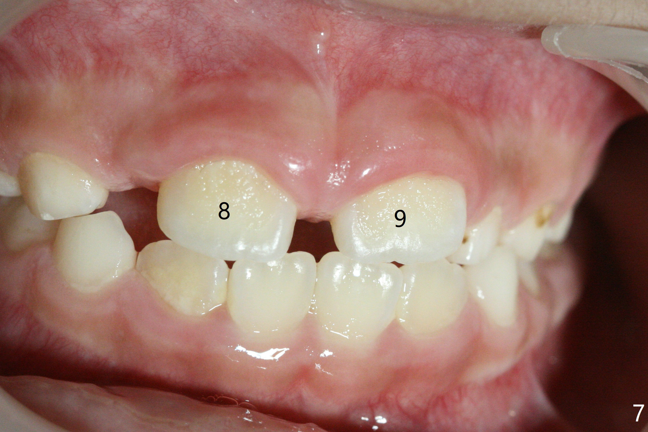
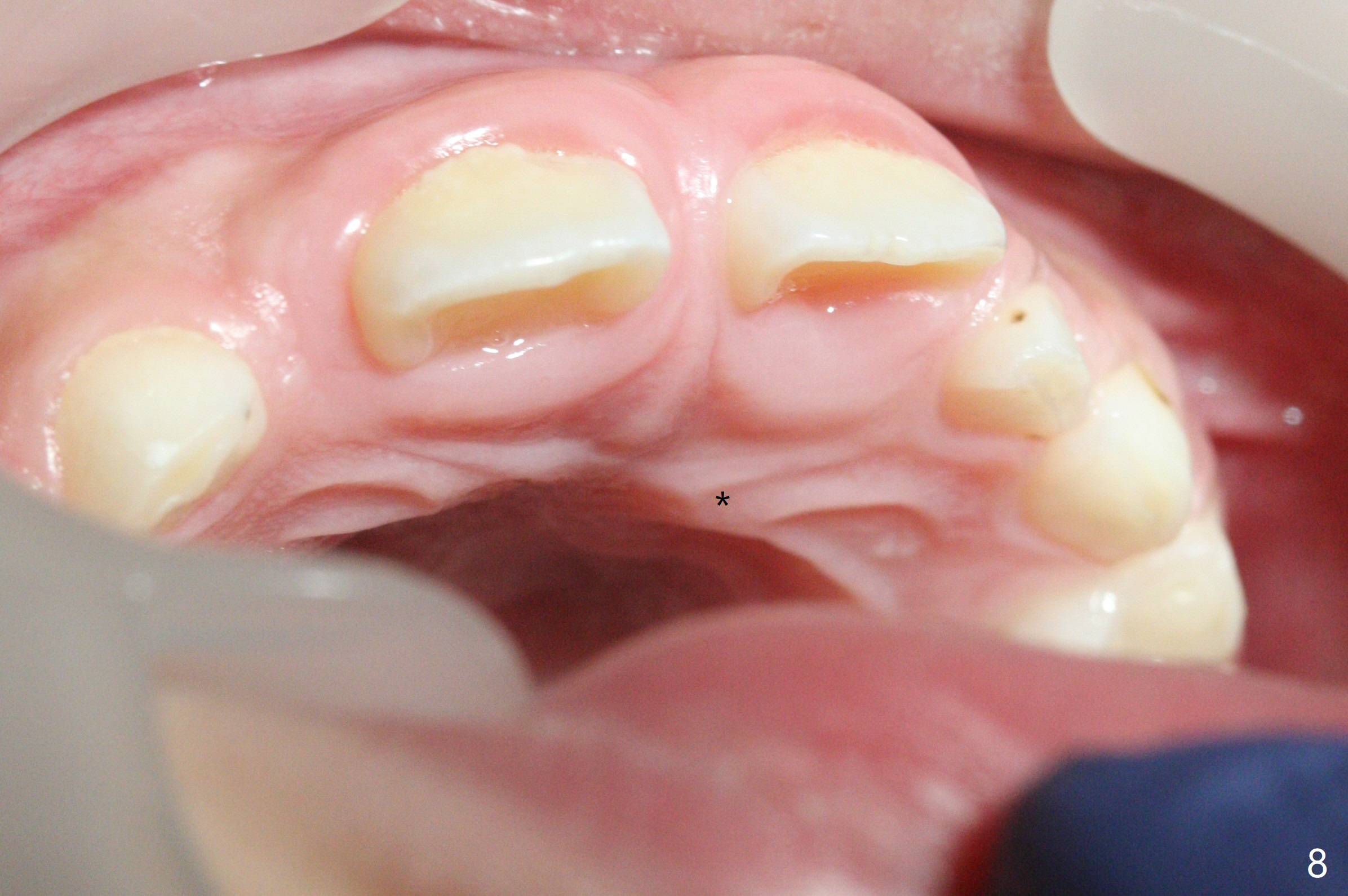
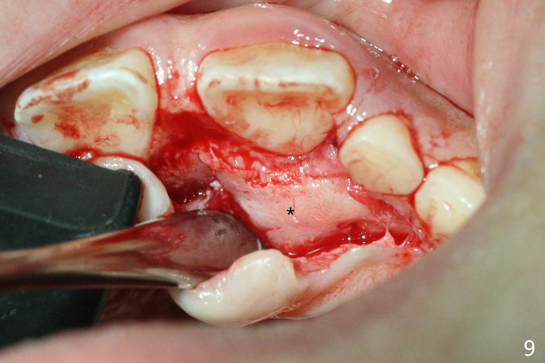
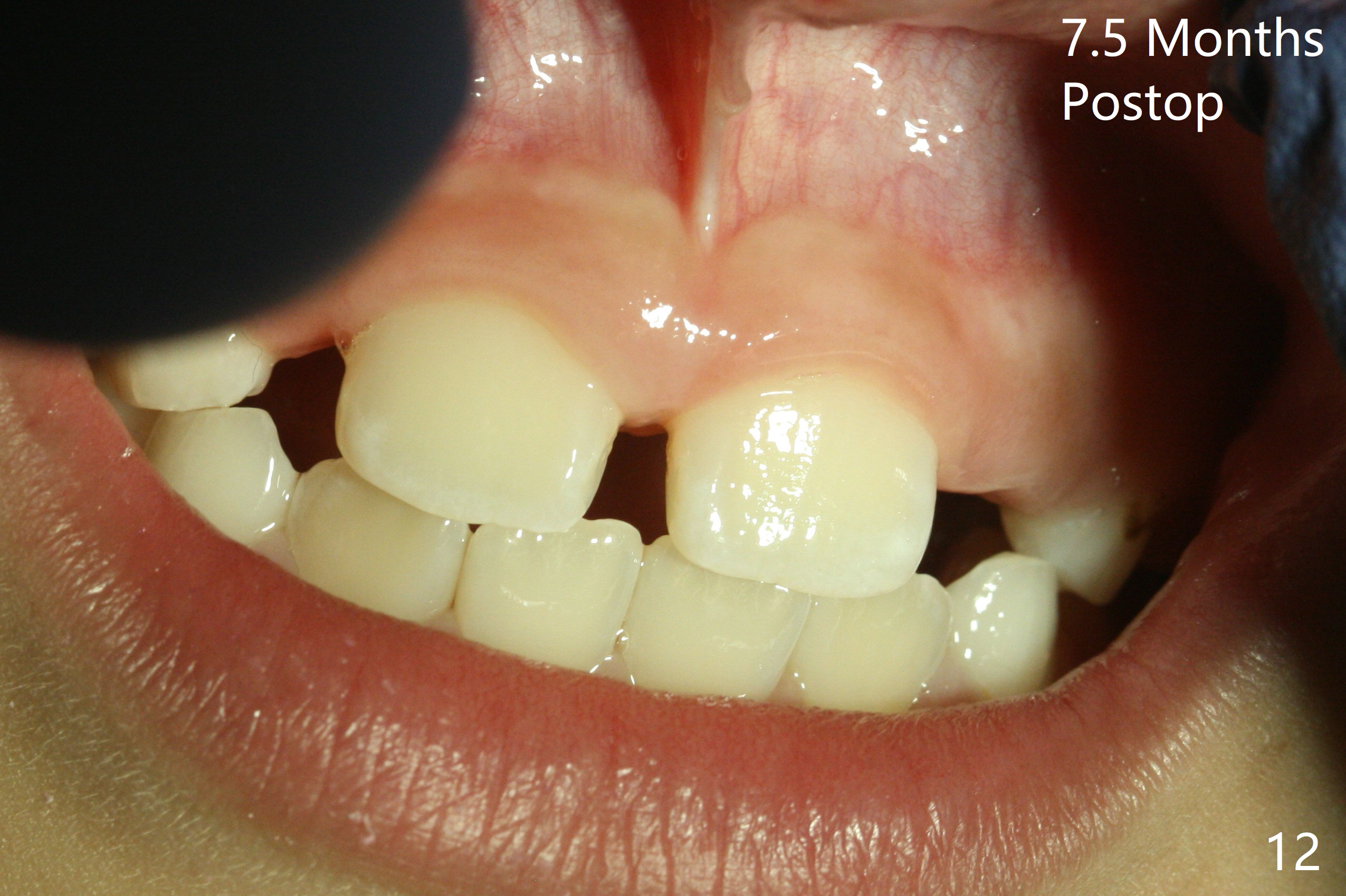
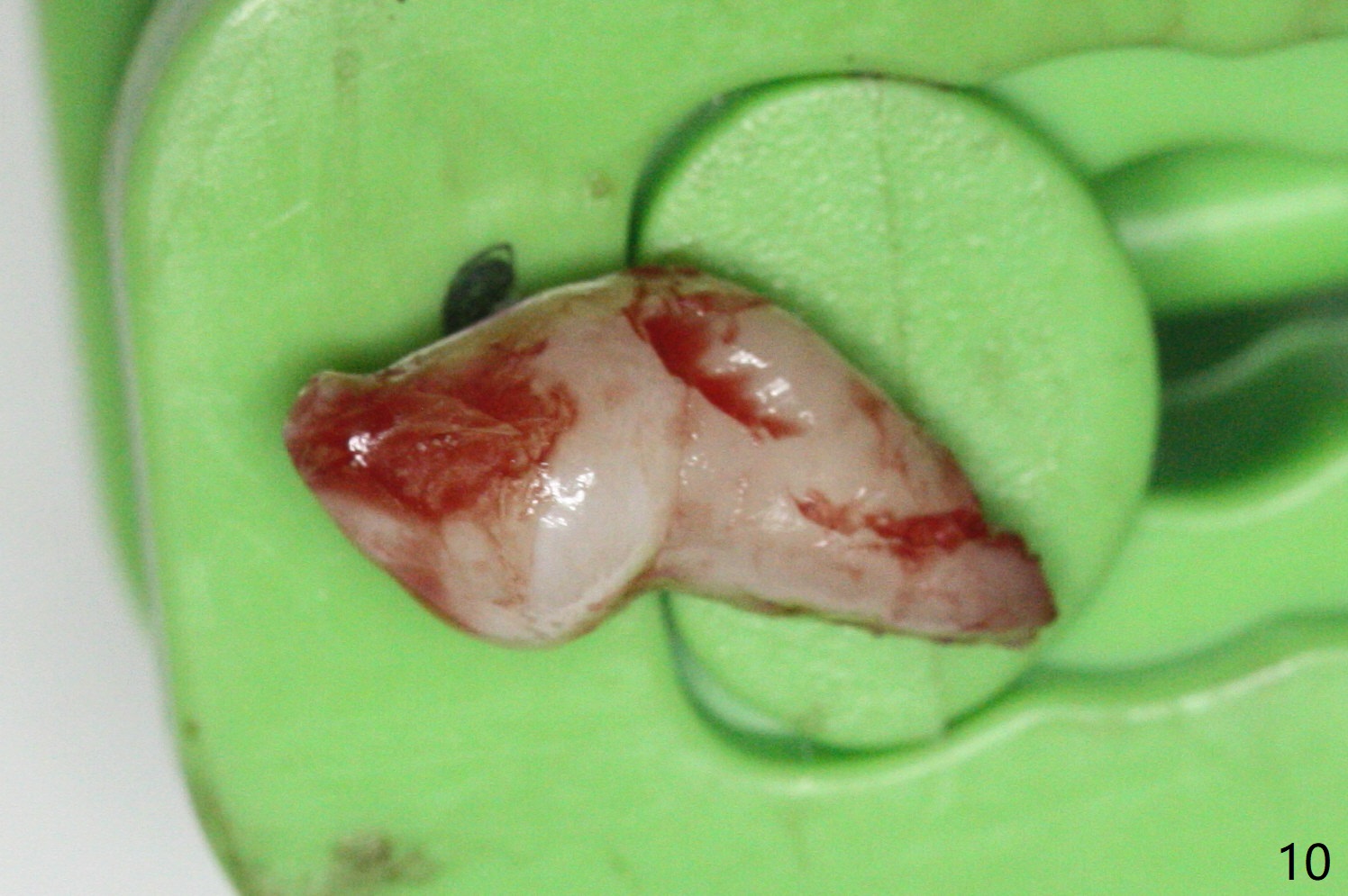
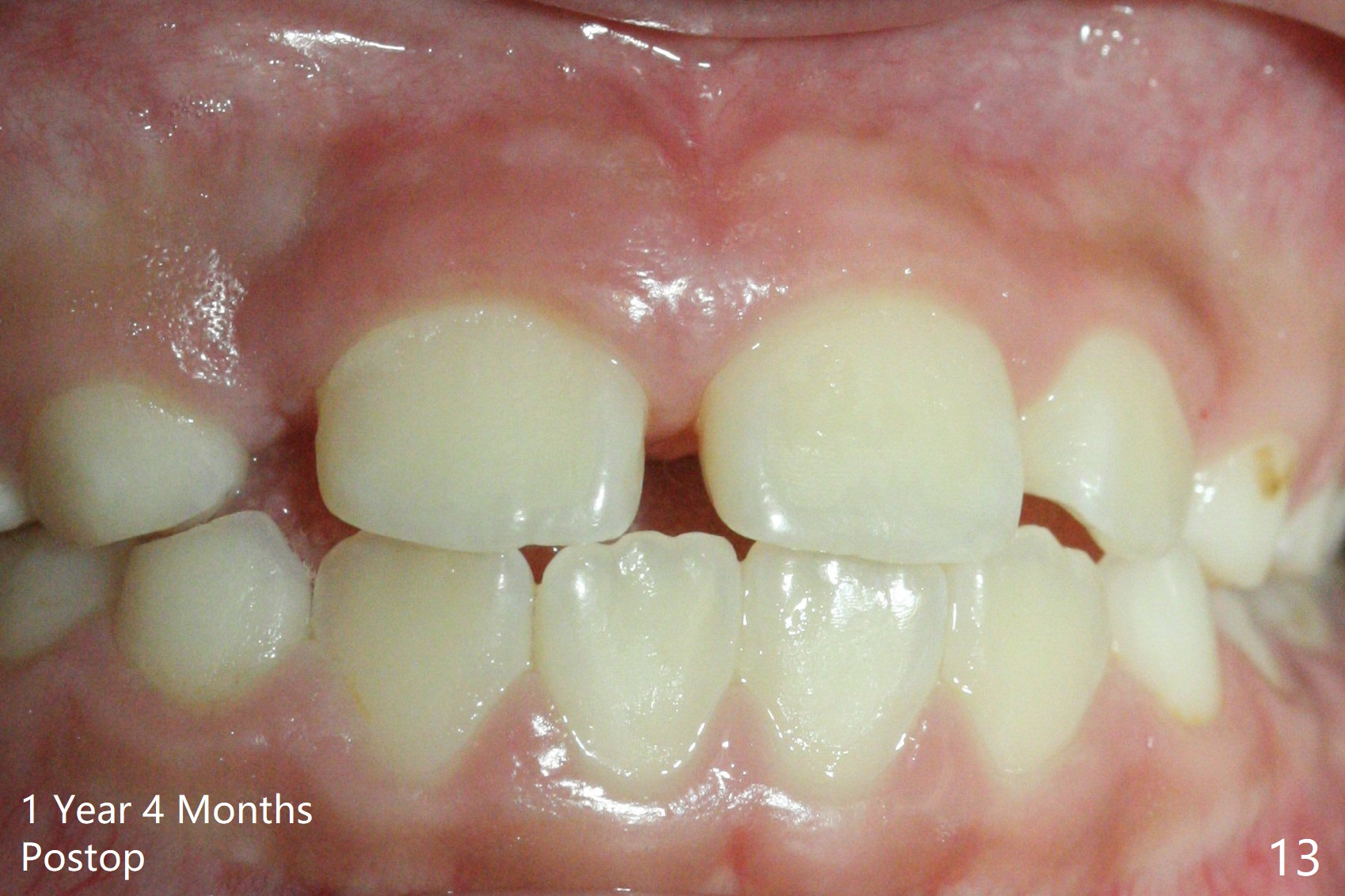
There is a large diastema between the upper central incisors (Fig.7). The left anterior palate is slightly elevated (Fig.8 *). In fact the cortex overlying the mesiodens (Fig. 9 *, 11 <) is to be removed with a surgical handpiece in order to extract the mesiodens (Fig.10). The left central shifts mesial in 7.5 months postop, whereas the right one remains in place (Fig.12). The tooth #7 is unerupted 1 year 4 months postop (Fig.13).
Return to Professionals Last Next
Xin Wei, DDS, PhD, MS 1st edition 01/14/2018, last revision 05/28/2019