|
|
|
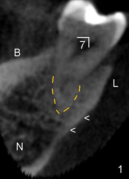
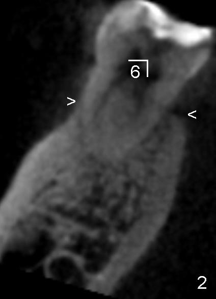
Fig.1,2 are coronal sections of CBCT through the distal root of the lower right 2nd molar (7) and the mesial root of the 1st molar (6), respectively. The patient is a 41-year-old Chinese male. The lingual concavity (<) is more pronounced in the 2nd molar (Fig.1) than that of the 1st one (Fig.2). The apex of the 2nd molar (yellow dashed line) is closer to the lingual concavity than to the inferior alveolar nerve canal (N). B: buccal; L: lingual.
Return to Submandibular Fossa
Xin Wei, DDS, PhD, MS 1st edition 04/14/2013, last revision 04/14/2014