|
|
|
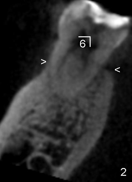
Fig.2 is a coronal section of CBCT through the mesial root of the lower right 1st molar (6). The buccal bone is lower and thinner than the lingual one (compare two arrowheads). In other word, the mesial root is located more buccally than lingually.
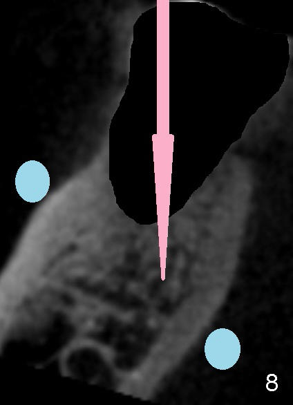
When the tooth is extracted, the osteotomy (Fig.8 pink arrow) should be slightly lingual with two fingers (blue circles) holding the ridge in case the drill deviates and perforates one of these plates.
In fact, osteotomy may need to be initiated on the lingual wall of the immediate socket of a 2nd molar according to local anatomy, revealed by CBCT.
Return to Submandibular Fossa
Xin Wei, DDS, PhD, MS 1st edition 04/14/2013, last revision 04/15/2014