|
|
|
|
|
|
|
|
|
|
|
|
|
||
Bony Changes of Dens Evaginatus After Apexification
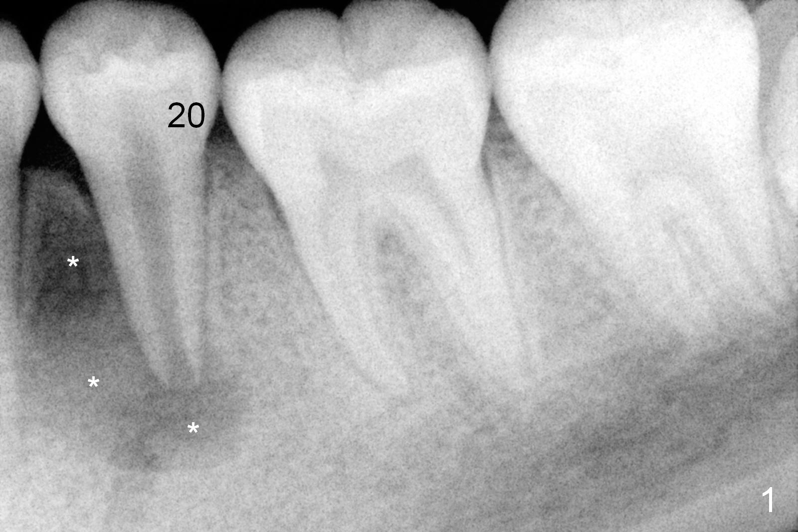
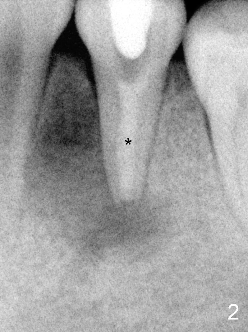
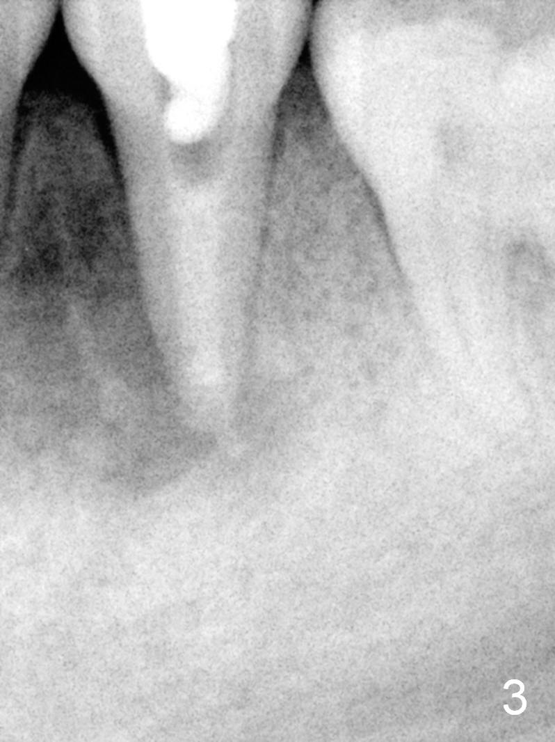
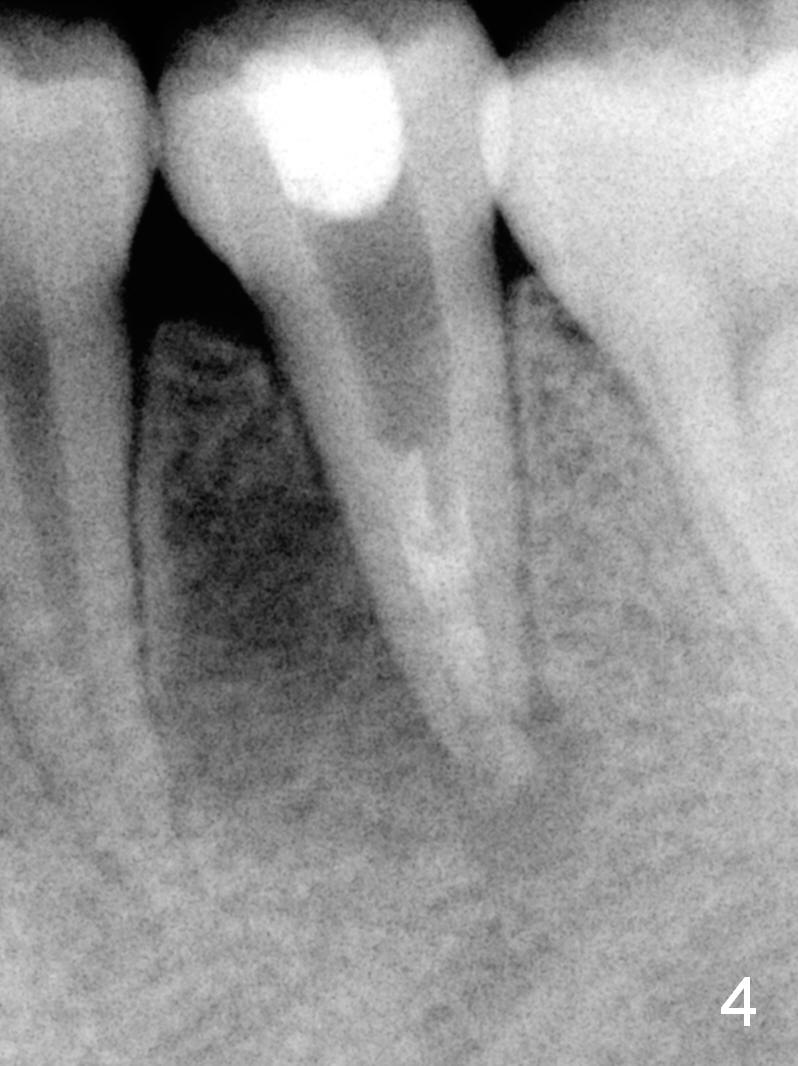
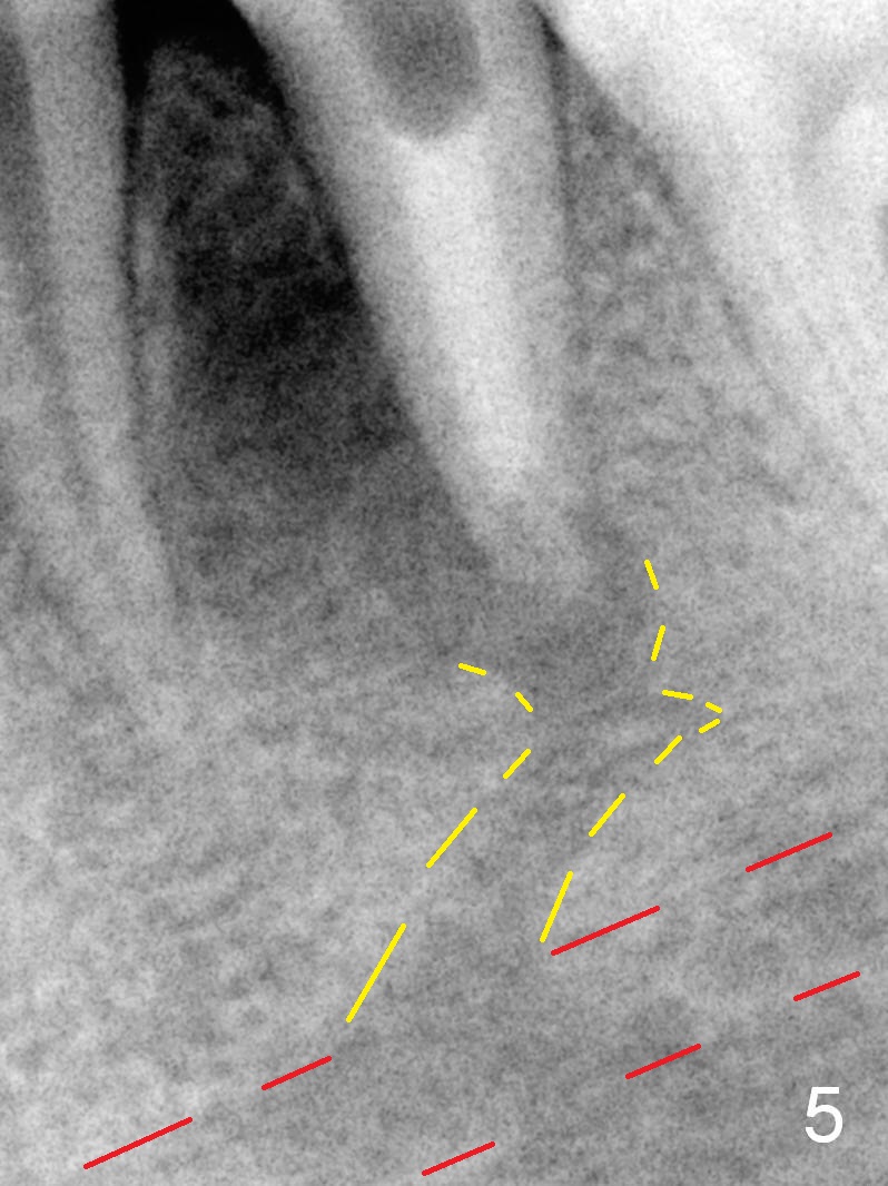
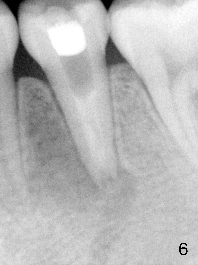
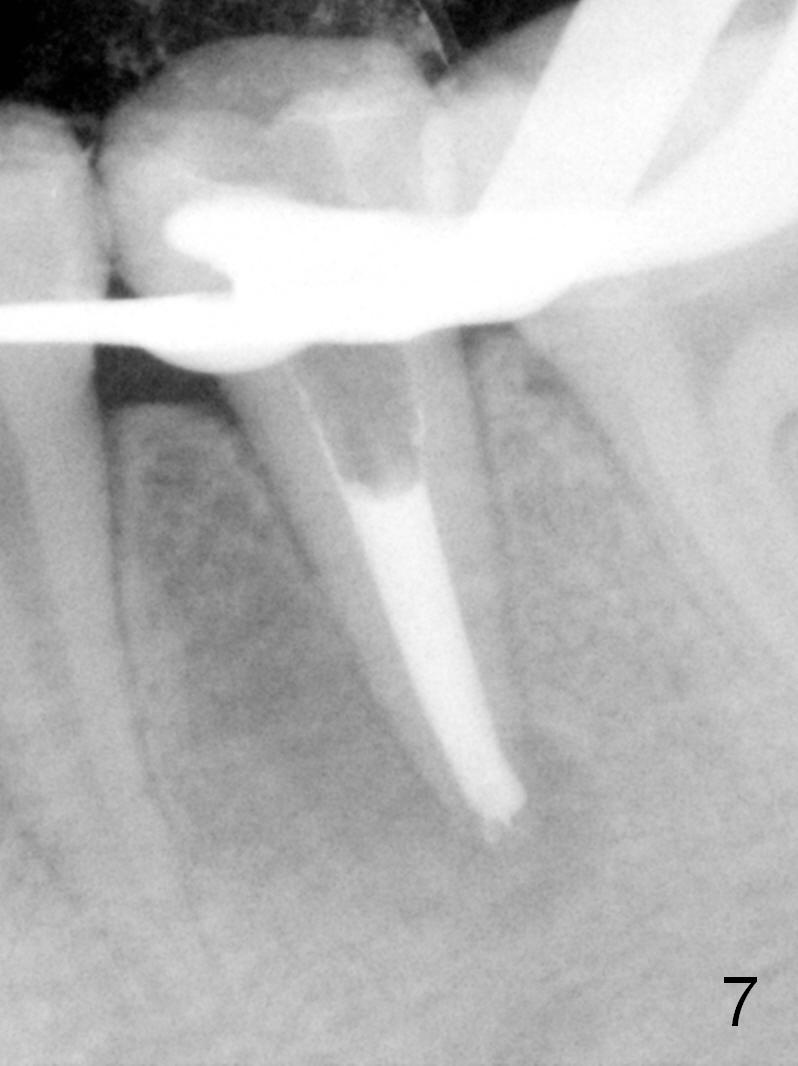
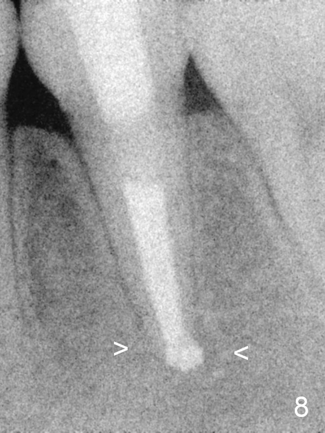
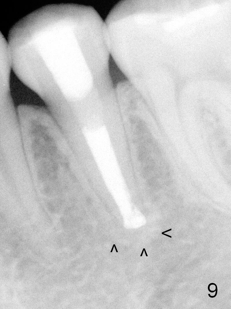
A 13-year-old girl has a buccal apical fistula at the tooth #20 with large periradicular radiolucency (Fig.1 *). After canal debridement, calcium hydroxide paste is placed in the canal (Fig.2 *). One month later, the fistula does not disappear with light percussion. New paste is placed (Fig.3). Two months later, the fistula disappears without percussion. The existing paste (Fig.4) is changed (Fig.5 (yellow: radiolucency; red: Inferior Alveolar Canal)). Another 2 months later, the fistula does not recur; as before, the paste density decreases, so does periradicular radiolucency (Fig.6). Root canal is packed (Fig.7). Six months postop, periradicular radiolucency continues to decrease, while the lamina dura at the apex is discontinuous (Fig.8 between arrowheads). Seven years postop, the lamina dura at the apex is seemingly intact (Fig.9). The patient remains asymptomatic at #20. In fact there is an acute infection at #29.
Return to Professionals
Xin Wei, DDS, PhD, MS 1st edition 10/09/2015, last revision 10/10/2015