
reason for RCT failure?
|
|
|
|
|
|
|
|
|
|
Why is missing canal not the reason for RCT failure? |
|
Missing DB Canal in Lower 1st Molar
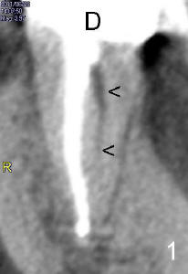
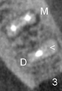
Sixty-year-old man remains symptomatic after RCT in the tooth #19. Clinical exam or X-ray does not show any significant finding. CBCT shows missing distobuccal canal (Fig.1: <), although there is no periapical radiolucency (PARL). In contrast, there are PARL (Fig.2: A) and radiolucency in the lingual (L) aspect of the mesial root (M). Axial cut shows again missing DB canal (Fig.3: <). Gutta percha in the distal canal is off the center. Note the distance between distal canals is narrower than the mesial ones.
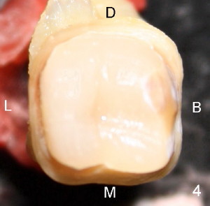
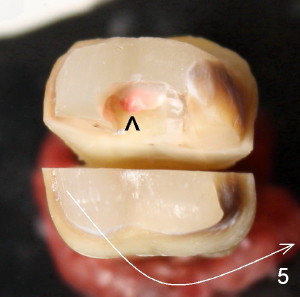
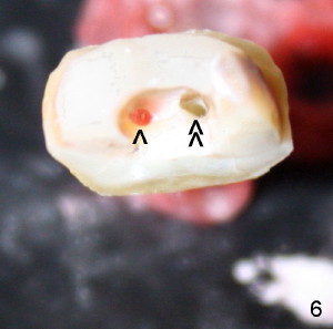
Finally the tooth is extracted due to development of mesiolingual abscess (mesial root fracture). Fig.4 is the occlusal view of the affected tooth after crown removal. Fig.5 show the same tooth after sectioning through buccal (B) to lingual (L) axis as shown in Fig.4. Then part of build up is removed. Gutta percha is again shown off the center (<), suggesting missing canal on the other side of the center. It is confirmed by further dissection (Fig.6: <<).
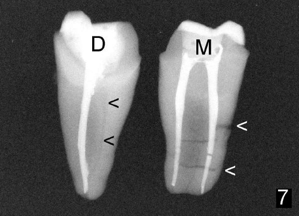
Let us return to Fig.5, turn the mesial portion of the tooth 180 degree as shown by a curved arrow and take PA (poor man CT, Fig. 7). Missing DB canal (black <) and mesial root fracture (white <) are further confirmed. Further question: why is the missing canal not the reason for failure of this tooth?
Xin Wei, DDS, PhD, MS 1st edition 08/18/2011, last revision 08/24/2011