|
|
|
|
|
|
|
|
|
|
|
|
|
|
|
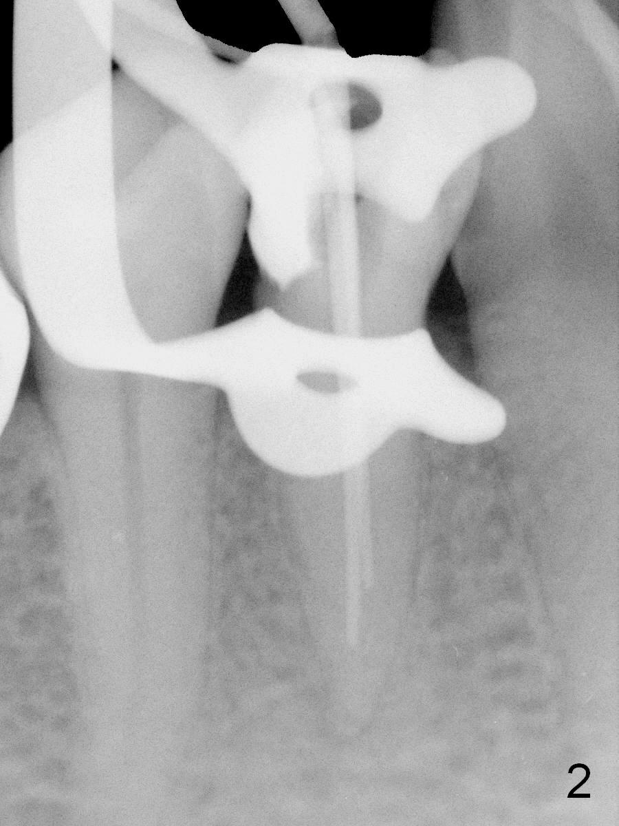
Extra Canal
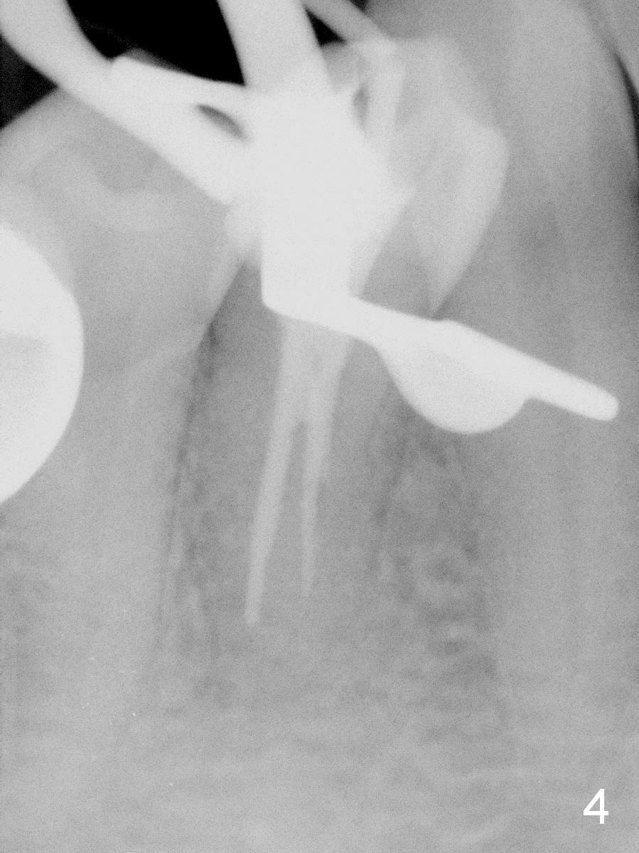
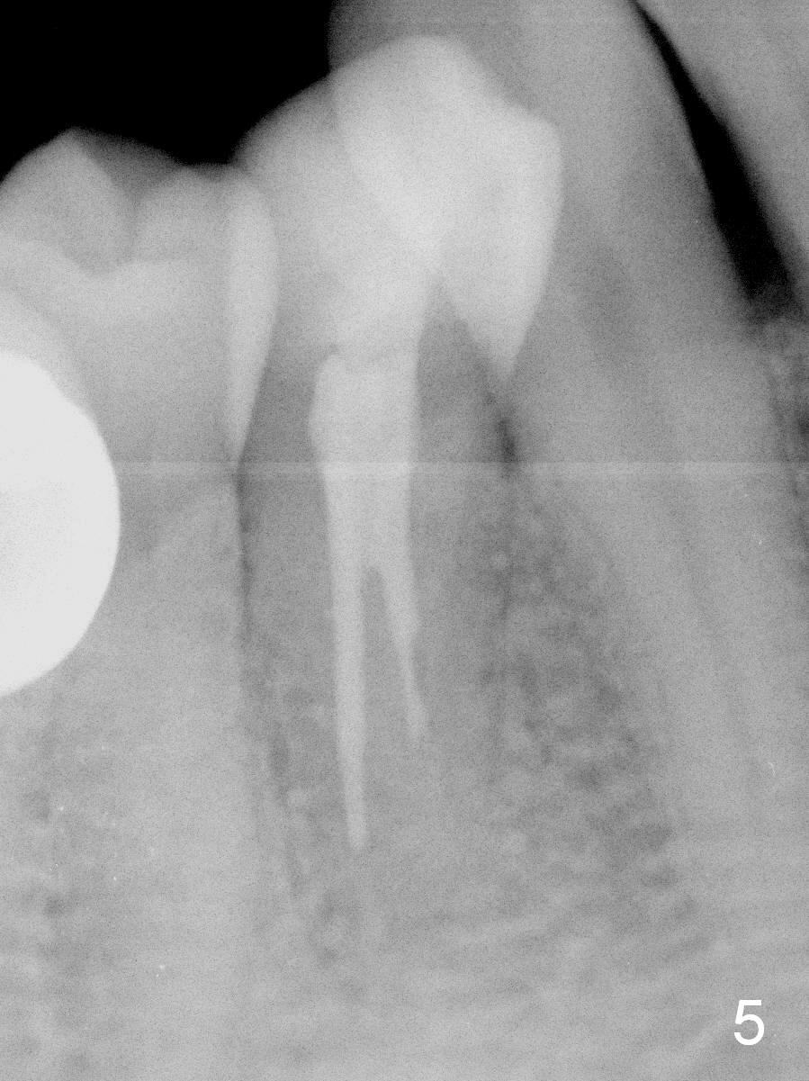
Further exploration finds a lingual canal (Fig.2), which is debrided until 30 hand file. When the canals are being filled with gutta percha and paste, the lingual canal is not filled (Fig.2). After removal of buccal gutta percha, the lingual canal is enlarged with 30/.04 rotary file and filled (Fig.4). The lingual canal is laterally condensed; composite build up is finished (Fig.5).
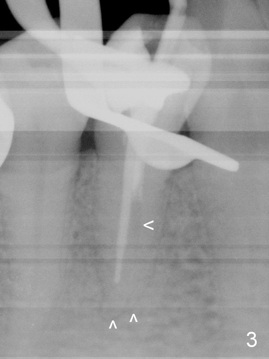
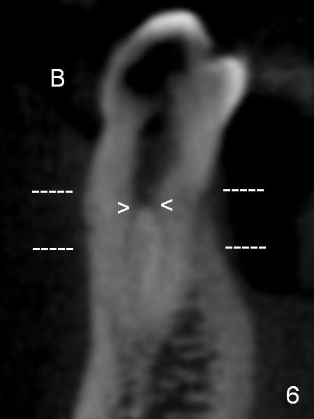
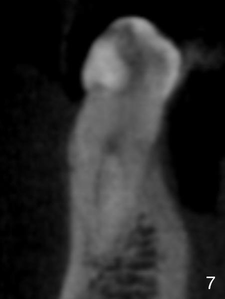
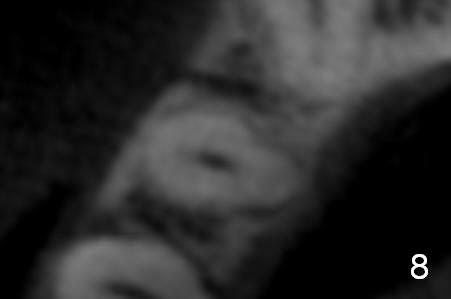
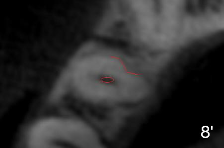
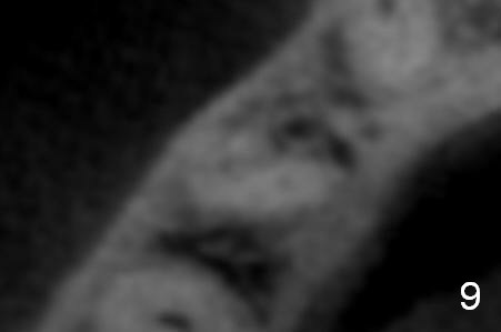
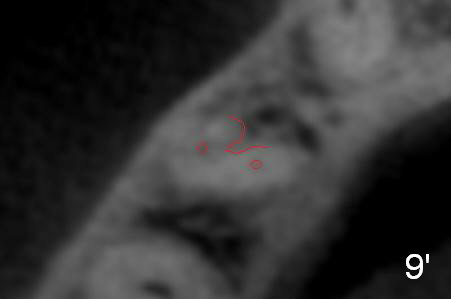
In fact CBCT has ben taken for #14 RCT and is reviewed prior to this case. If CT were reviewed, finding the extra canal would be easier (Fig.6 coronal section). It appears that the apical canals are blocked (Fig.7). The canal is split at the middle of the root, as shown in Fig.8,8' (axial upper section, as shown by the upper dashed line in Fig.6) and in Fig.9,9' (axial lower section, as shown by the lower dashed line in Fig.6). The buccal canal should not have been debrided with 40/.04 rotary file (30/.04 would have been better). In all, the tooth has two fused roots (Fig.3,9,9').
CBCT should be reviewed for RCT if available.
Return to
Professionals
Xin Wei, DDS, PhD, MS 1st edition 07/02/2016, last revision 07/02/2016