|
|
|
|
|
|
|
|
Dental Education Lecture: Have Complete Teeth
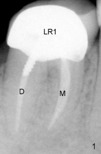
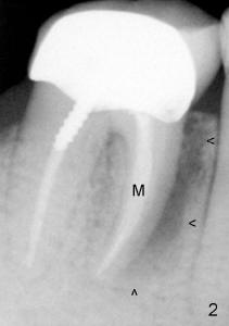
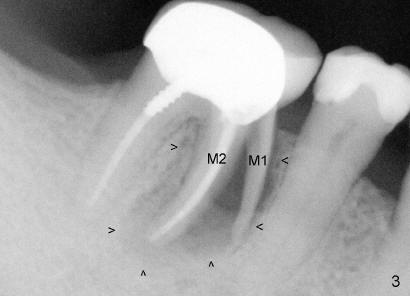
Mr. Chan has been our patient for five years. Today we discuss his lower right 1st molar (LR1 in Fig.1). The lower 1st molar usually has two roots: mesial (meaning near midline of our body) and distal (away from midline). Mr. Chan's lower right 1st molar has had root canal (where D and M are labeled) and crown (where LR1 is labeled). This X-ray was taken 4 years ago. Two years later, there was sign of bone infection around the mesial root (indicated by arrowheads in Fig.2), but Mr. Chan appeared to be alright. Since then he has had light pain occasionally. Today he returned to our clinic for cleaning. Routine X-ray shows that the mesial root of the lower right 1st molar splits into two parts (M1, M2 in Fig.3) with increased bone infection (arrowheads, as compared to Fig.2). The molar is extracted after his consent and finally replaced by an implant.
Do you know the main reason that one of roots of this molar splits?
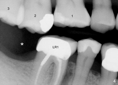
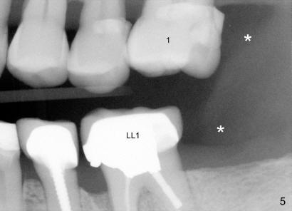
In fact this molar (LR1) is the only molar in the lower right area of his mouth (Fig.4). Mr. Chan has lost a 2nd molar (*). In contrast, there are three molars in the upper jaw (1,2,3). The third molar is a wisdom tooth, which is the least important. The single remaining molar (LR1) sustains almost all of chewing force. Although the crown protects the tooth (above the gum line) from breakdown, it does not protects the root from splitting, particularly after root canal. Equally interesting is that the upper and lower 2nd molars are also missing on the left side (Fig.5: *). The lower left 1st molar with crown and root canal has had some problem for a while (LL1). Sooner or later this molar will be taken out. If the three missing 2nd molars had been replaced earlier, these two lower 1st molars would have been alright. It seems that every one of our teeth is important. It must be fixed as soon as possible when it is gone.
Xin Wei, DDS, PhD, MS 1st edition 04/27/2011, last revision 08/21/2012