|
|
|
|
|
|
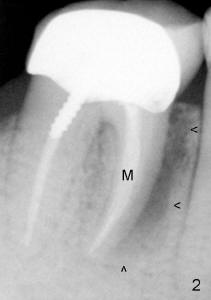
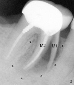
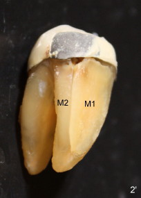
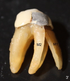
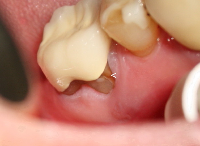
The tooth was extracted in two pieces (Fig.3'). The separate piece (M1) is placed where it belongs (Fig.2'). It appears that the meisal root must have had a crack two years ago when X-ray shows that there was bone infection (arrowheads in Fig.2). Mr. Chan has severe gum receding (Figure below, taken immediately before extraction). When the root has a crack, bacteria in his mouth enter inside the tooth to cause bone infection. As the infection gets worse and the patient keeps chewing, the separate piece of the mesial root (M1) moves from its original place (Fig.3), back to main article
Xin Wei, DDS, PhD, MS 1st edition 04/29/2011, last revision 09/29/2012