|
|
|
|
|
|
|
|
|
Large Implant
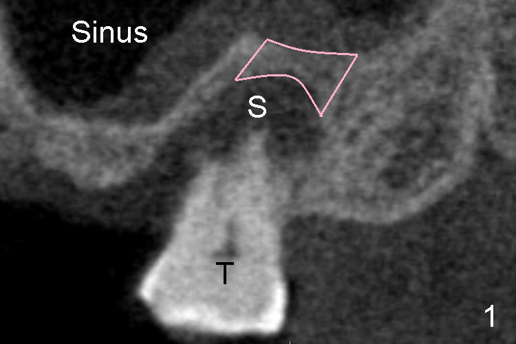
Mr. Shi develops severe gum disease (periodontitis) around an isolate molar tooth on the top (Fig.1 T). There is so much bone loss that the socket (S) is much larger the root. The tooth is too loose to support upper partial denture. We plan to take the loose tooth out and replace with a super-large implant (8 mm in diameter) at the same time. The diameter of most implants is usually less than 4 mm.
Over the top of the upper molar is an empty space, called sinus (Fig.1), where we often develop sinus infection after common cold. Between the socket and the sinus is a bony structure called sinus floor (pink outline in Fig.1).
Below the bottom molar tooth is a big nerve, which we try every means to avoid when placing an implant (1 2 3). On the other hand, we have intention to push an implant into the sinus if necessary as will be described next (1 2).
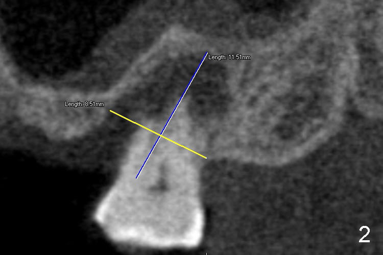
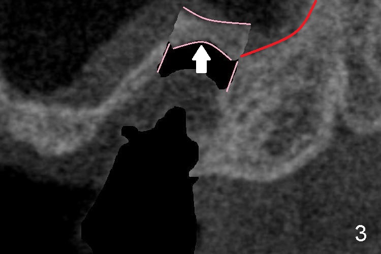
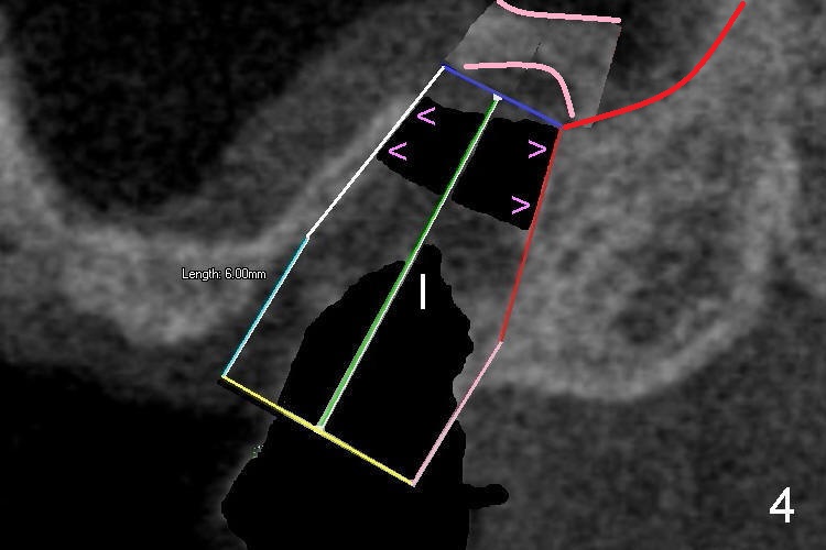
Fig.2 is a CT scan (like Fig.1). Before extraction, we measure the socket dimensions: ~8 mm wide, and 11 mm deep. After extraction, the implant of the socket size is inserted (8x11 mm), but is loose. We have to use a series of bone instruments to push the sinus floor up (Fig.3 arrow) so that a longer implant is placed (I: 8x14 mm). The reason of longer, the better is that the upper portion of the implant (Fig.4 >) is tightly engaged into the space between the two straight lines shown in Fig.3. The implant is very stable.
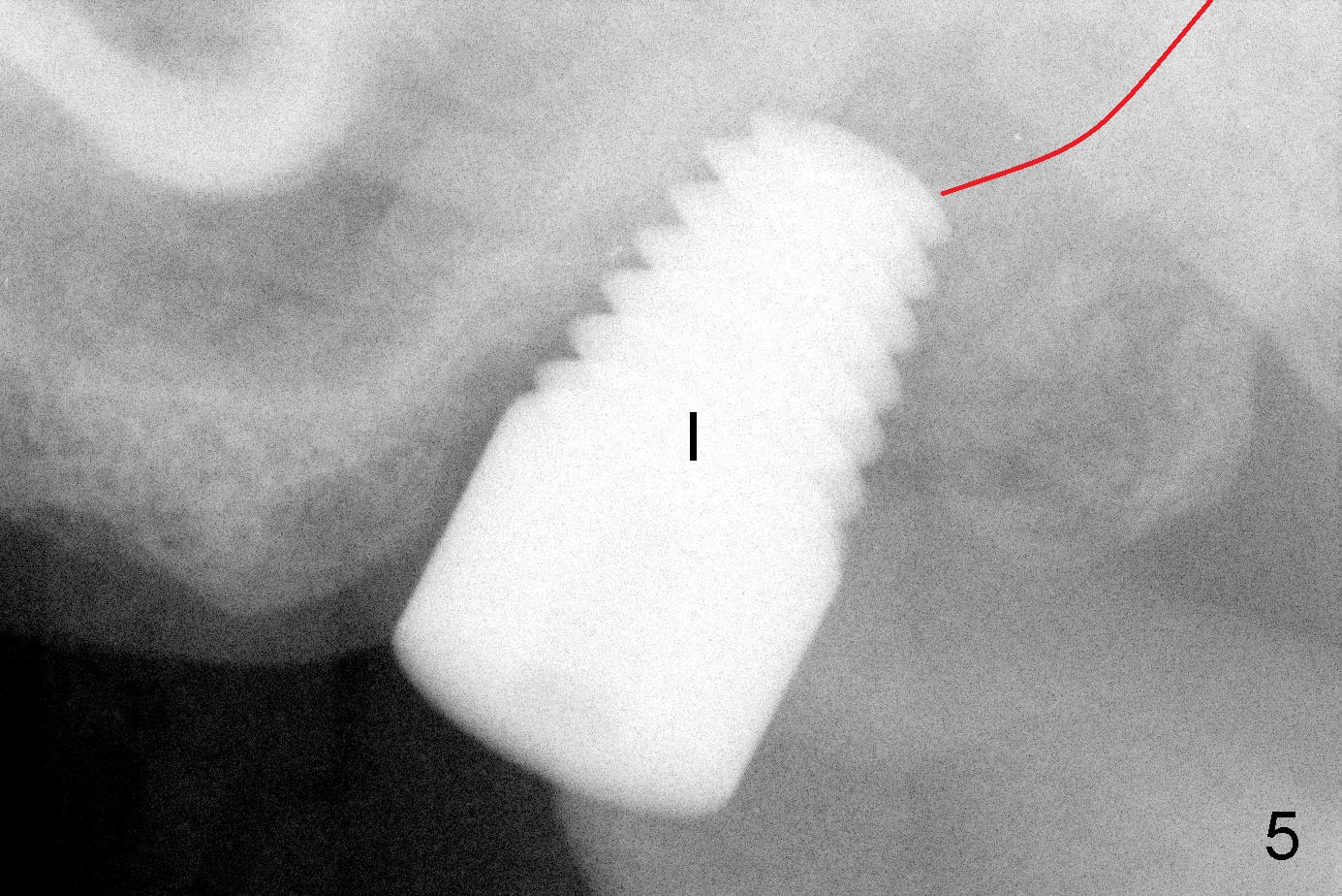
Fig.3 and 4 diagrammatically show how to lift the sinus floor and how to place the longer implant, whereas X-ray in Fig. 5 shows the real image of implant placed. Red curved line in Fig. 3-5 indicates the upper border of the sinus floor.
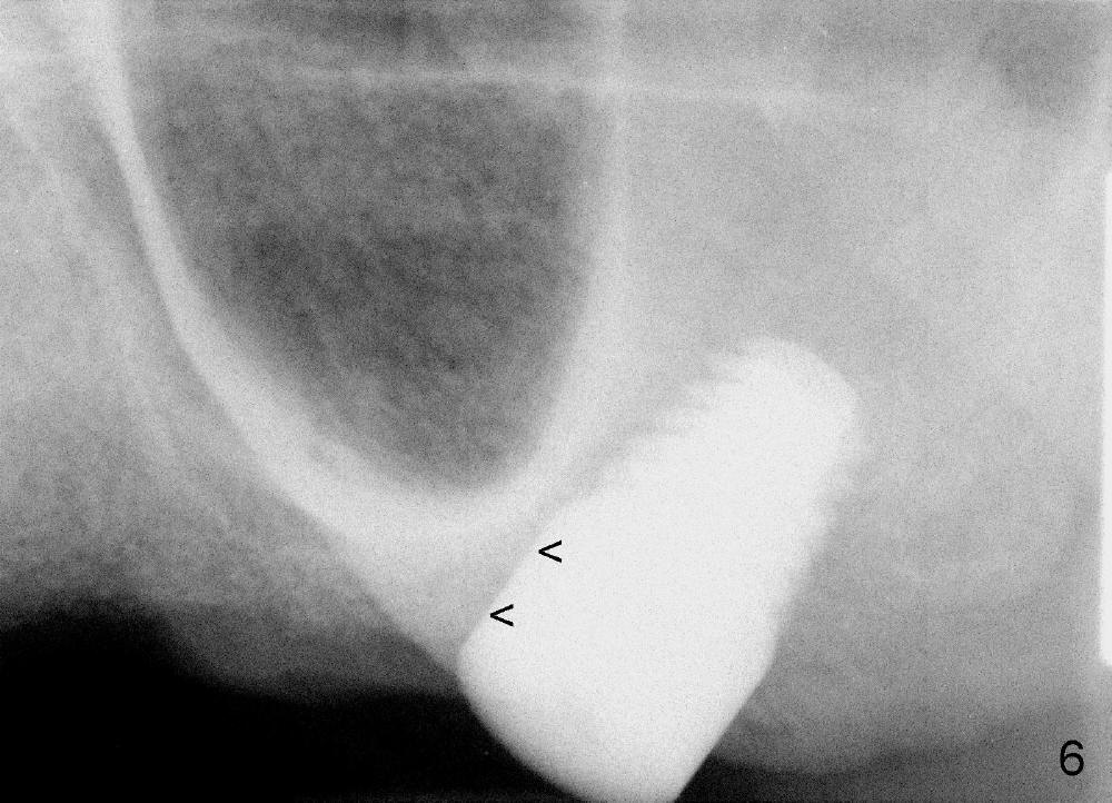
Four months later, there is no gap between the implant and the bone (Fig.6 <), as compared to Fig.5. This indicates that the implant has healed with the bone and that the implant is ready to have a crown.
Xin Wei, DDS, PhD, MS 1st edition 09/01/2012, last revision 09/30/2018