|
|
|
|
|
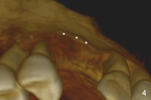
Fig.4: The buccal plate (*) is thin, as compared to the palatal one (see below).
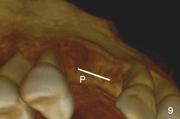
Retrospectively, bone expansion should have been initiated (Fig.9 white line) close to the thicker palatal plate (P). The coronal end of the osteotomes should have been directed palatally. The implant should have been short to reduce the chance to perforate the buccal plate apically.
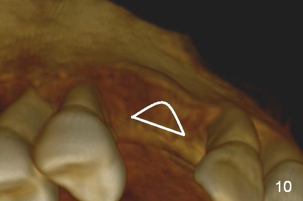
It would be much better to use a D implant (Fig.10).
Xin Wei, DDS, PhD, MS 1st edition 03/24/2015, last revision 11/17/2019