|
|
|
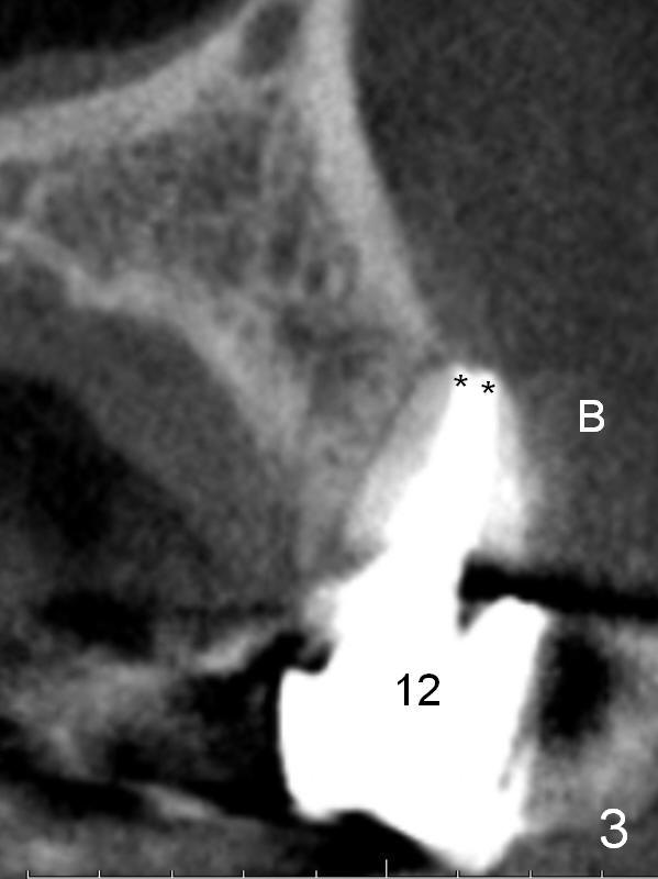
CBCT shows the pathology not completely resolved (Fig.3, coronal section, 2012). Root canal morphology is special. The buccal and lingual canals fuse except apically (**). B: buccal.
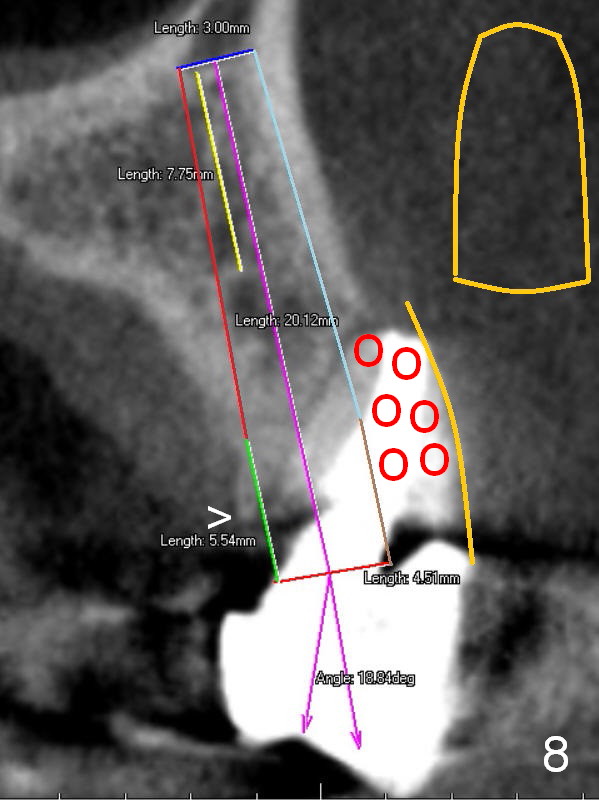
CBCT shows that there is enough bone to place a 4.5x20 mm tissue-level implant (Fig.8). This is more appropriate, considering the large radiolucency, the age of the patient (possibly osteoporosis), and the maxilla (bone softer than that of the mandible). The implant will be supported by approximately 8 mm solid bone apically (yellow line).
An angled abutment is expected, as large as 20 degree (purple angle).
After preparation of the implant and abutment and fabrication of an immediate provisional, a cone-shaped Osteo-tape (orange outline)is inserted against the buccal socket wall (most likely completely soft tissue), followed by bone graft (red circles, to the level of the palatal crest (>)).
Xin Wei, DDS, PhD, MS 1st edition 07/12/2015, last revision 01/19/2018