|
|
|
|
||
|
|
|
|
|
|
|
|
|
|
|
|
|
|
|
|
||
|
|
|
|
||
|
|
||||
Keep Bone Graft and Membrane in Place Following Immediate Implant at Upper Bicuspid
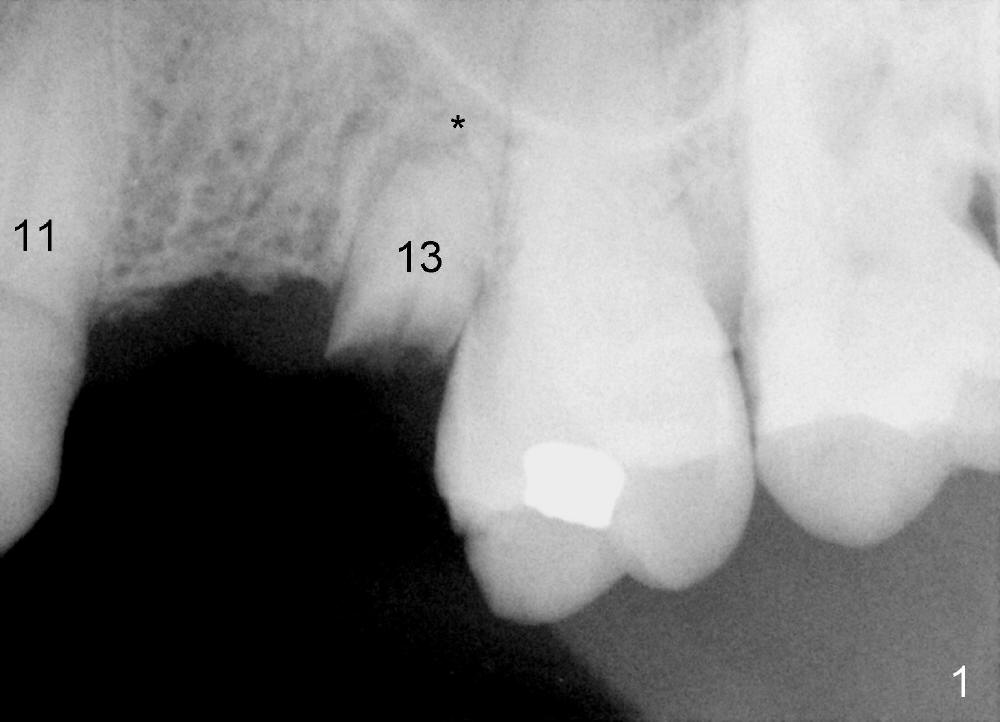
A 77-year-old man dislodged a bridge involving the teeth #11-13 5 years ago (Fig.1). It was recemented. Recently it is dislodged again. The patient agrees to have a definitive treatment to support the lower right quadrant reconstruction.
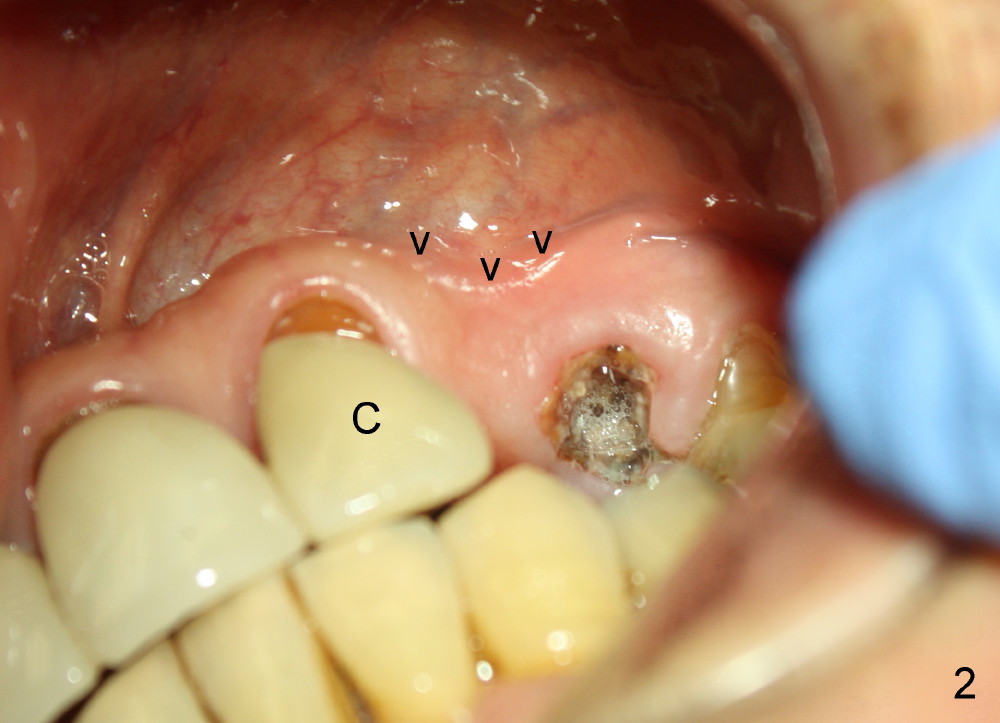
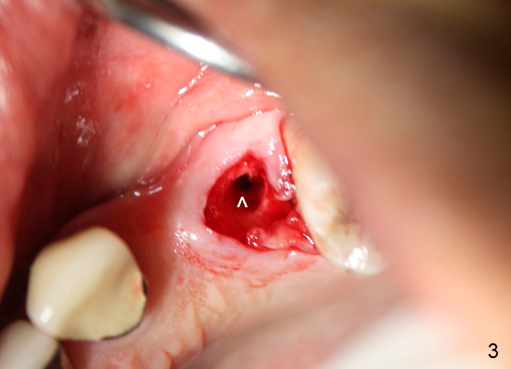
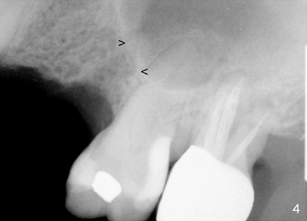
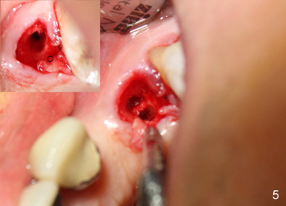
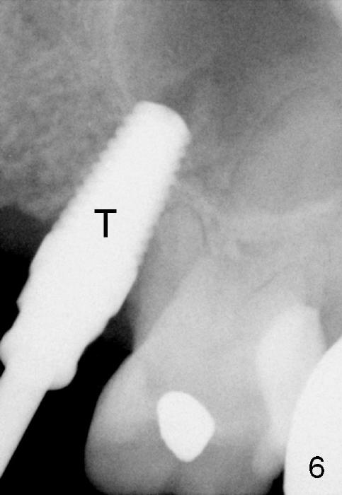
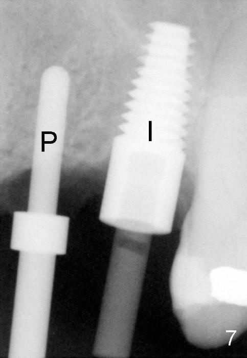
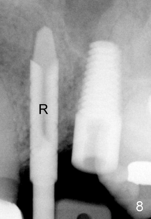
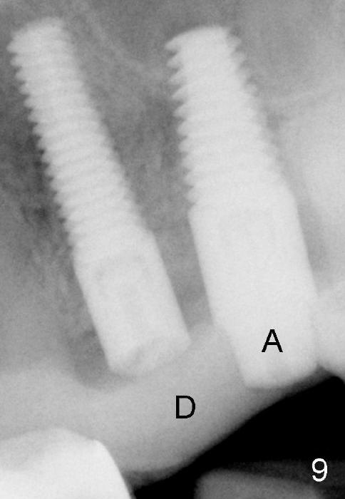
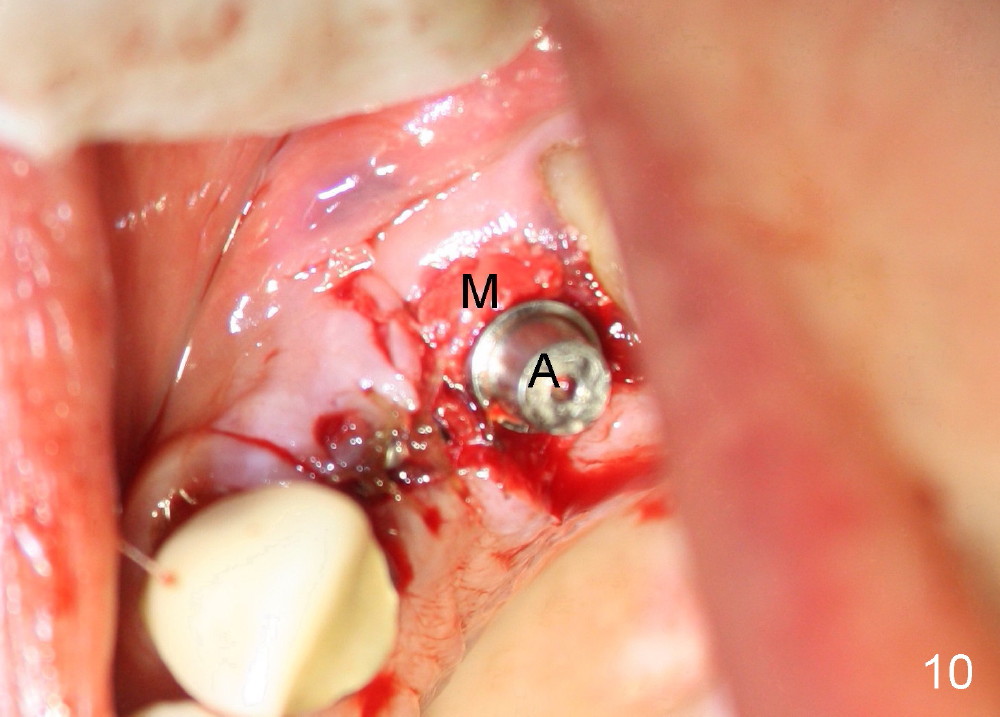
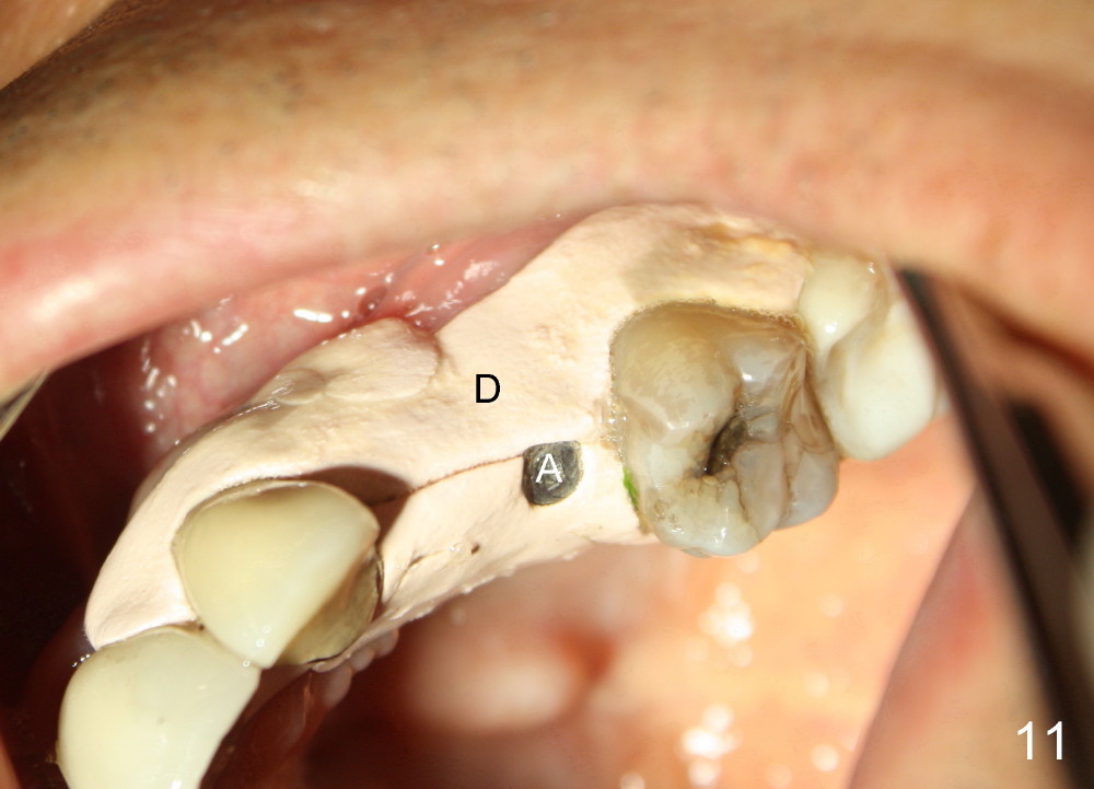
The anterior retainer is kept as a crown at #11 (Fig.2: C), whereas the residual root at the site of #13 is extracted with apical perforation (Fig.3 ^). It is confirmed by Fig.4 (<). To close the perforation, an osteotomy is initiated on the palatal wall of the socket with a 2 mm osteotome (Fig.5 insert: circle). As the osteotomy is being enlarged by a 3 mm osteotome, the bone between the original socket and the osteotomy is being pushed buccally. The former is being closed (Fig.5). The osteotomy is finished with combination of osteotomes and reamers. Fig.6 shows that a 5x14 mm tap is inserted at the site of #13 and that the sinus floor is lifted. In fact the sinus membrane is partially torn at the osteotomy, which is repaired by insertion of Colla-form Dressing (Impladent), followed by autogenous bone (harvested from reamers) mixed with Osteogen (Impladent). A 5x14 mm implant is placed at the site of #13 with insertion torque more than 60 Ncm (Fig.7: I). An incision is made at the site of #12 to start osteotomy with insertion of a parallel pin (Fig.7 P). A 3 mm reamer is kept in place for position confirmation (Fig.8 R). Due to ridge atrophy (Fig.2 arrowheads), a much smaller, but longer implant is placed at the site of #12 (Fig.9: 4x17 mm). The autogenous bone harvested from #12 osteotomy is placed in the buccal gap of #13, followed by insertion of Colla-form Dressing (Fig.10 M). To protect the membrane, a short abutment (4x3 mm) is temporarily placed (Fig.9,10 A) and perio dressing (Fig.9 D) is applied around the abutment and the interproximal areas of the neighboring teeth. Usually perio dressing dislodges around 1 week postop, particularly for a large edentulous space. In this case, the dressing is quite stable 11 days postop: the abutment (Fig.11 A) appears to contribute to retention of the dressing (D).
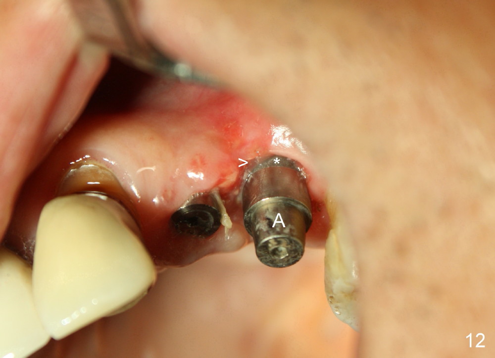
The perio dressing dislodges 18 days postop (Fig.12). By that time, the extraction wound has healed. The exposed rough surface of the implant (*) is most likely to be covered by fresh and healthy granulation tissue (>) underneath the gingiva. Subsequently the abutment (A) is removed, since it has finished its temporary function.
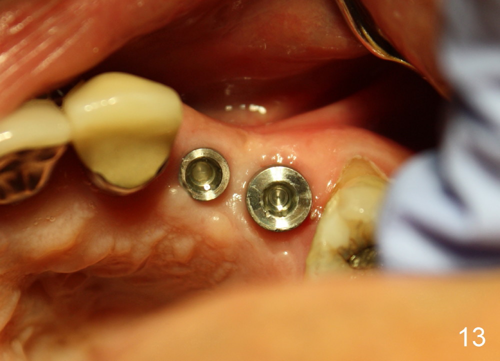
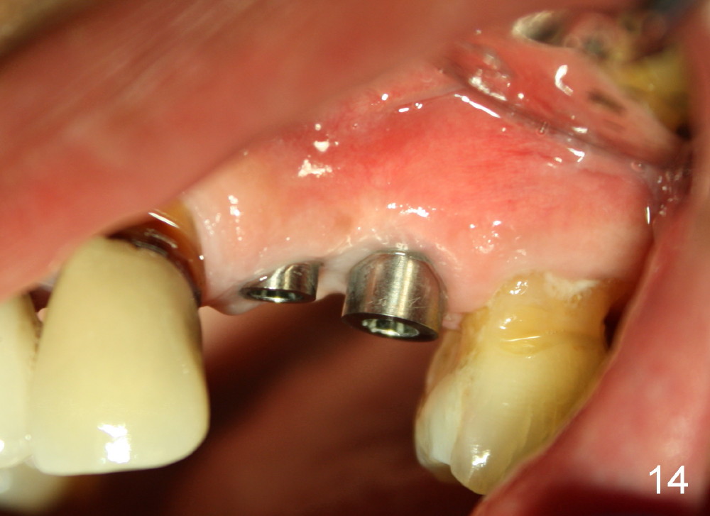
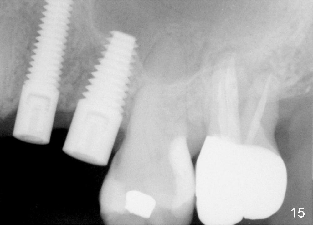
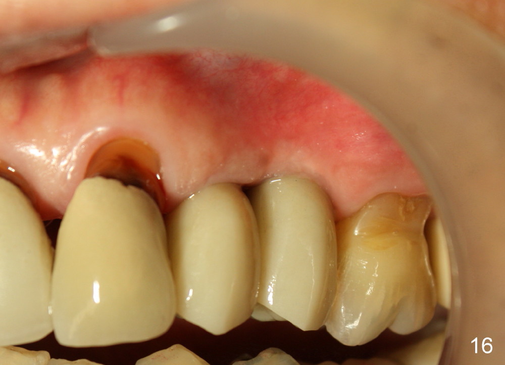
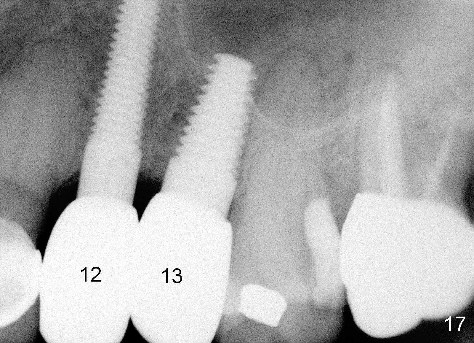
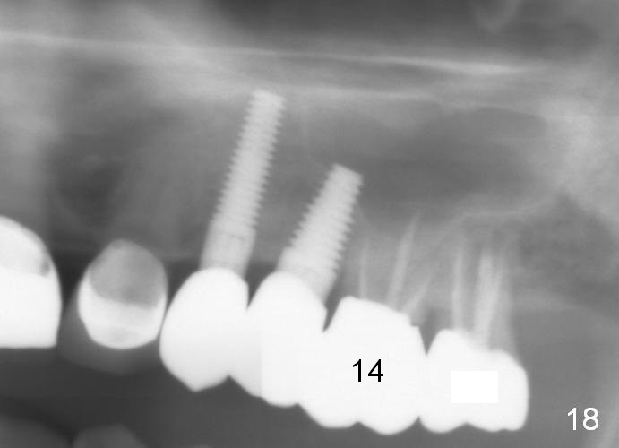
Three month follow-up shows that the gingiva and bone heal around these two implants (Fig.13-15). Crowns are cemented 4.5 months postop (Fig.16). There is no or minimal bone loss 9 months post cementation (Fig.17, as compared to Fig.15). The bone is stable around the implants 18 months post cementation (Fig.18 panoramus). Root canal therapy is done at #14 between the last follow up appointments.
Return to Immediate Implant for Upper Bicuspids Bone Graft
Xin Wei, DDS, PhD, MS 1st edition 05/06/2013, last revision 01/19/2018