|
|
|
|
||
|
|
|
|
|
|
|
|
|
|
|
|
|
|
|
|
||
Implant after Socket Preservation
Six months after socket preservation, a 49-year-old lady returns for #14 implant placement. Preparation will be the same as a recent case for sinus lift. Use a new sinus lift kit if available.
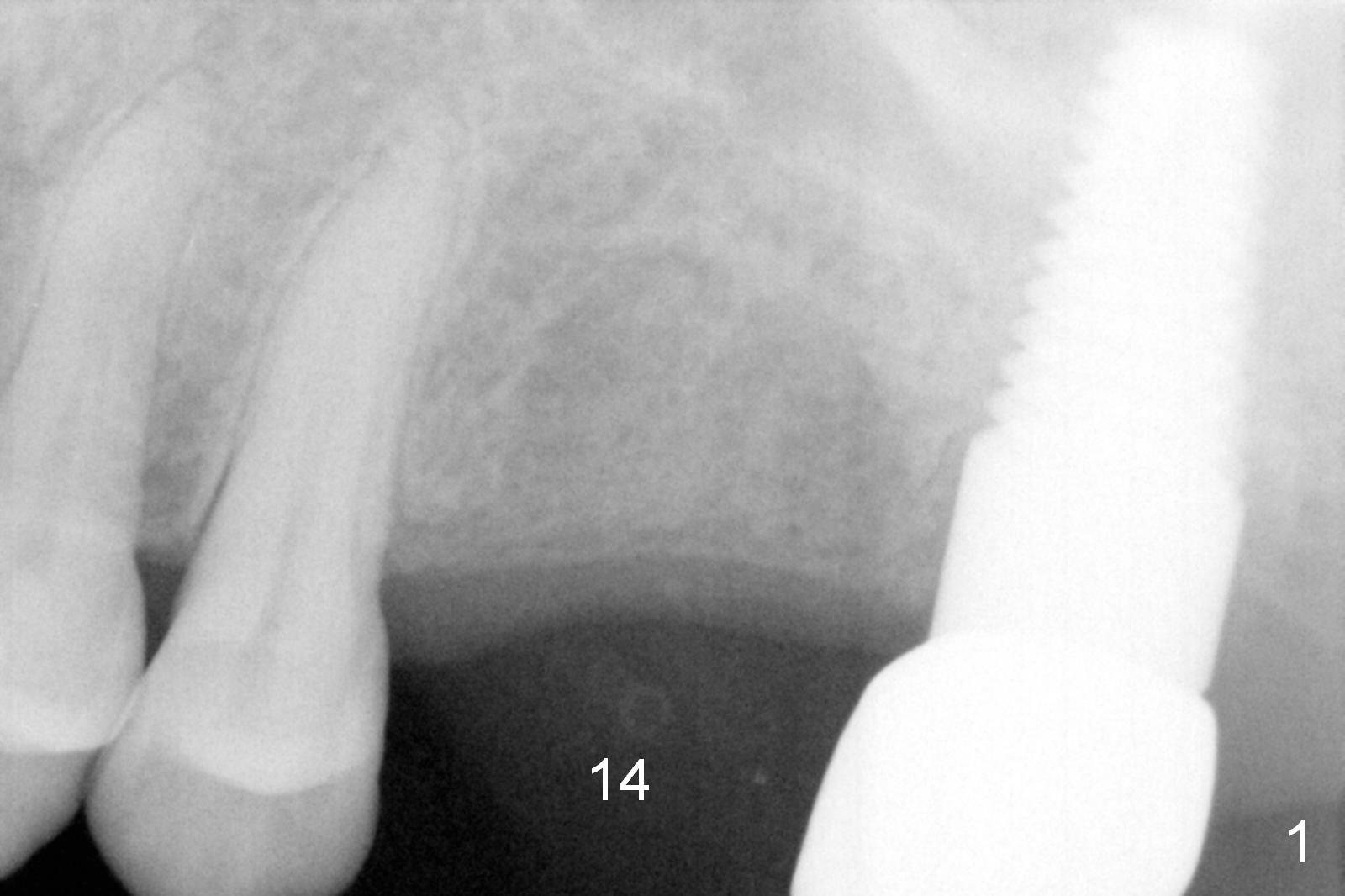
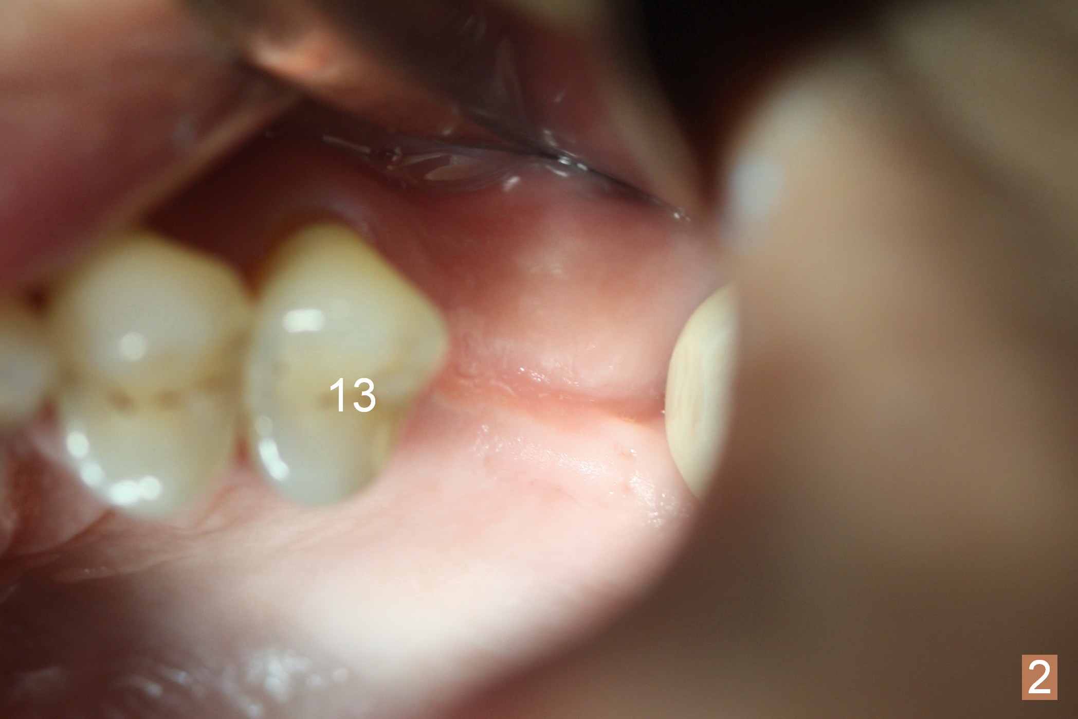
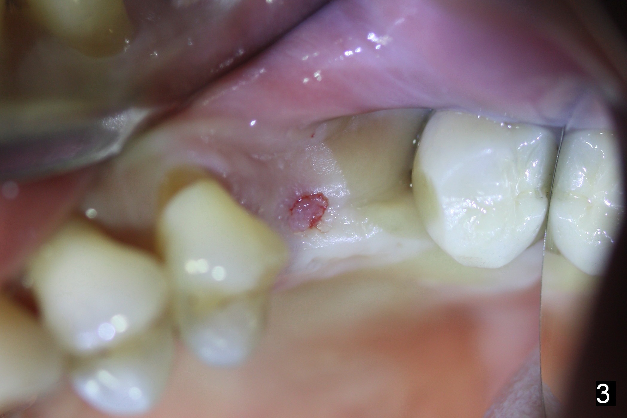
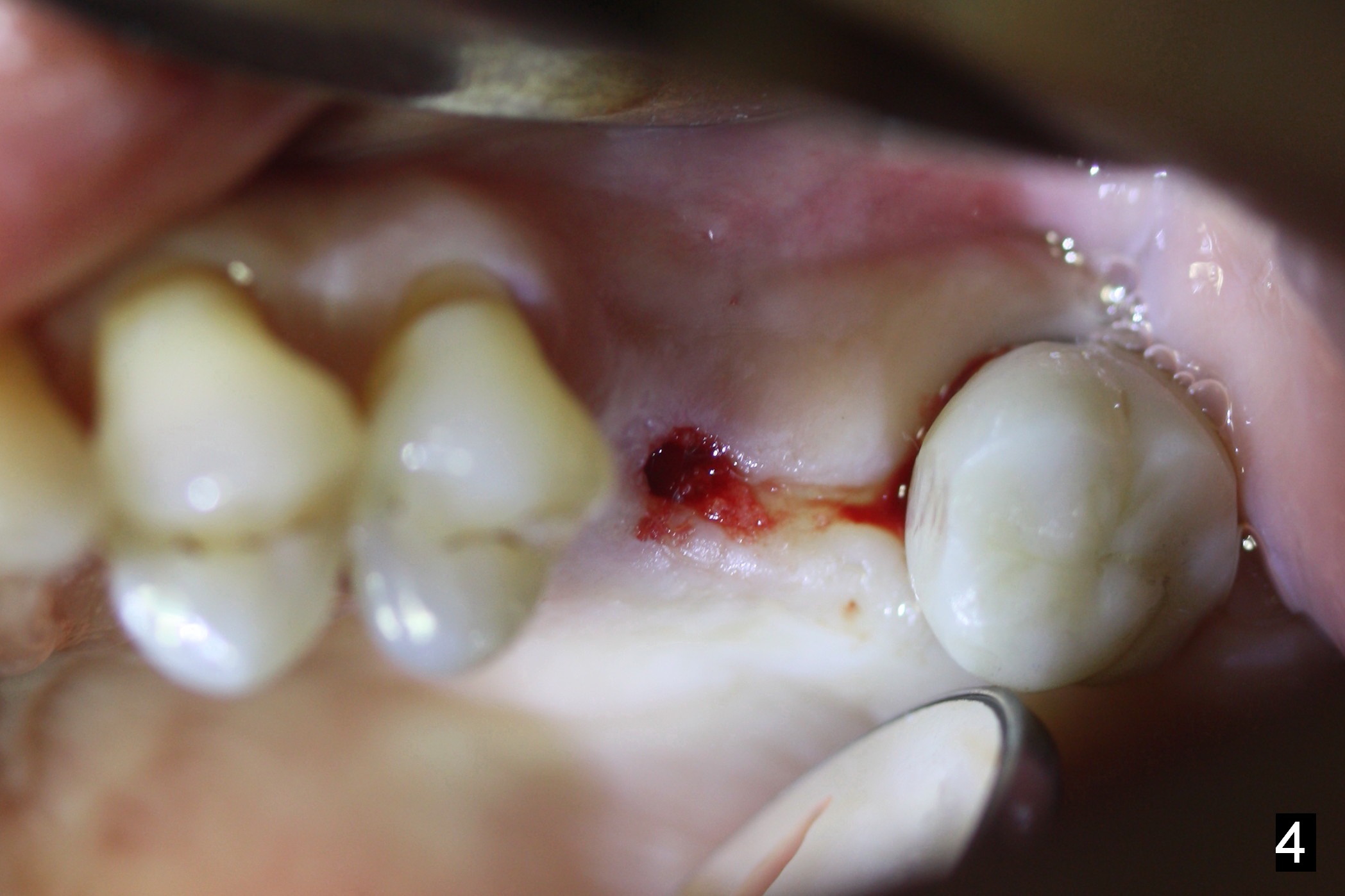
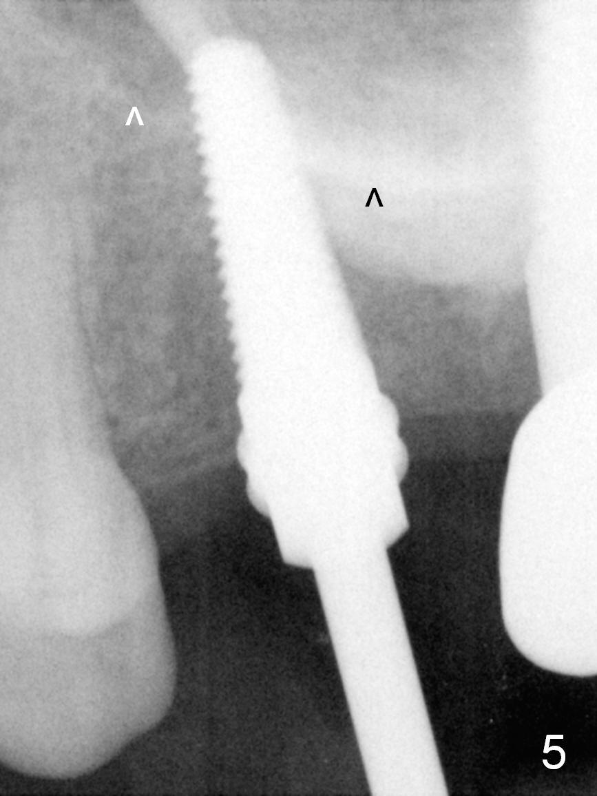
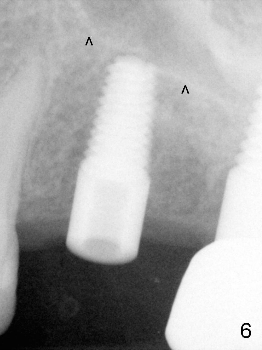
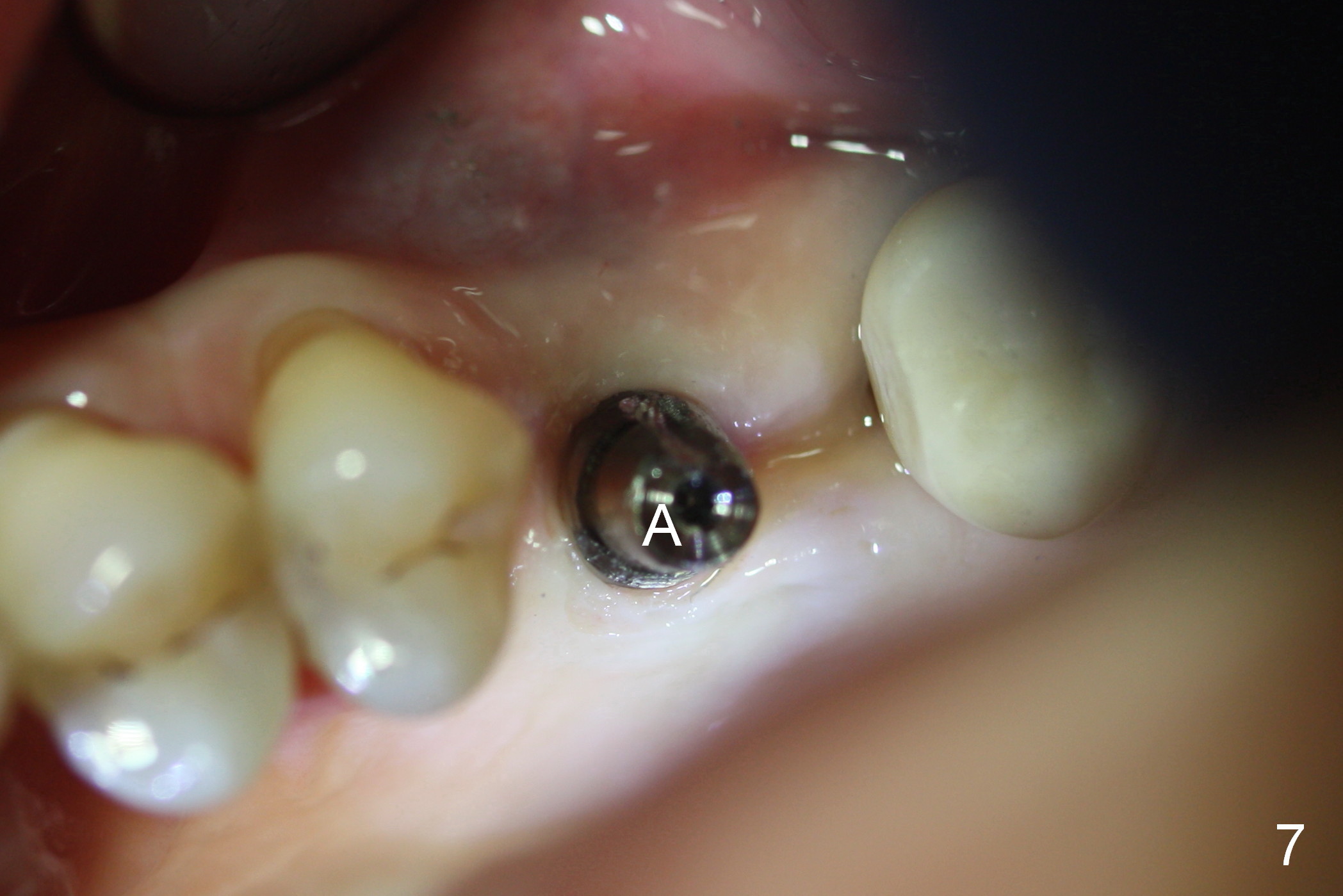
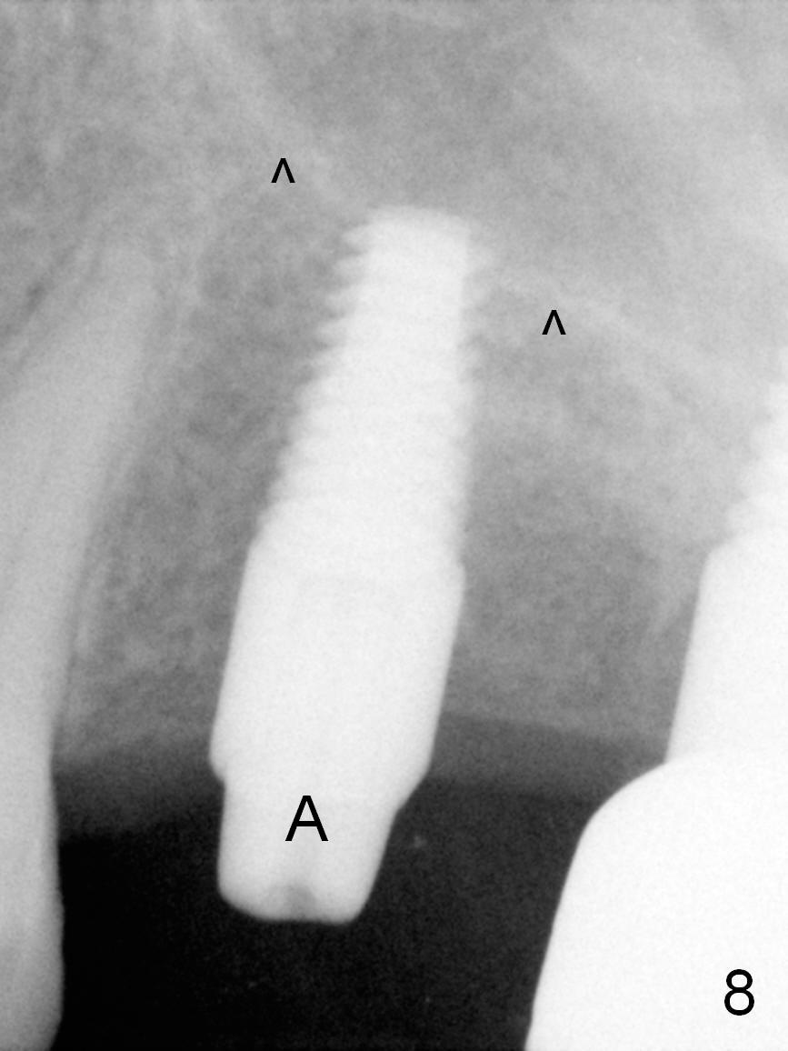
Preop PA shows bone height 13 mm (Fig.1). There is ridge atrophy buccally (Fig.2-4,7, drawback of delayed implant). A 2/3 mm trephine bur is used for access (Fig.3) and collection of the gingiva and underlying osseous tissue for 10 mm from the gingiva (Fig.4). The osteotomy is extended with 2 mm pilot drill and 2.5 mm reamer for 13 mm, followed by insertion of a 4.5x17 mm tap for 14 mm (Fig.5). Mineralized Cortical allograft and Osteogen is placed for sinus lift before placement of a 5x14 mm implant (Fig.6). After placing the implant deeper, a 4x3 mm abutment is placed (Fig.7,8 A) for an immediate provisional.
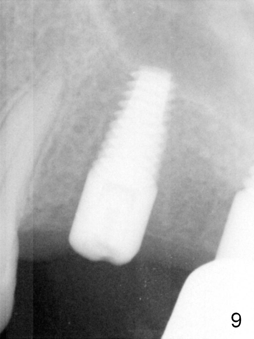
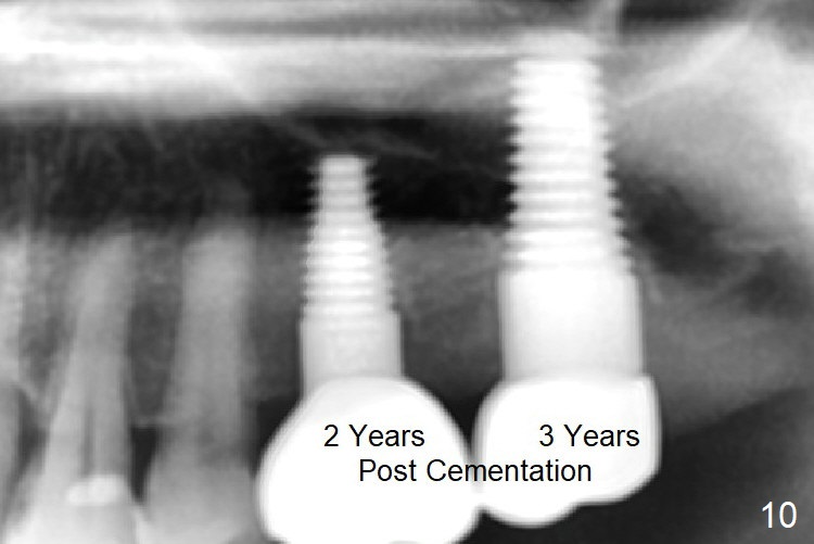
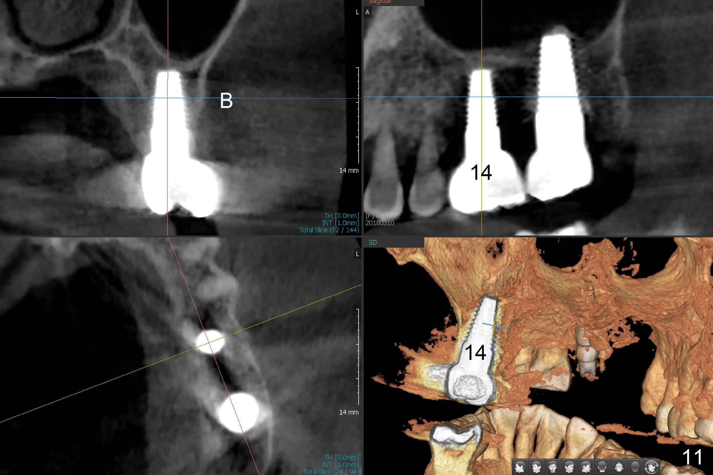
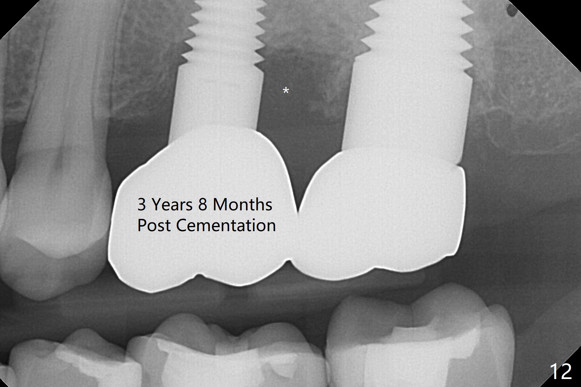
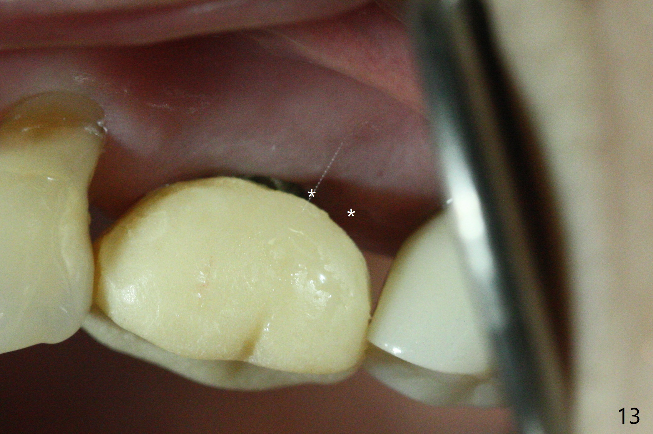
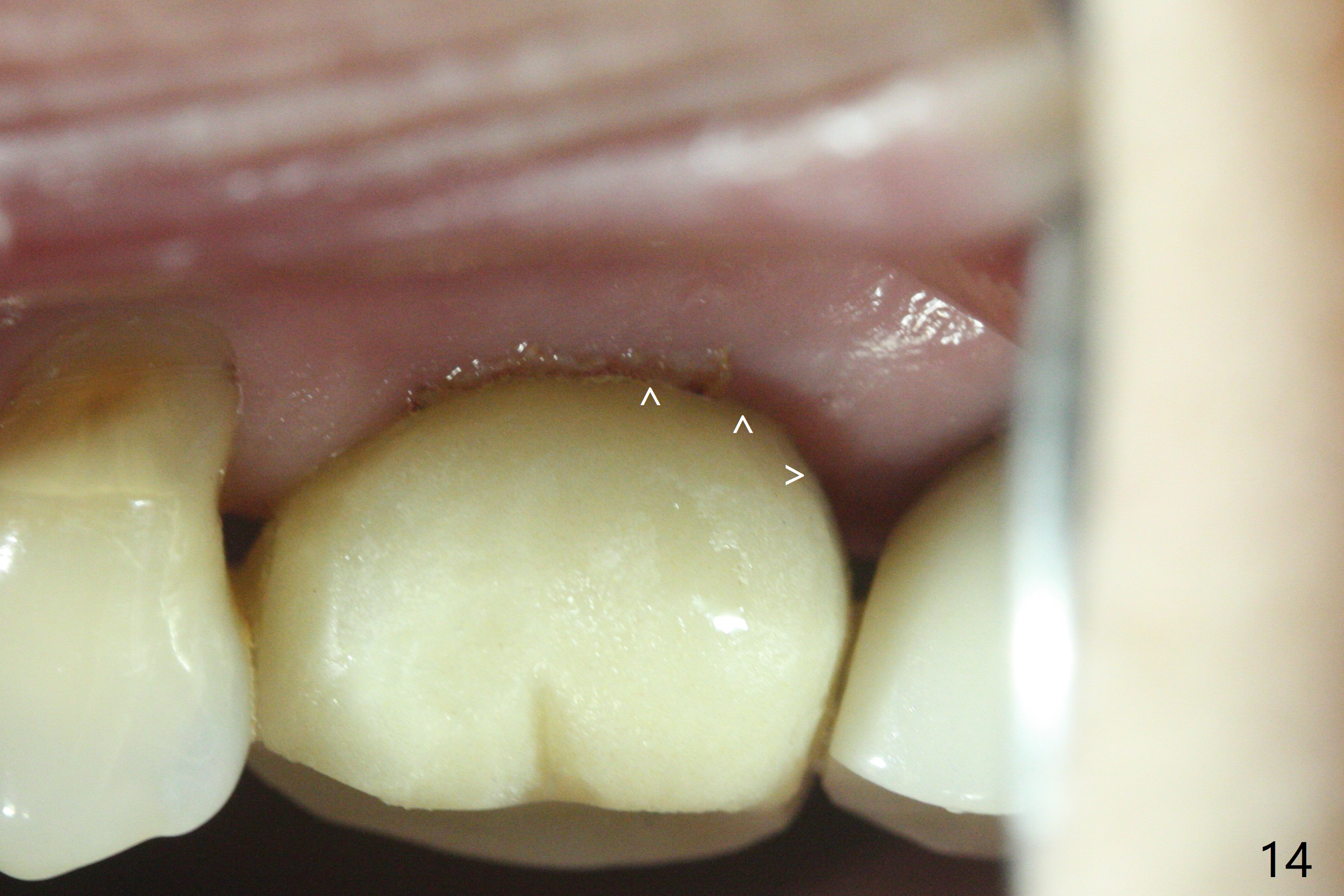
The patient returns for restoration 3 months 1 week postop. Her chief complaint is bleeding that morning. The provisional is loose. The margin of the provisional is probably not smooth. The distal gingiva is erythematous and edematous. There is excessive hemorrhage when gingival retraction cords are inserted. The abutment is removed and a healing screw is inserted. Three weeks later, the gingiva around the implant is healthy. There is no bone loss surrounding the implant (Fig.9). The case would develops into periimplantitis if the definitive restoration were processed when there was early sign of infection. There is no bone resorption 2 years post cementation (Fig.10) CBCT confirms no buccal placement (Fig.11). There are deep distal pockets of the implant with pain and bleeding on probing at #14 three years 8 months post cementation, consistent with bone loss (Fig.12 *). The latter is associated with the loose contact between the crowns. A provisional is fabricated for #14 with normal proximal contact after crown removal. The gingival inflammation subsides in a month, although there is food impaction, most likely associated with enlarged gingival embrasure (Fig.13 *). After gingivectomy, a temporary crown is remade with extension of the margin (Fig.14 ^) to minimize the gingival embrasure.
Return to Upper Molar Immediate Implant, #2, 6 ,15 Xin Wei, DDS, PhD, MS 1st edition 12/30/2015, last revision 12/30/2019