|
|
|
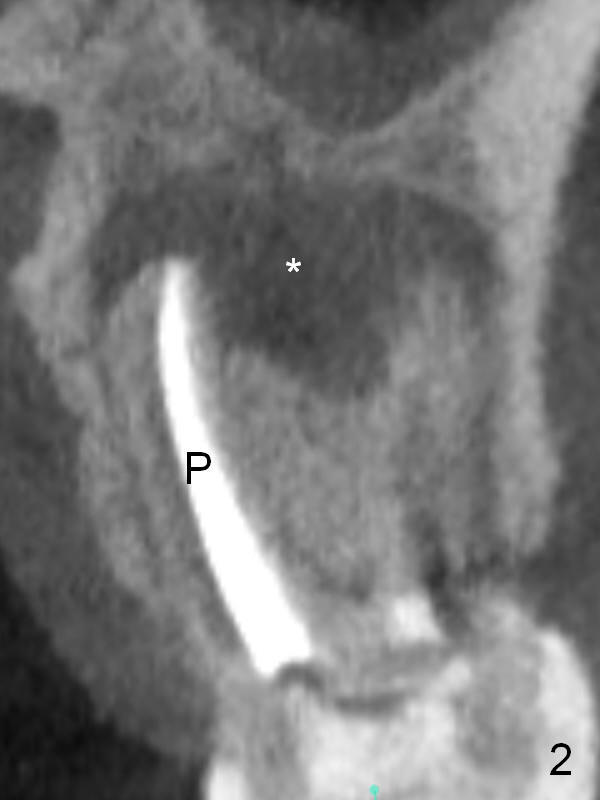
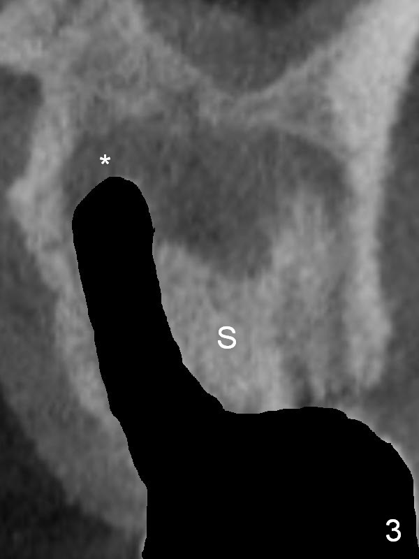
Fig.2 is a coronal section of CBCT of the affected tooth showing a large apical lesion (*). When the tooth is removed (Fig.3), limited amount of granulation tissue is able to be removed apical to the palatal root (P in Fig.2).
Xin Wei, DDS, PhD, MS 1st edition 02/17/2017, last revision 02/17/2017