,%20Vera,%20parallel.jpg)
|
|
|
|
|
|
|
|
|
|
|
|
Study Septal Dimension Before Osteotomy
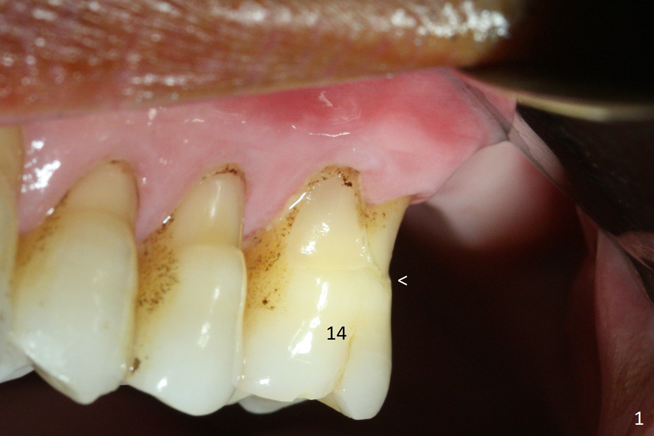
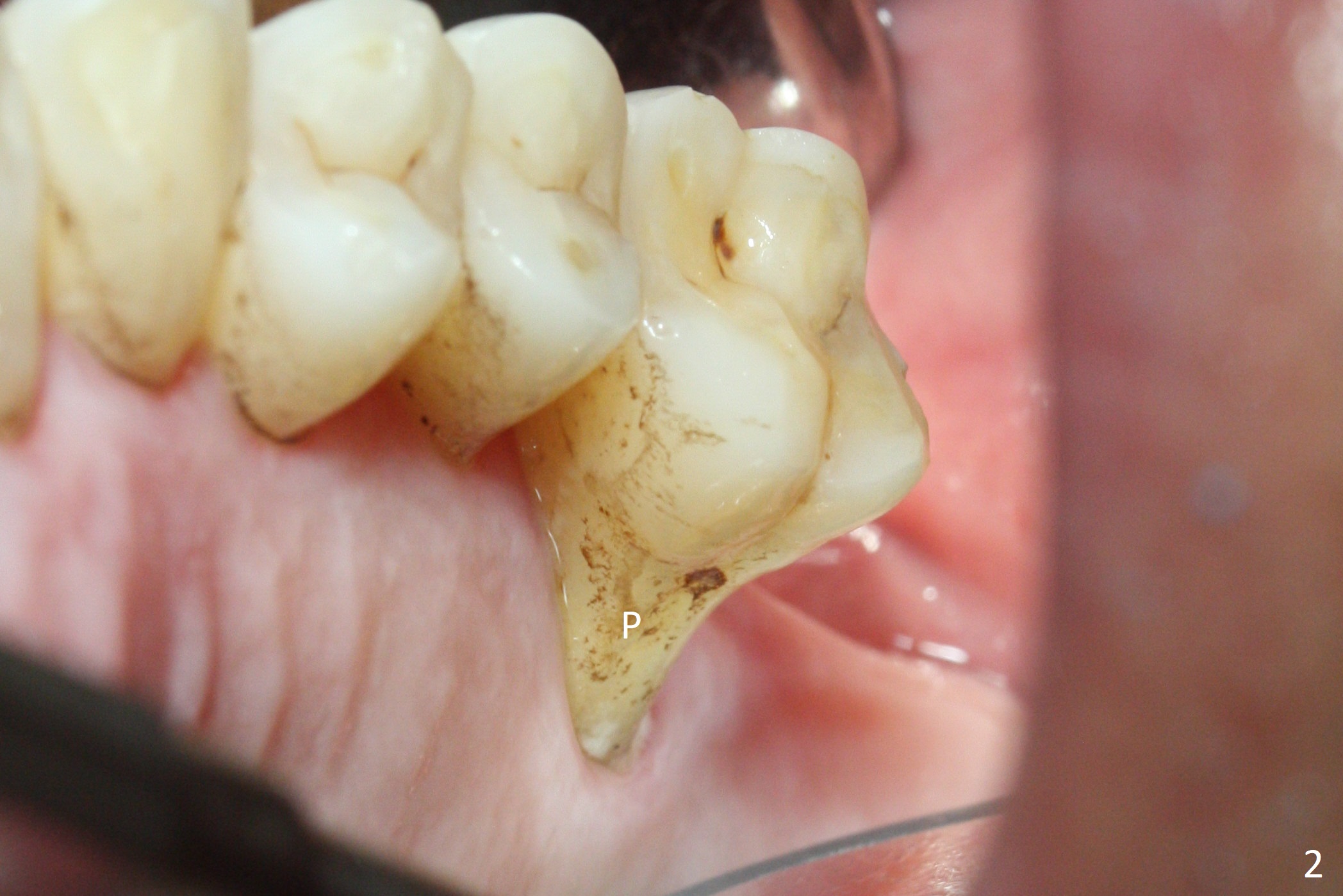
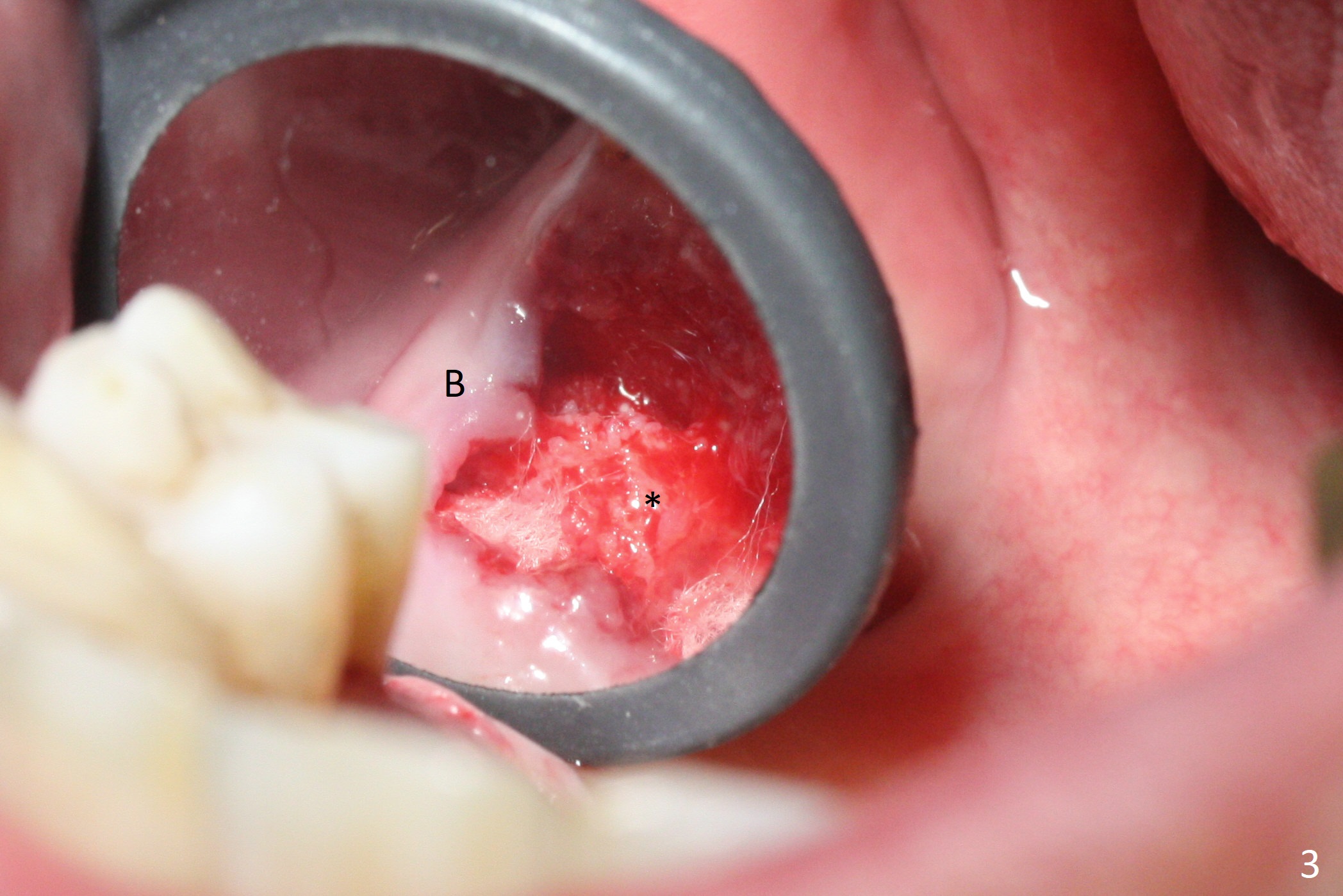
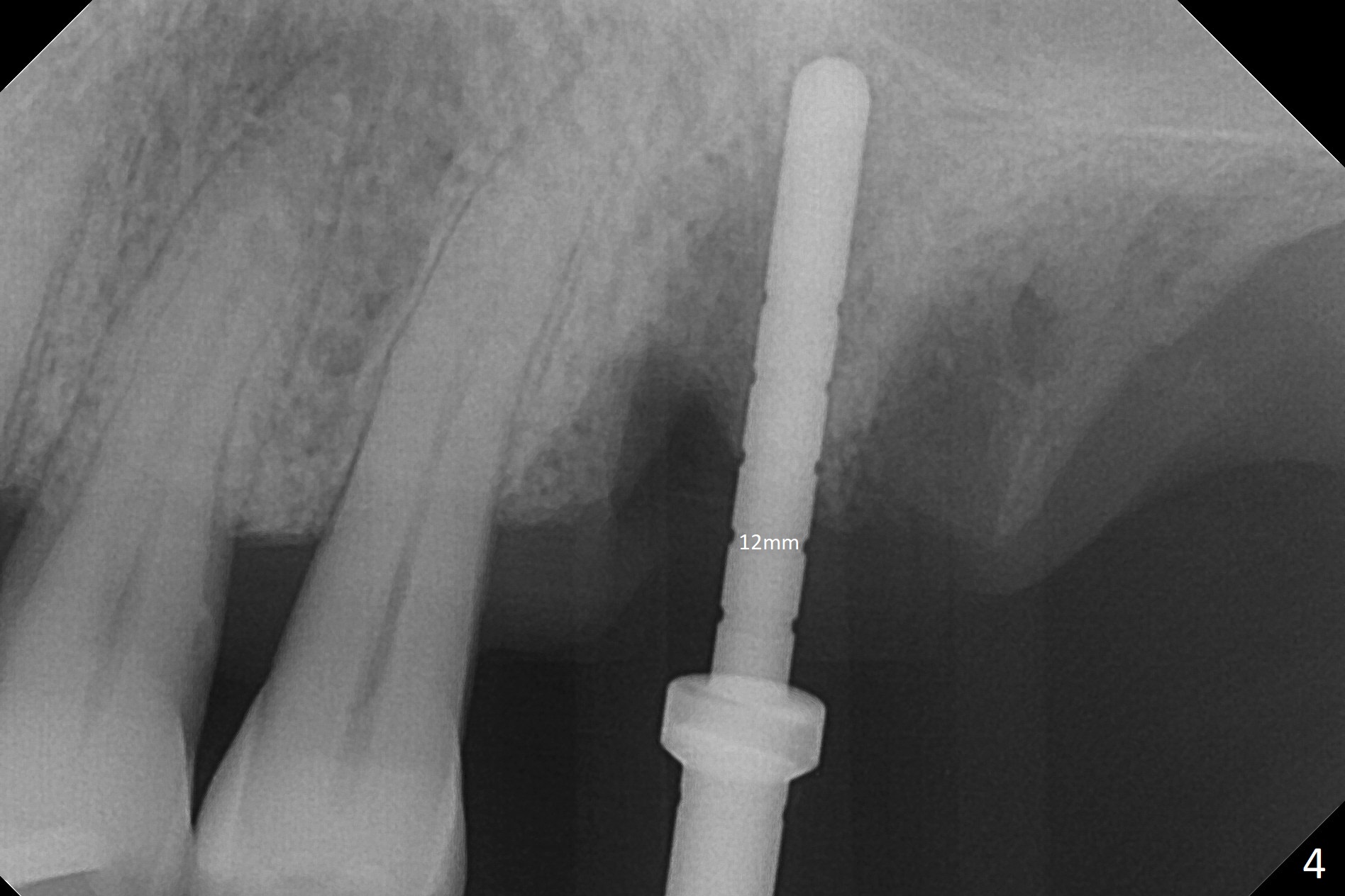
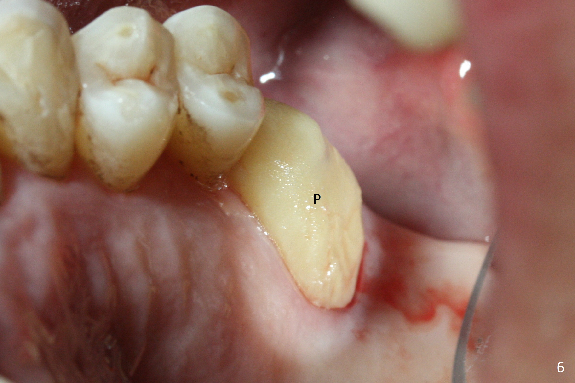
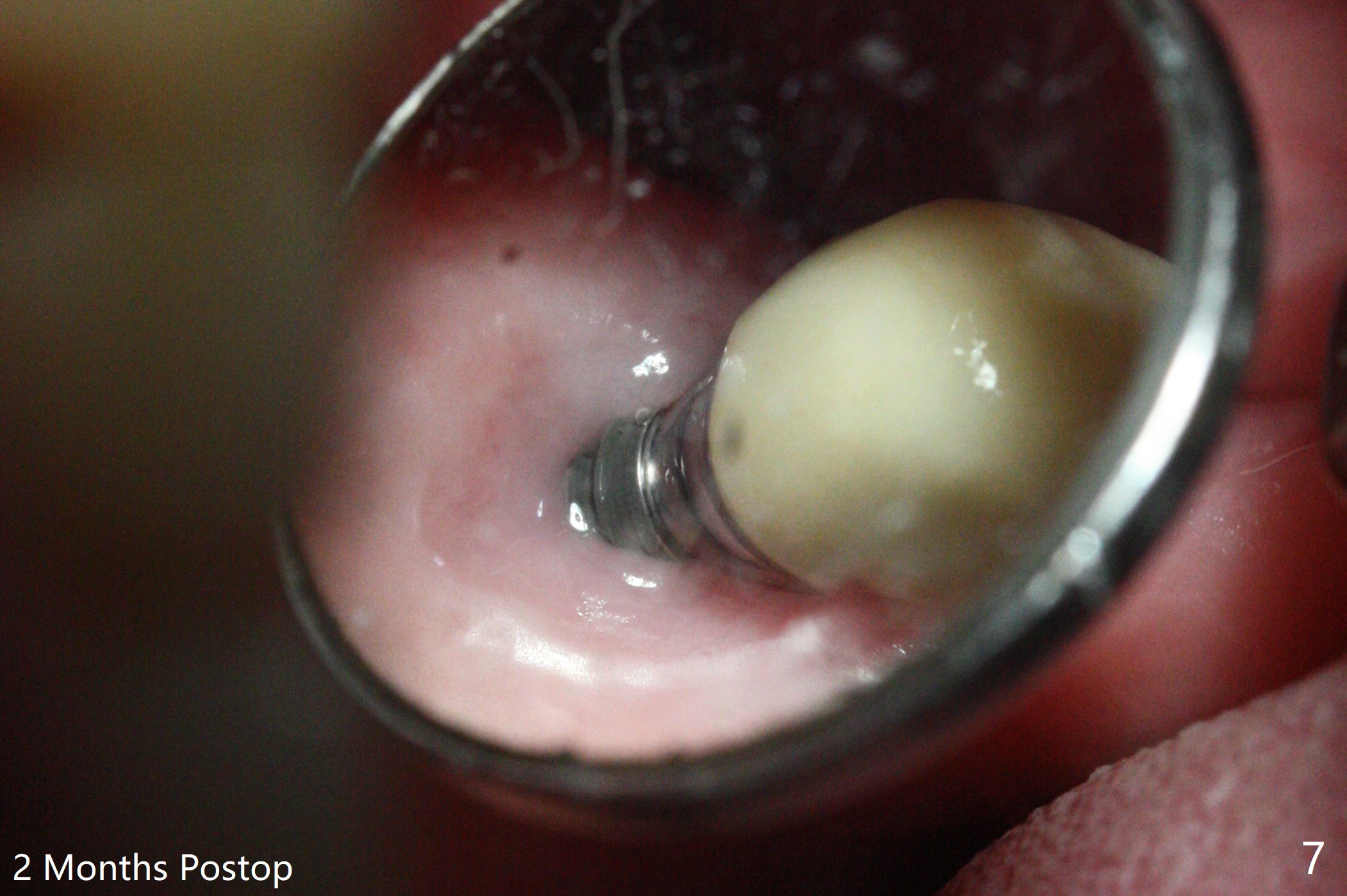
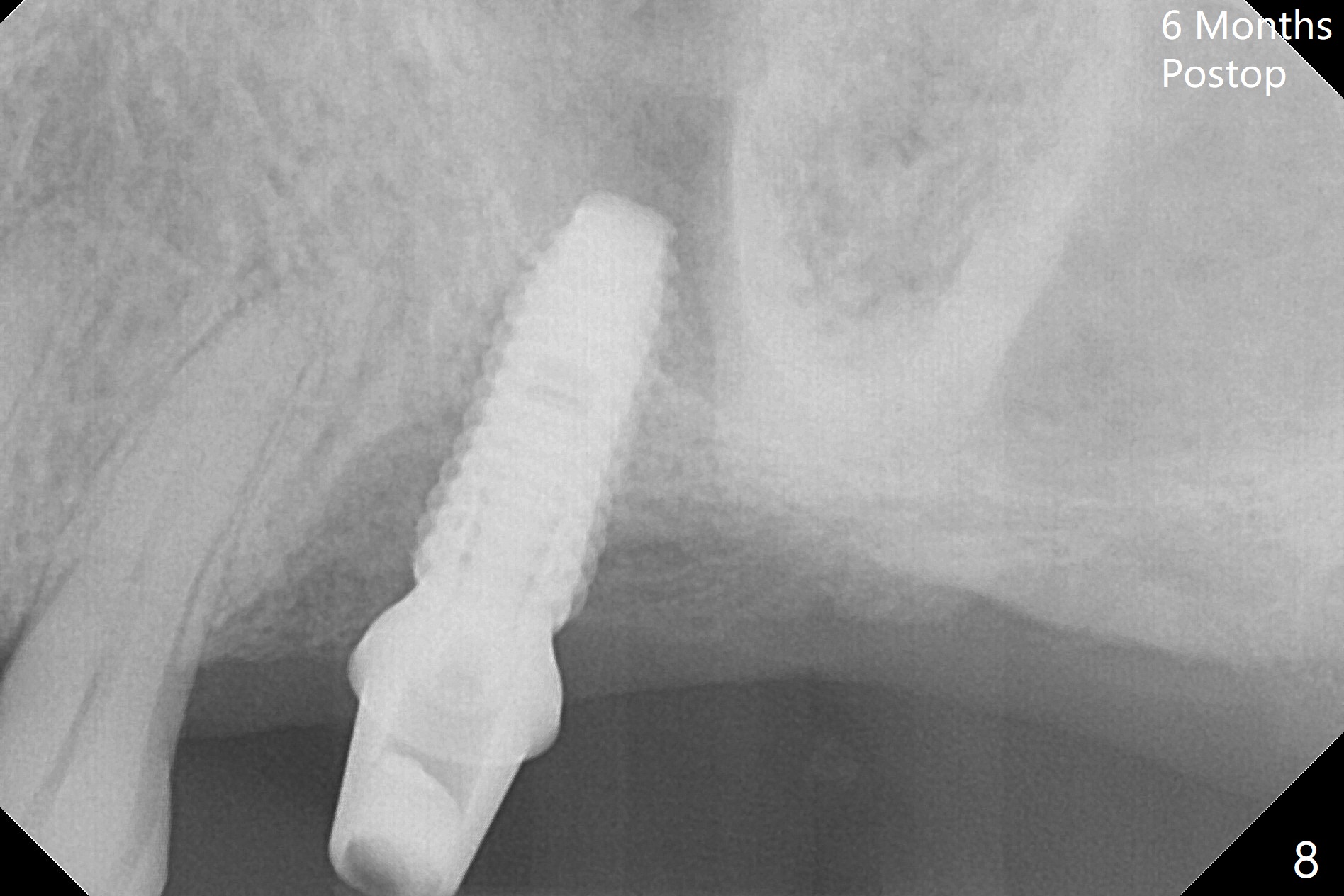
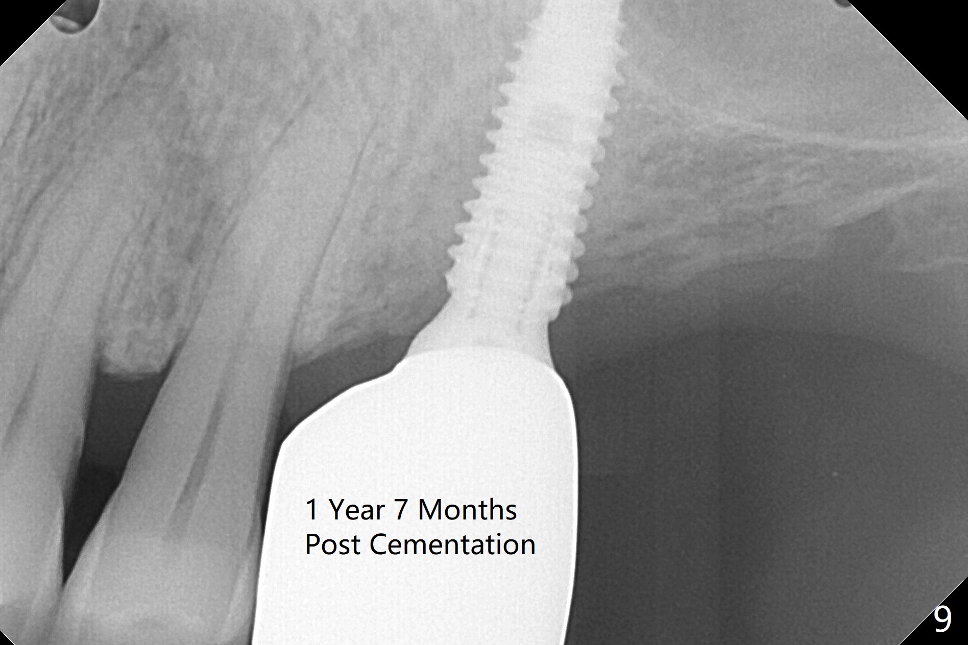
Buccal gingival recession at #14 (Fig.1) is less severe than the palatal one (Fig.2). Without raising the buccal gingiva (Fig.3 B), osteotomy in the septum is established apparently in the middle socket (Fig.3 *, 4). As the osteotomy increases with 3.8 mm drill, the palatal wall of the osteotomy starts to be perforating. When a 4.5x11.5 mm implant is placed, palatal threads are exposed, to which autogenous bone and Vera Graft are placed (Fig.5 *). After placement of a 5.5x5(3) mm abutment, an immediate provisional (Fig.6 P) is fabricated to cover the sockets. If the septal dimension were studied carefully by raising the buccal gingiva slightly, the osteotomy could be initially more buccal so that the palatal thread exposure could be less. When the provisional is removed 1.5 months postop, the implant is exposed palatally. The margin of the provisional is modified so that the implant can be cleaned by the patient using Water Pik. In fact, the healthy gingiva seems to be attached to the implant threads 2 weeks later (Fig.7). The distal implant threads remains exposed 6 months postop; it appears that the distal socket wall has resorbed (Fig.8). The sockets heal 1 year 7 months post cementation (Fig.9).
Return to
Upper
Molar Immediate Implant, Prevent
Molar Periimplantitis (Protocols,
Table), Armaments
Xin Wei, DDS, PhD, MS 1st edition 11/10/2017, last revision 12/25/2019