|
|
|
|
Osteotomy in Healed Site
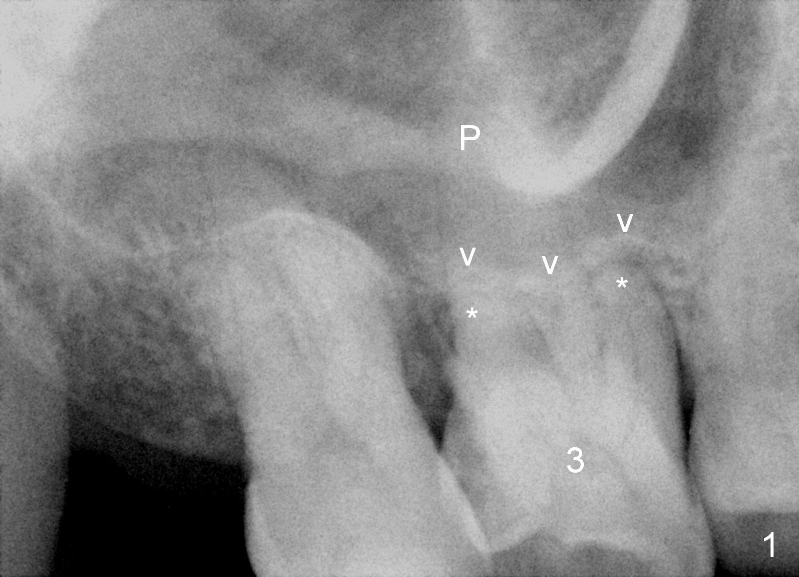
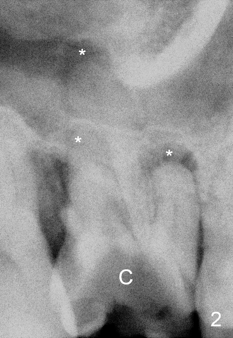
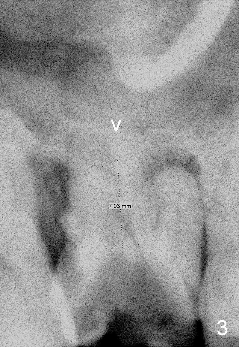
A 43-year-old man will return for #3 implant placement, 2.5 years after extraction. Pre-extraction PAs (Fig.1,2, taken 5 and 2.5 years ago) show that the palatal apex (Fig.1 P) is much higher than the buccal ones (*). The tooth became non-salvageable because of caries (Fig.2 C) in 2.5 years with periapical radiolucency (*). After extraction, the bone available for implantation is probably below the sinus floor (Fig.1,3 arrowheads), 7-9 mm.
Take preop PA for design. The biggest difference between the latest PA and the previous ones is probably the mesial shifting of the tooth #2.
Also take photos to show the buccal plate atrophy. Most likely use a scalpel to make a 9 mm long incision and initial osteotomy (to test whether the bone is soft or not), followed either by osteotomes or bone expanders. After making the incision, measure the thickness of the gingiva. Insert Tatum taps for stability confirmation. Place a UF implant and cemented abutment. Fabricate an immediate provisional if necessary.
Return to Upper Molar Immediate Implant
Xin Wei, DDS, PhD, MS 1st edition 08/27/2015, last revision 09/02/2015