|
|
|
|
|
||
|
|
|
|
|
||
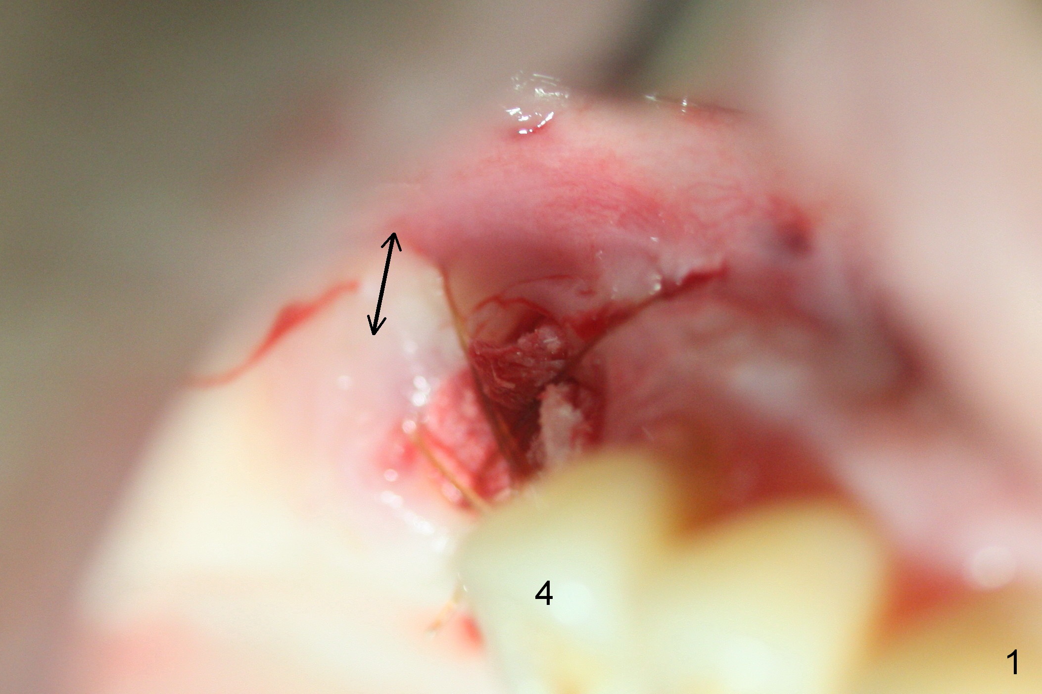
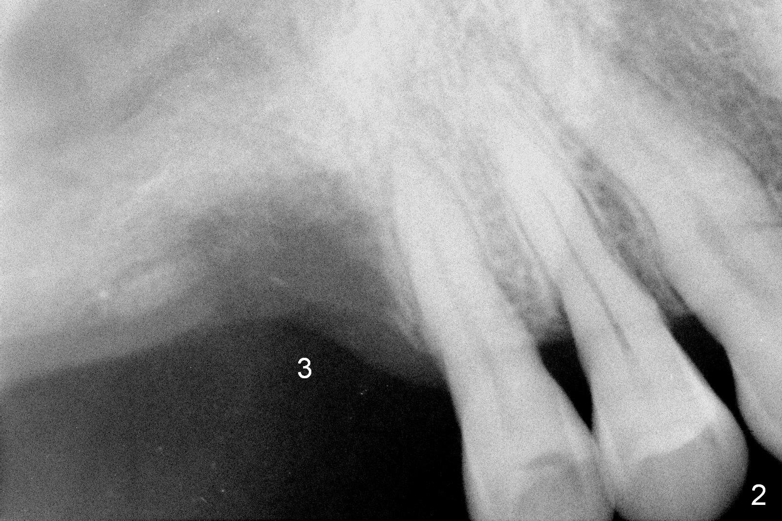
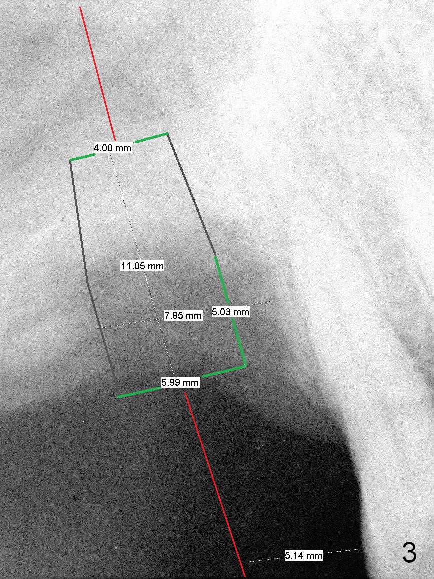
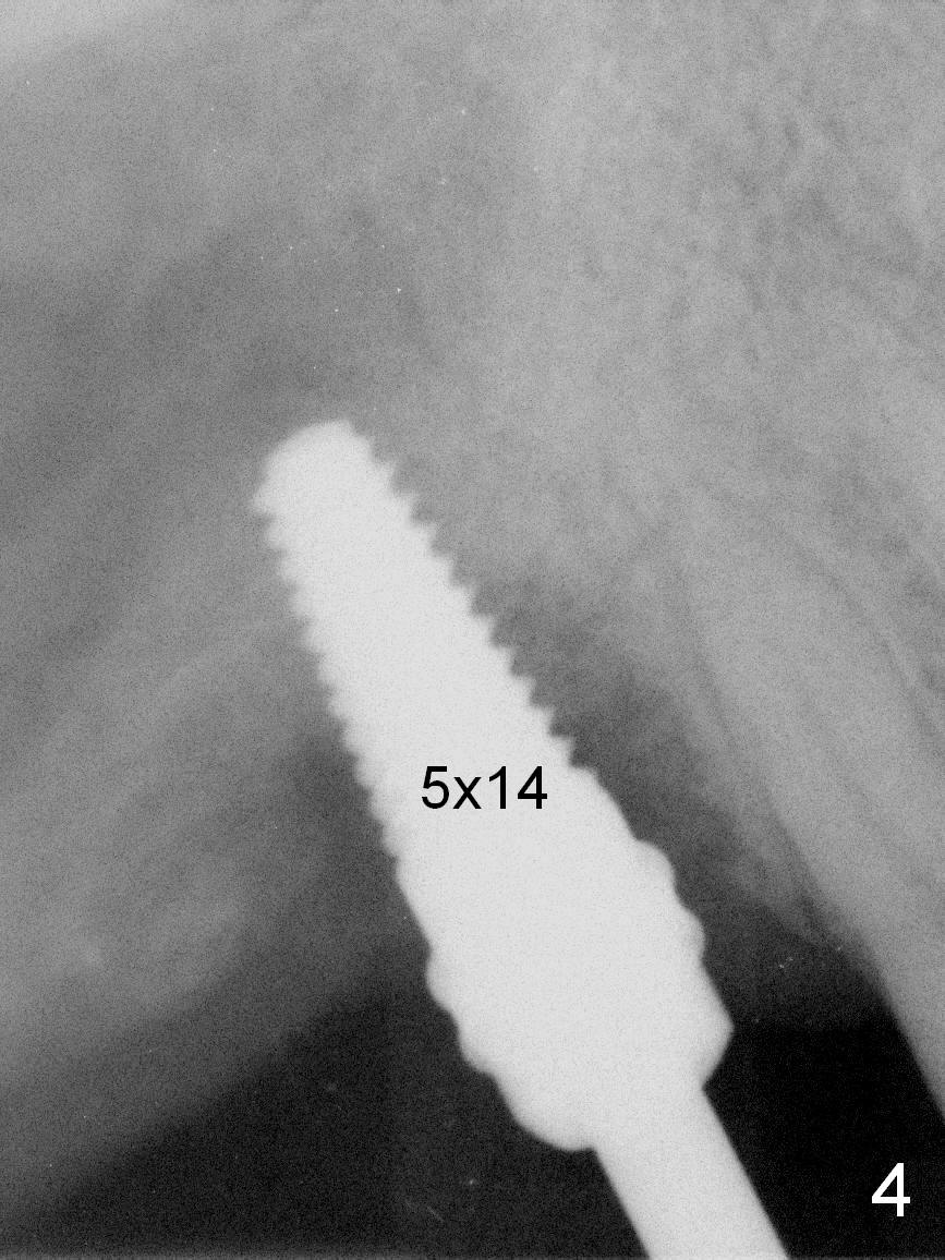
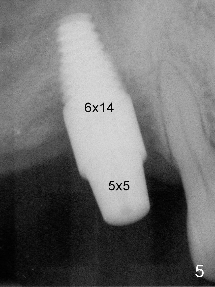
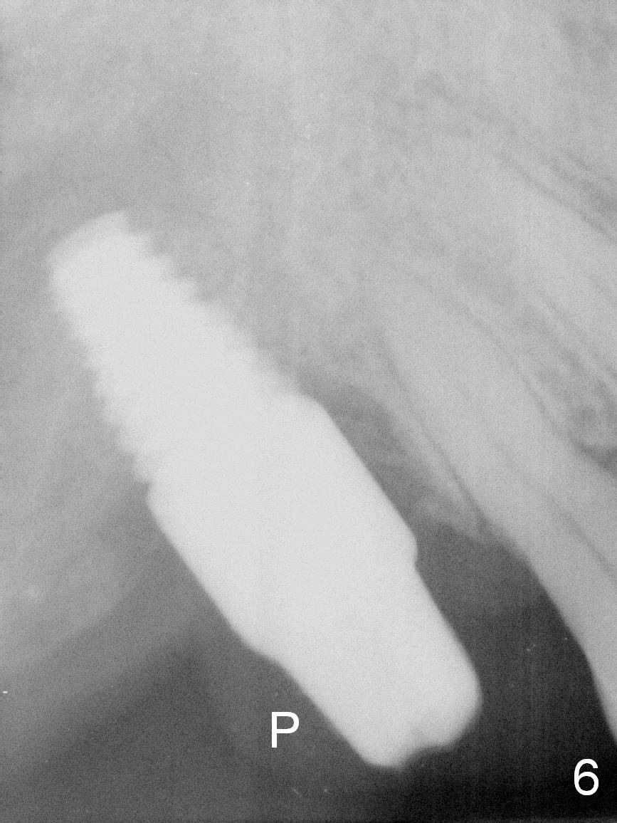
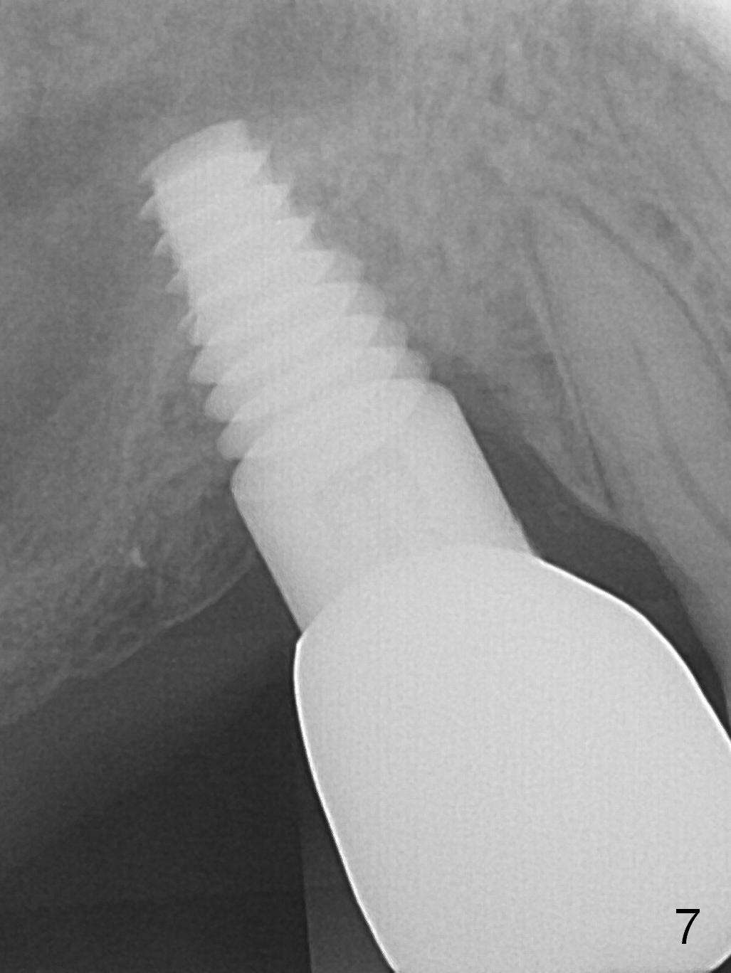
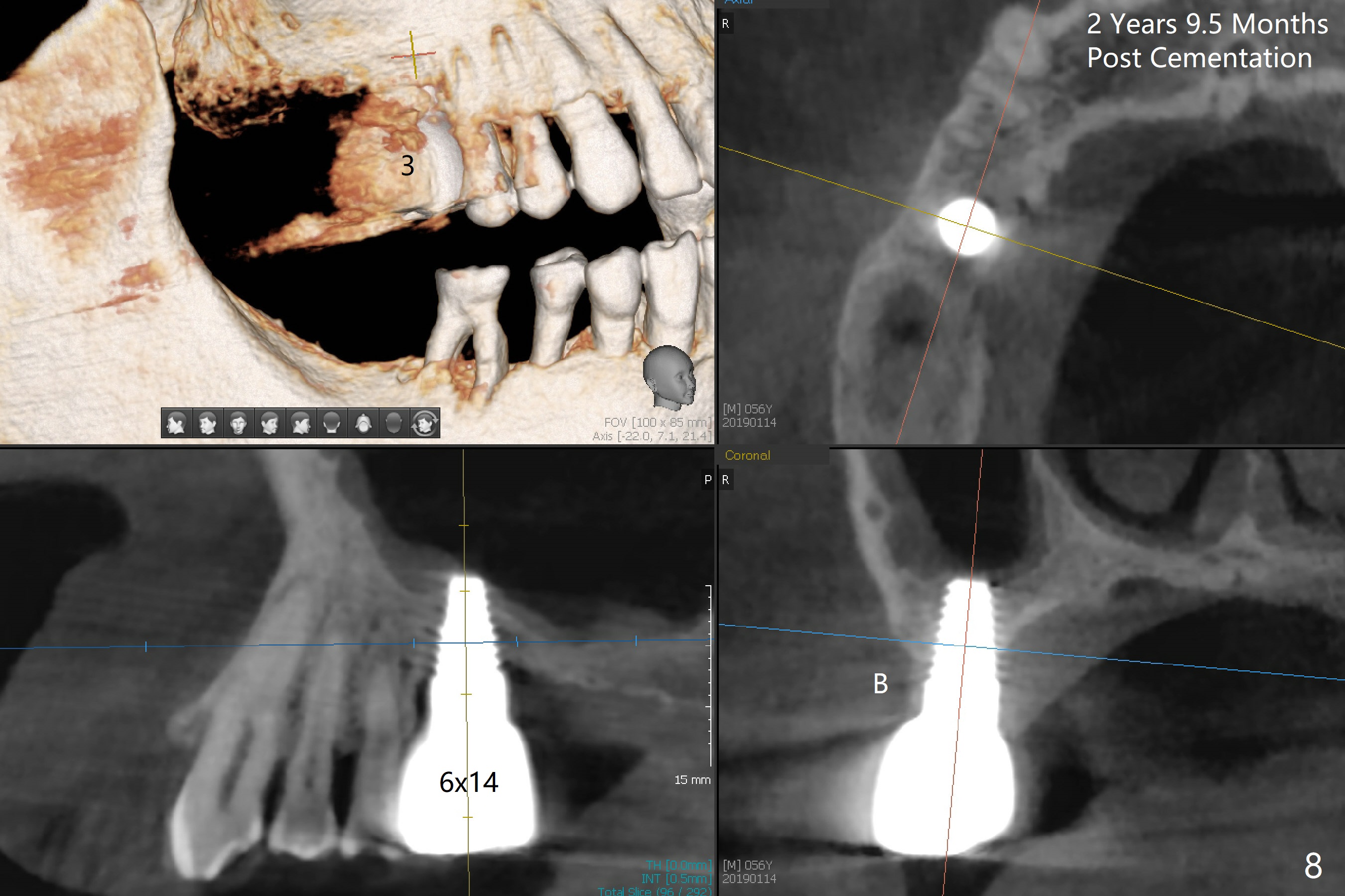
One month post immediate implantation at #3, the implant has lost stability. After the implant removal, socket (osteotomy) preservation is done (Fig.1). Note the limited buccal plate height (double arrows), which may be the main reason for implant failure. The patient returns for follow up 3.5 months post preservation. Bone height is approximately 7 mm (Fig.2). Clinically the buccal plate remains low. Use #15 blade to start bone expansion, followed by 1 and 1.5 mm microosteotomes, RT2,3 and taps (4.5 and 5). The incision should be as palatal as possible. The implant does not need to be large. Primary stability is priority. Use 4.5 mm implant spacer when 1 mm microosteotome is used; 4 for RT2 (rounded tapered osteotome); 3.5 for RT3. If the RT2 feels stable, insert a parallel pin to make sure that the axis of the osteotomy is parallel to that of the neighboring teeth (Fig.3 red line). Also make certain that the coronal end of osteotomes and taps is in the center of the occlusal table. Surgery proceeds as planned above except that RT4 has to be used prior to insertion of taps (Fig.4). A 6x14 mm tissue-level implant is placed, followed by placement of a 5x5 mm abutment (Fig.5). An immediate provisional is fabricated. There appears to be no bone growth 4 months postop (Fig.6), although clinically the implant and the immediate provisional (P) stable. Bone density seems increase once the permanent restoration is in place (Fig.7 (9 months post cementation)). With immediate implant (although fails), a larger and longer (6x14 mm) one is placed at #3 than that at #14 (5x11 mm) of the same patient. There is no gross bone loss 2 years 9.5 months post cementation (Fig.8).
Return to Upper Molar Immediate Implant 19 Xin Wei, DDS, PhD, MS 1st edition 10/10/2015, last revision 12/26/2019