,%20Vera%20and%20autogenous.jpg)
|
|
|
|
|
|
|
|
|
|
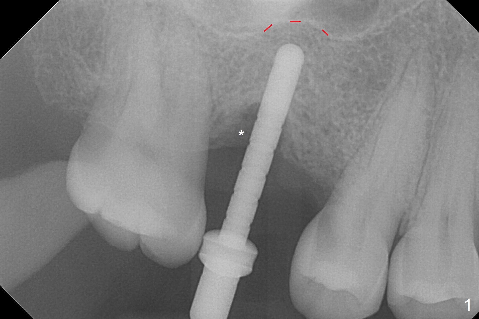
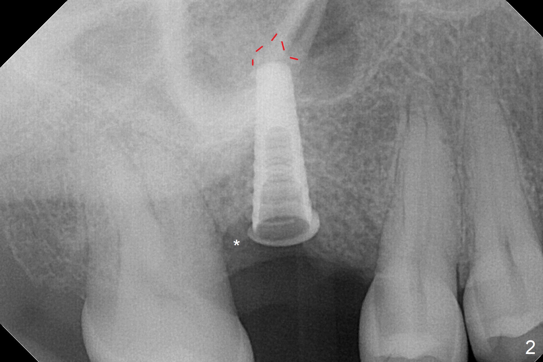
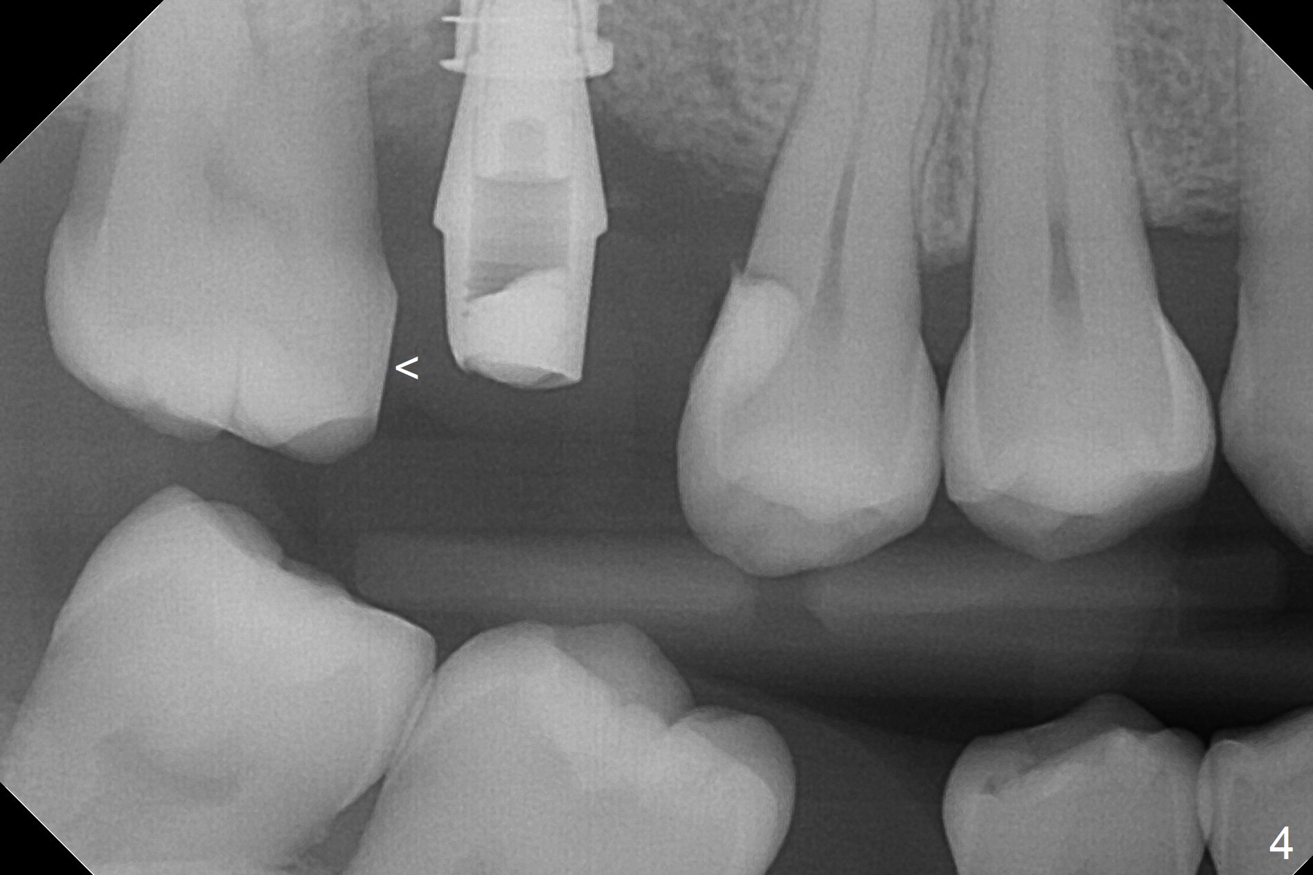
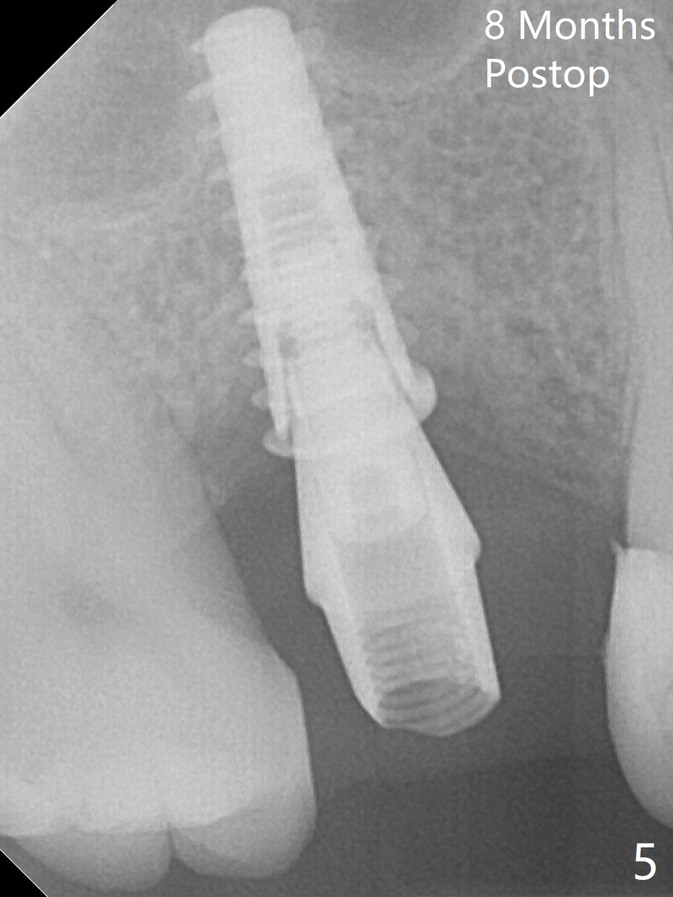
Sinus Lift with Implant
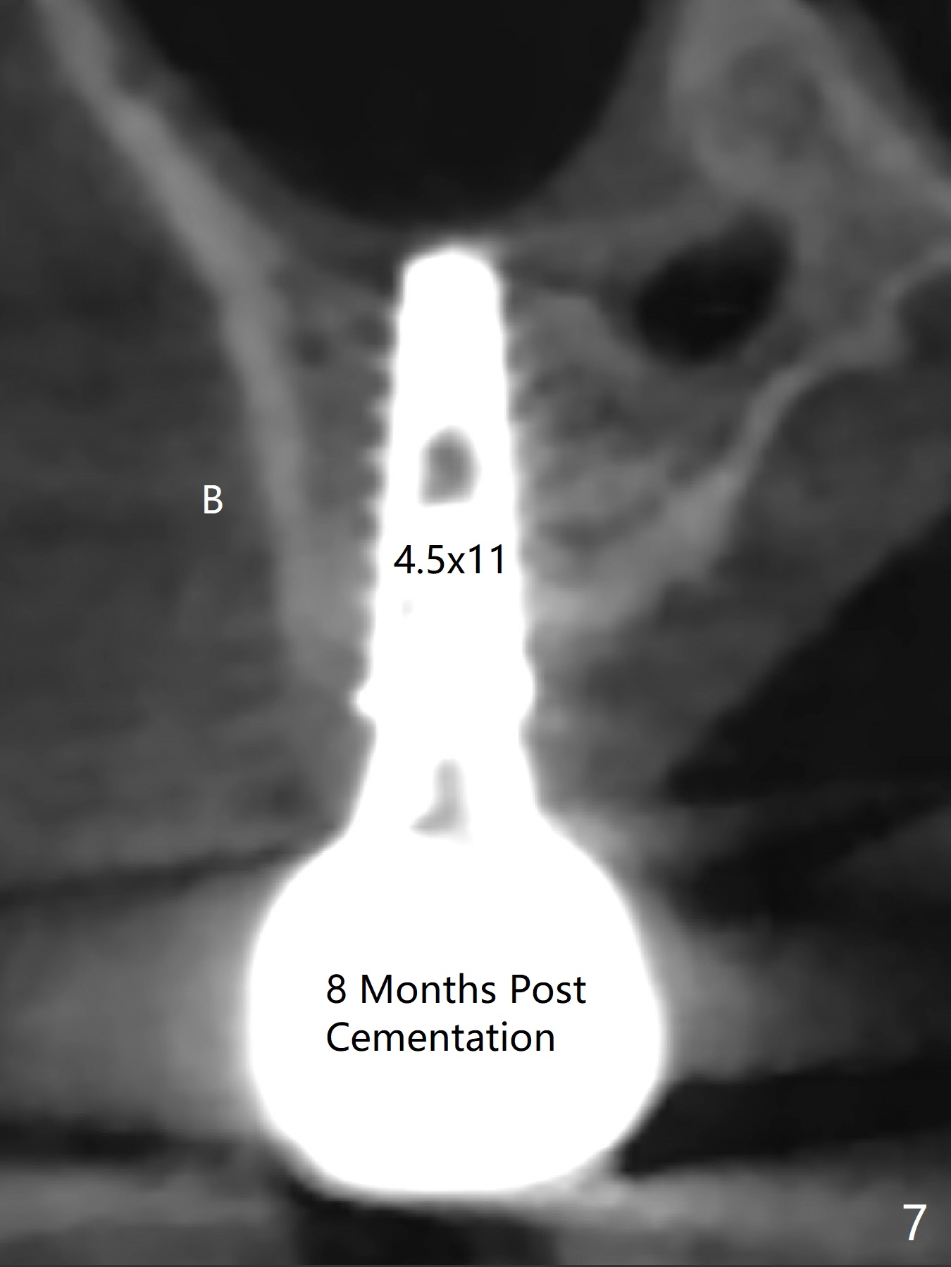
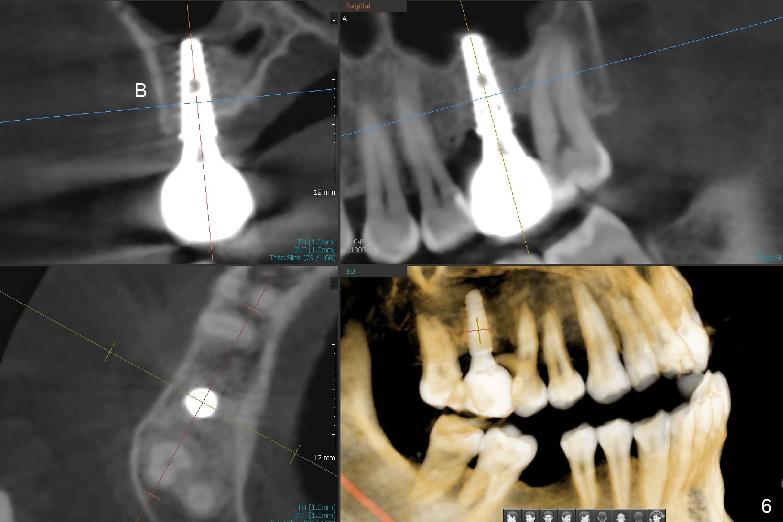
There are 2 sockets at #3 when the residual roots are extracted; osteotomy is established in the palatal one for 11 mm (gingival level; Fig.1), ~ 2 mm from the sinus floor (red dashed line). Following the last drill (3.8 mm) for 11 mm, a 4.5x11 mm dummy implant is placed 2 mm subgingival (Fig.2). In fact the sinus lift (red dashed line) is done by implant insertion, since the bone plug after Magic Drill is present in the apical portion of the osteotomy when the last drill is finished prior to implant placement.. After the dummy implant is removed, the definitive one (the same size) is placed with >50 Ncm, followed by placement of a 4.5x4(3) mm abutment and VeraGraft mixed with autogenous one (Fig.3 *). The most coronal portion of the socket is sealed with collagen plug. The latter is fixed in place by an immediate provisional. The mesial surface of the tooth #2 is reduced (Fig.4 <) prior to provisional fabrication. The implant is placed distal, which could be corrected by pushing the 2nd molar distal orthodontically. There is mild crestal bone loss nearly 8 months postop (Fig.5). An angled abutment (5x15 degrees, 3 mm cuff) is used before impression. CT is taken immediately post cementation (Fig.6 (8.5 months postop)). CT taken 8 months post cementation shows that the 4.5x11 mm implant barely passes the sinus floor (Fig.7). Return to Upper Molar Immediate Implant, Prevent Molar Periimplantitis (Protocols, Table), IBS, 30 19 Xin Wei, DDS, PhD, MS 1st edition 09/13/2017, last revision 01/31/2019