|
|
|
|
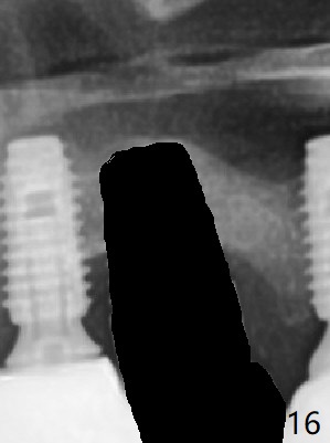
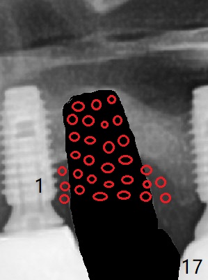
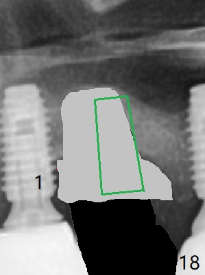
It appears that the implant should be removed (Fig.16), followed by bone graft, particularly mesial to #1 implant and PRF (Fig.17 red circles). When the defect heals (with increased bone height, Fig.18 greyish area), place a smaller implant (green) with guide.
Return to Soft Tissue Regeneration Last Next
Xin Wei, DDS, PhD, MS 1st edition 10/12/2018, last revision 12/16/2018