|
|
|
|
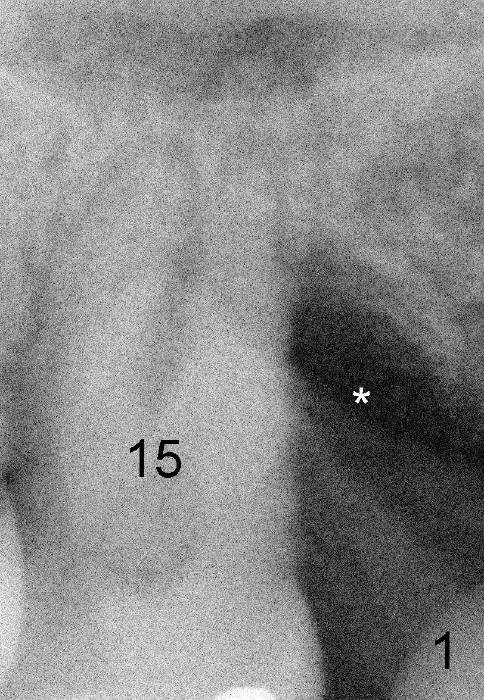
Fig.1 (preop PA): There is a large defect distal to the upper left 2nd molar (#15).
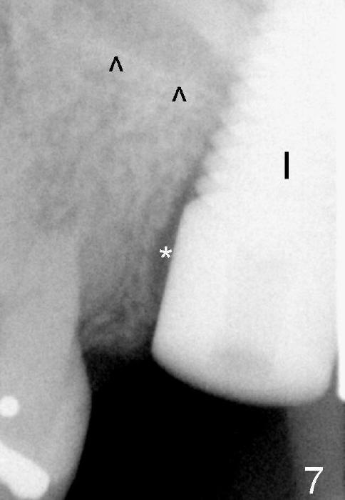
Fig.7 (immediately postop): There is a small gap mesial to the 7x17 mm implant. ^: sinus floor.
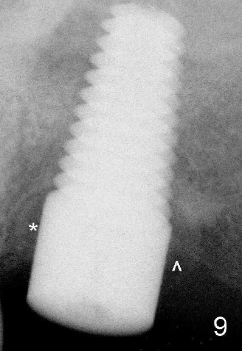
Fig.9 (4 months postop): Both mesial (*) and distal (^) defects have been repaired.
Return to New World
Xin Wei, DDS, PhD, MS 1st edition 04/27/2013, last revision 01/19/2018