,%20graft_t.jpg)

|
|
|
|
|
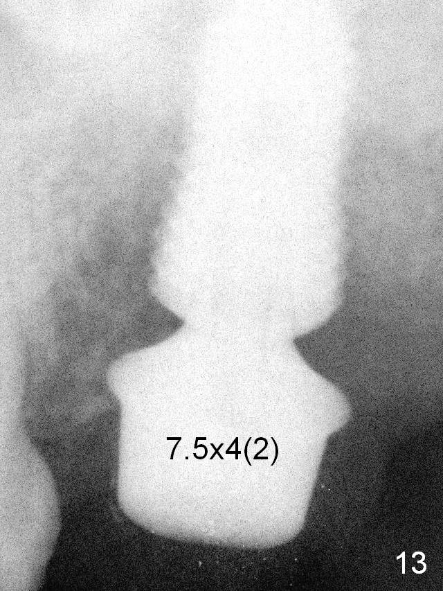
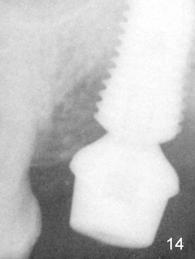
Fig.11 shows 6x12 mm implant (I), 7.5x4(2) mm cemented abutment (A) and bone graft (*). An immediate provisional is fabricated to close the socket completely. Fig.13: 9 months postop. Although there is bone growth mesially radiographically, the mesiobuccal implant (coronally) has no bone contact clinically. Bone graft is placed after debridement with Titanium brush. The abutment is changed from 7.5x4(2) to 7.5x4(3) mm. Fig.14: 1 month 20 days post 2nd grafting. Note large bone particles are placed deep and next to implant threads.
The patient complains of tenderness superficial to the mandibular body (probably supramandibular lymph node). Exam shows purulent discharge from the buccal sulcus of the site when probing, consistent with bone loss 3 years 2 months post cementation (Fig.15).
Septal Slope of Upper 2nd Molar Last Next Xin Wei, DDS, PhD, MS 1st edition 10/16/2019, last revision 03/05/2021