 |
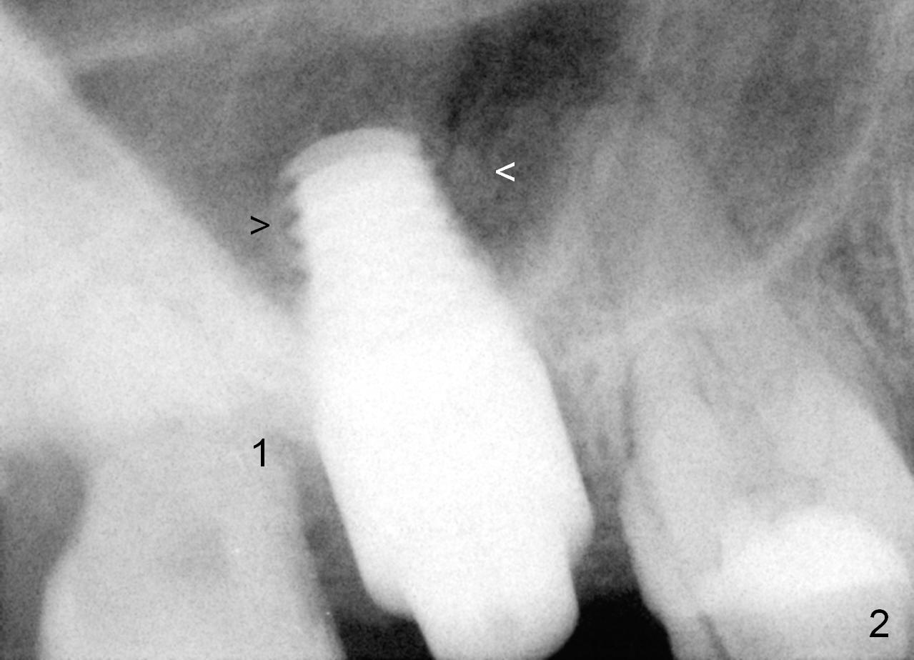
More important with the 3rd molar present, the immediate provisional at the site of #2 is more stable. The prominent pathology associated with the tooth #1 is the enamel pearl in the mesial root surface (*), which is removed after #2 extraction (Fig.2: #1).
As planned, a 7x14 mm tissue-level implant is placed in the middle of the socket (mesiodistally (Fig.2) and buccopalatally (not shown) with insertion torque >60 Ncm. What is not ideal is that the implant is placed too deep into the sinus, although the apical portion of the implant is covered by the apparently lifted sinus floor (arrowheads; no graft used for sinus lift).
Xin Wei, DDS, PhD, MS 1st edition 05/01/2015, last revision 10/21/2019