|
|
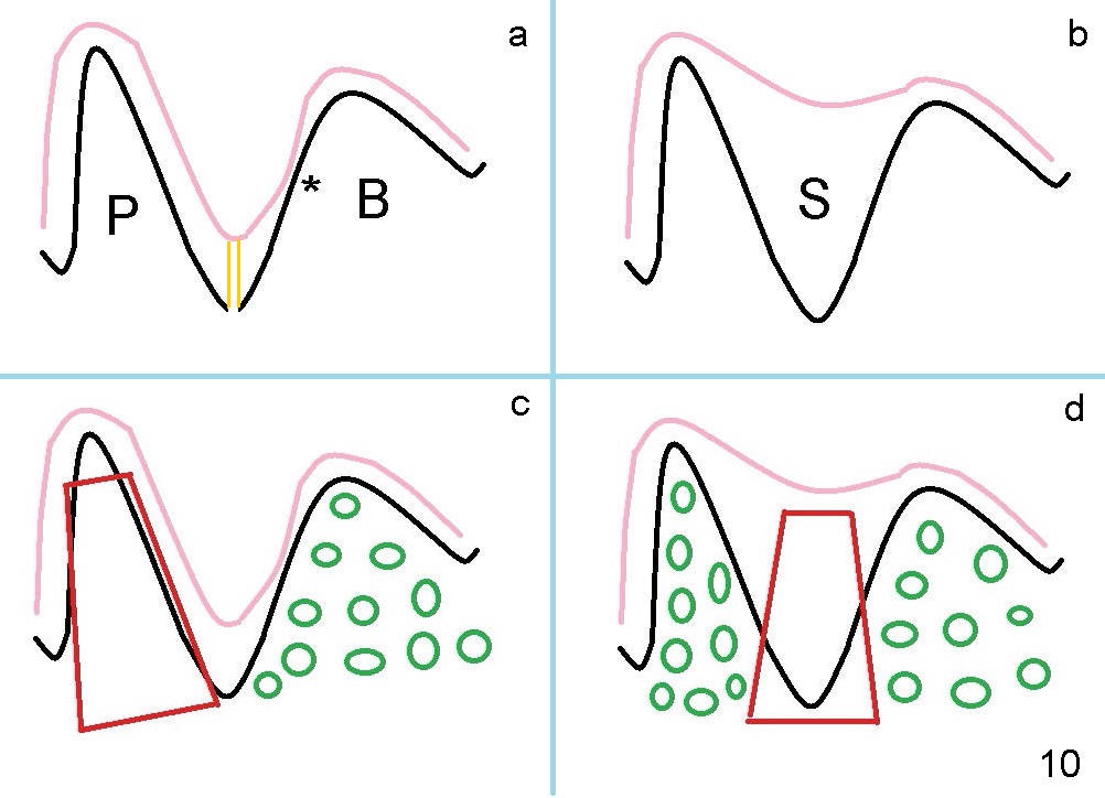
Fig.10a is an illustration of a section through the sockets of the affected tooth buccopalatally. Black line represents the lamina dura of the sockets (P: palatal; B: buccal), whereas the pink the sinus floor. * is an area clinically looking thin, probably due to approximation of these two cortices. The buccal socket is large and shallow. The buccal plate is particularly short. When 2 mm pilot drill is being used approximately 4 mm in the septum (double yellow lines), it appears to have entered the sinus. In contrast, the palatal socket is deep and narrow. It is the most appropriate to place an implant (red in Fig.10c) there. Bone graft is placed in the buccal socket (green circles).
If the sinus floor (Fig.10b: pink line) is flat over the septum, there is enough bone height in the septum to support an implant (Fig.10d: red line). It is easier to restore. Bone graft (green circles) is placed in the buccal and palatal sockets.
Three Implantation Sites Last Next
Xin Wei, DDS, PhD, MS 1st edition 12/05/2014, last revision 11/14/2019