|
|
|
||
|
|
|
|
|
|
|
|
|
|
Sinus Floor Downward Extension Between Roots of Upper 2nd Molar
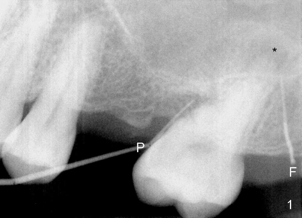
A 44-year-old man presents with a fistula distolingual to the tooth #15 (Fig.1). Two pieces of Gutta percha are inserted into the fistula (F) and the mesial pocket (P). The first gutta percha points to the apex (with periapical radiolucency *). The tooth #15 has no restoration. There is possibility of root fracture. The tooth has guarded to poor prognosis considering perio and endo complex as well as occlusal trauma (mesial tilting and shifting). The patient insists upon saving the tooth. Osseous surgery will be performed as an exploratory one to confirm whether there is root fracture.
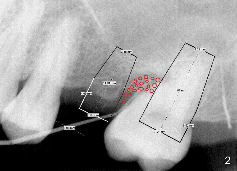
If the tooth is deemed non-salvageable, an immediate implant will be placed as distally as possible (Fig.2 in its original place, 7x14 mm). A delayed implant will be placed at the site of #14 (5x11 mm). Bone graft (red circles) will be placed between the implants.
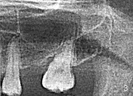
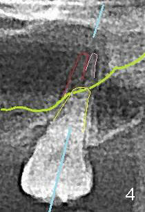
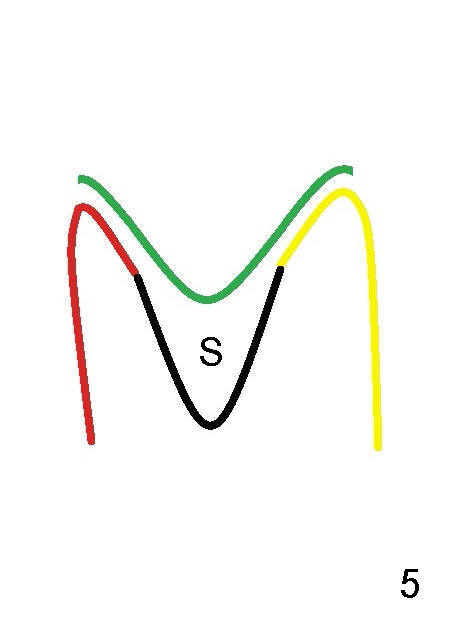
This most recently taken PA (Fig.1,2) and panoramic X-ray (Fig.3,4, taken 3 years ago) show that the sinus floor (green line in Fig.4) is more coronal than the fused buccal roots of #15 (red and pink). Fig.5 is an illustration of a coronal section of #15 socket (at a plane shown as a blue line in Fig.4). Green line: sinus floor; red: buccal socket; yellow: palatal socket; black: septum (S).
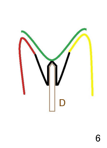
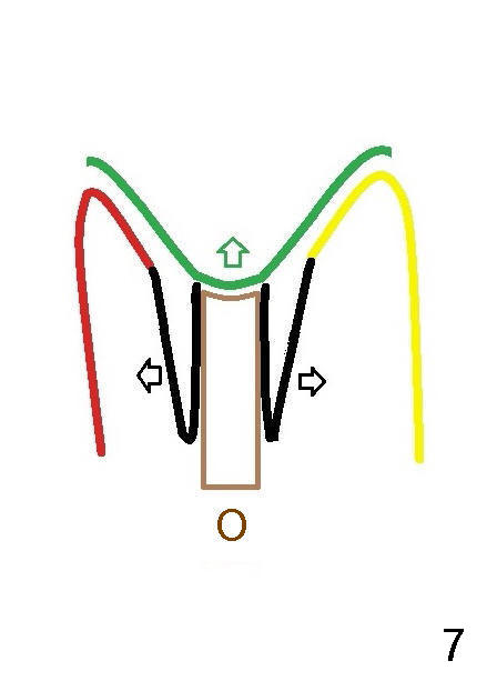
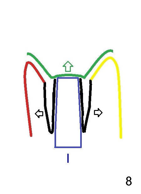
If the septum is found wide enough after extraction, osteotomy is initiated at the septum using a pilot drill (Fig.6 D, ~ 1 mm from the sinus floor), followed by osteotomes (Fig.7 O) to expand the septum (black arrows) and lift the sinus floor (green arrow). Finally a large implant (Fig.8 I) is placed with further bone expansion (black arrows) and sinus lift (green arrow). The remaining buccal and palatal sockets decrease due to septal bone expansion and will be closed by bone graft.
A bone-level implant may be used instead (6.9x10 mm). The largest abutment is 7.8 mm in diameter. There will be no or minimal prep. An immediate provisional will be fabricated to cover the sockets.
Return to Upper Molar Immediate Implant
Xin Wei, DDS, PhD, MS 1st edition 12/27/2014, last revision 12/27/2014