|
|
|
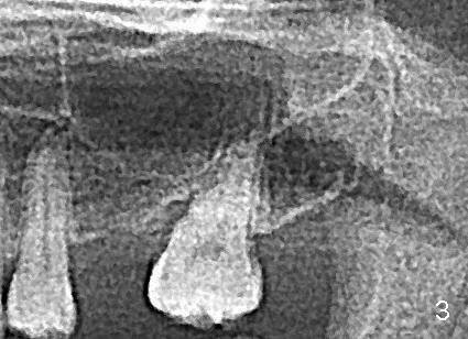
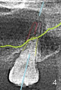
This most recently taken PA (Fig.1,2) and panoramic X-ray (Fig.3,4, taken 3 years ago) show that the sinus floor (green line in Fig.4) is more coronal than the fused buccal roots of #15 (red and pink). Yellow line: the palatal root; blue line: coronal section of the tooth for illustration.
Xin Wei, DDS, PhD, MS 1st edition 12/27/2014, last revision 12/27/2014