|
|
|
|
||
|
|
|
|
||
|
|
|
|
||
Anatomy & Pathology of the Upper Molars as Related to Immediate Implant
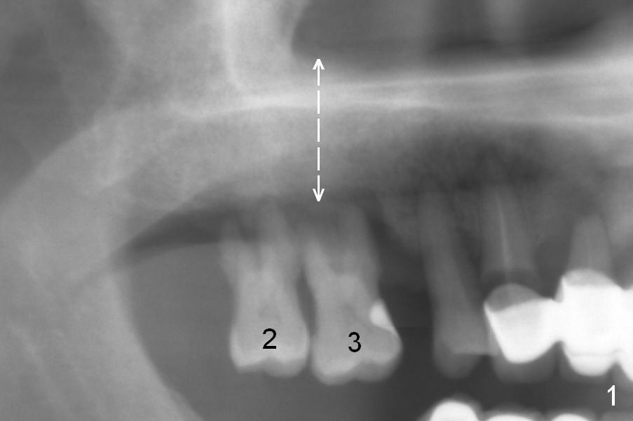
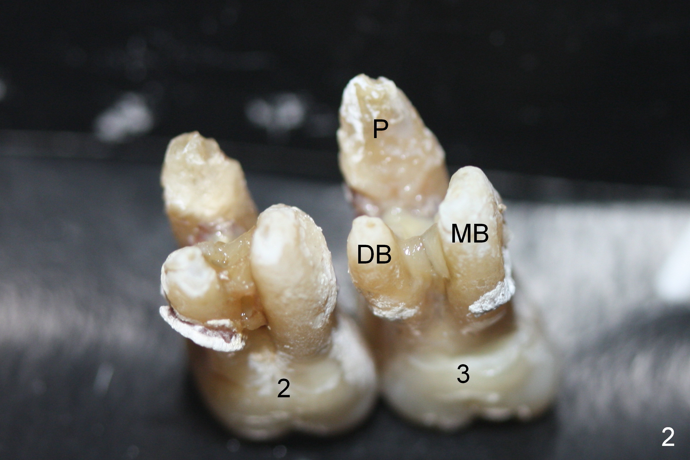
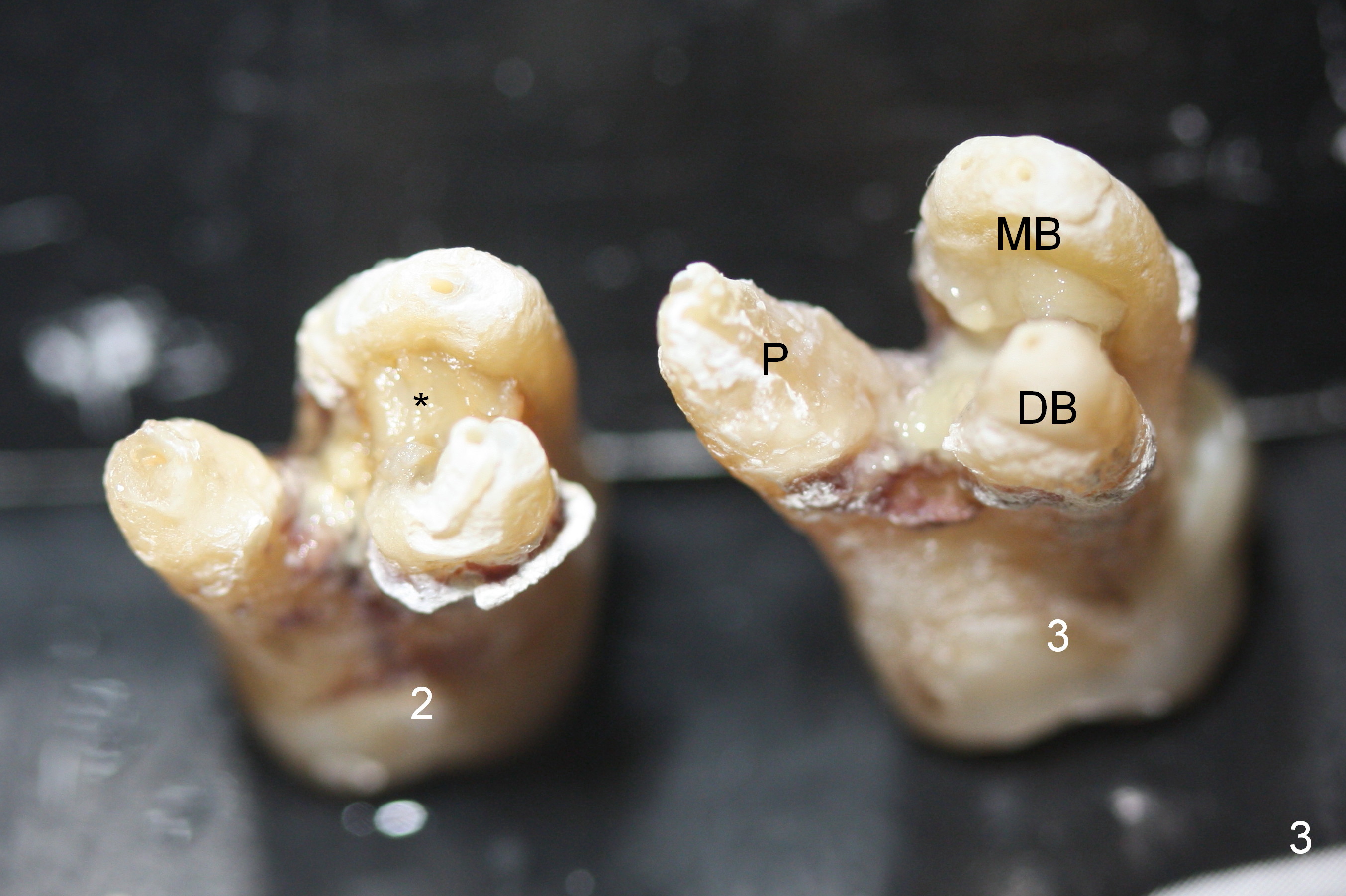
The maxillary 1st molar has 3 widely separated roots (Fig.1-3: #3) with a wide septum in between, as compared to the 2nd one (#2). The septum is a suitable site of immediate implant. When the septum is destroyed by pathology, the immediate implant has to be big.
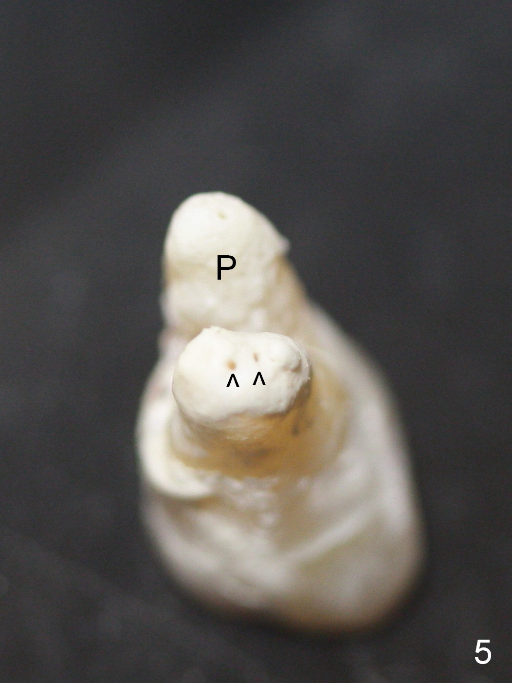
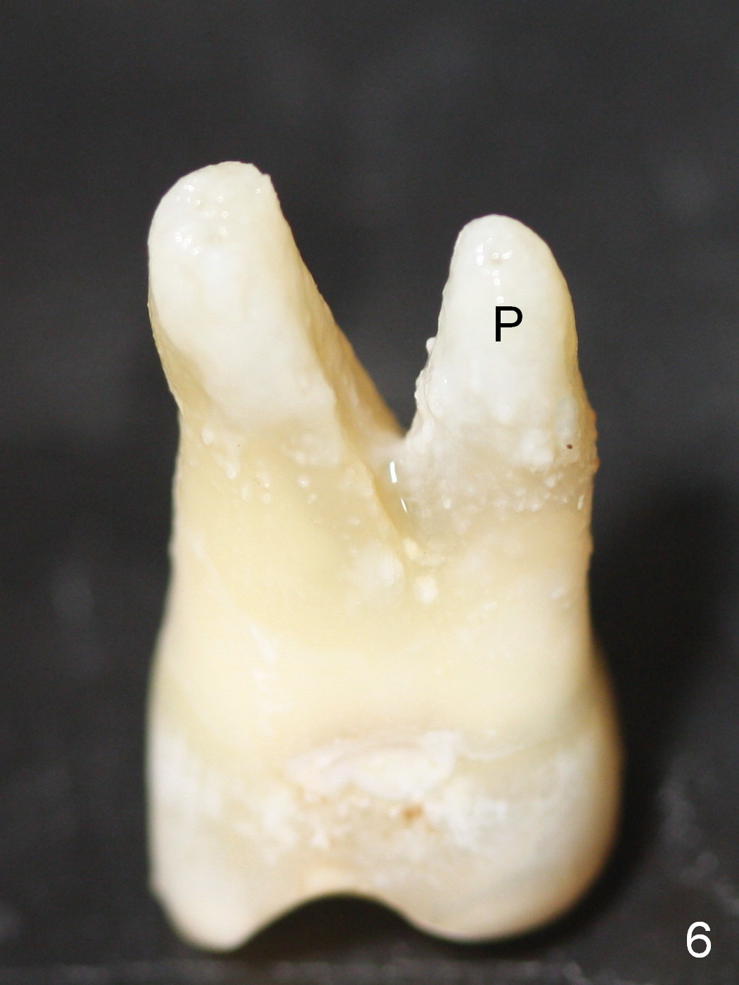
The furcation of the upper 2nd molars is variable. For example, the buccal roots of the 2nd molar may be fused (Fig.4-6). The septum may be destroyed due to infection such as in this case (Fig.4). An immediate implant could be placed in the septum or the buccal and palatal sockets, depending upon the integrity of the buccal and palatal plates.
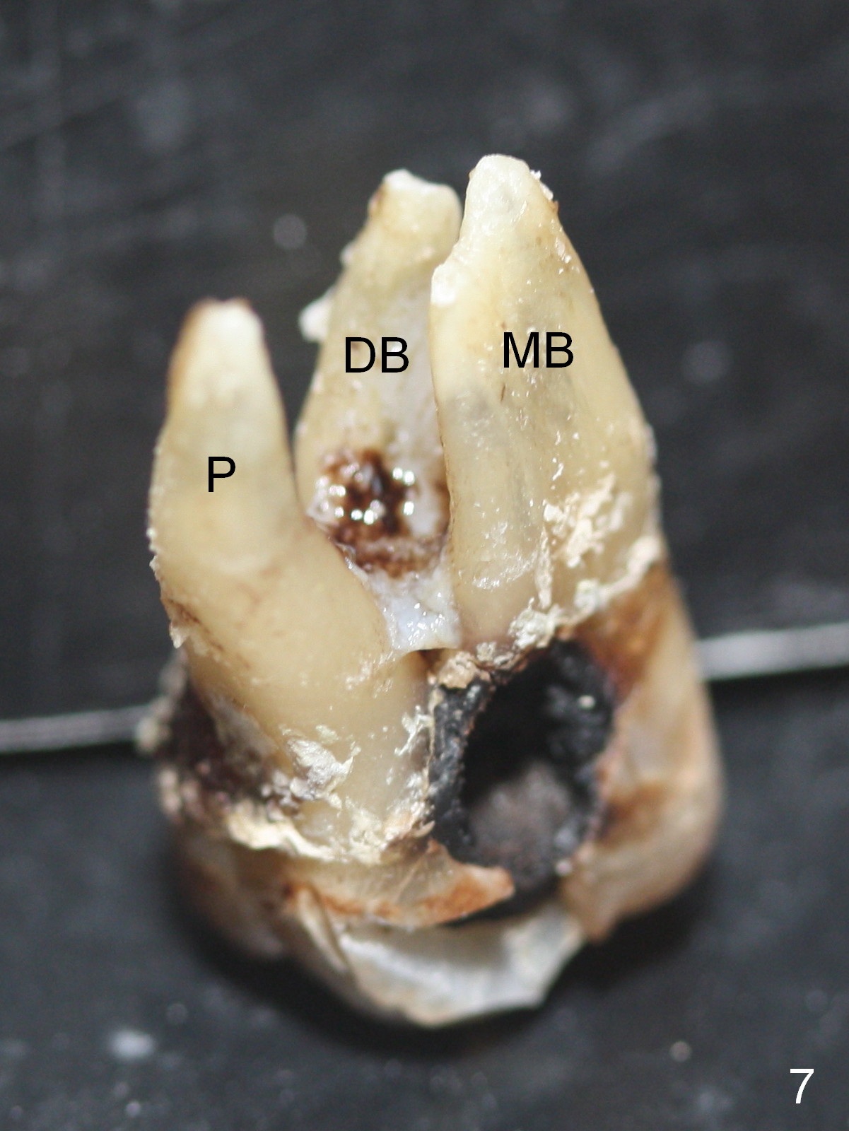
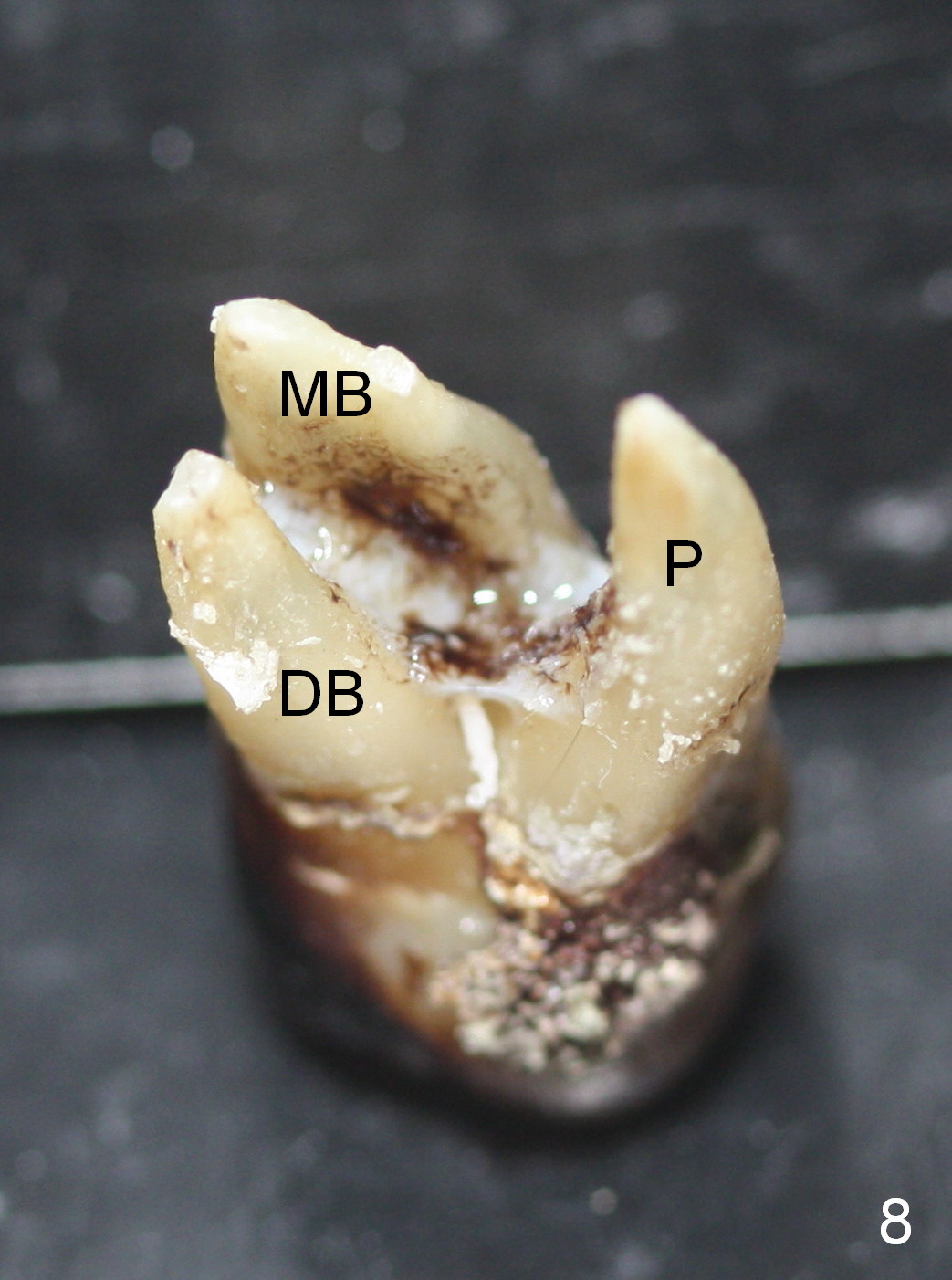
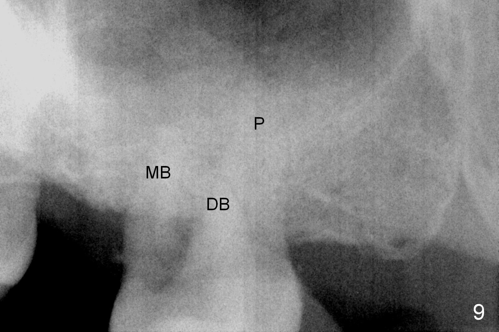
By comparing the root morphology of an extracted tooth (Fig.7,8: #15) with that of X-ray (Fig.9) repeatedly, we can develop an ability to visualize the septum prior to surgery.
Return to Upper Molar Immediate Implant
Xin Wei, DDS, PhD, MS 1st edition 06/28/2015, last revision 01/19/2018