|
|
|
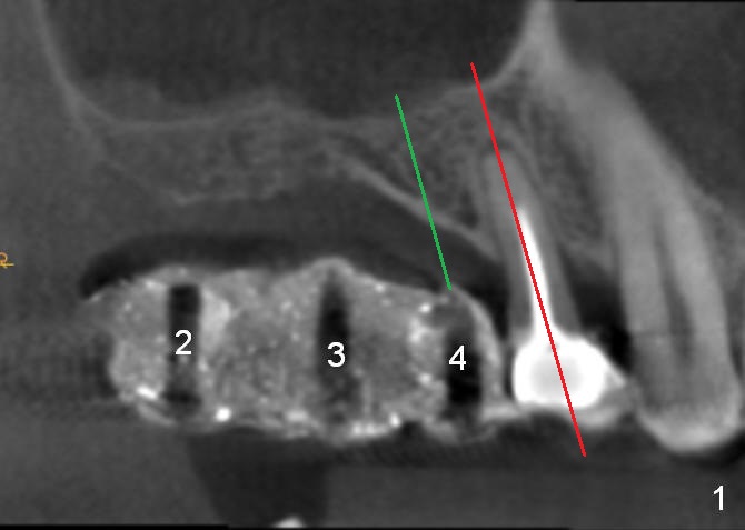
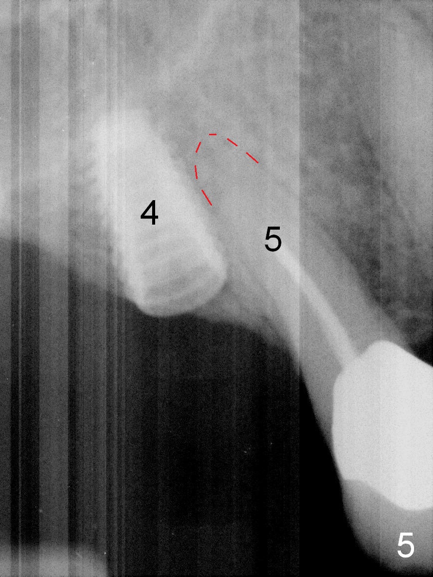
Fig.1: CBCT sagittal section. #2,3,4: CBCT scan stent. Red line: the long axis of the tooth #5, to which the trajectory of the implant at #4 (green line) should be parallel. The fact is ignored preop. The patient presents to surgery immediately after taking CBCT. There is no enough time for analysis, which leads to approximation of 4.5x8.5 mm implant at #4 to the apex of the neighboring tooth (Fig.5 red dashed line).
Implant Placed Between Sinus and Palate Next Fig.6
Xin Wei, DDS, PhD, MS 1st edition 01/22/2016, last revision 03/03/2018