.jpg)
|
|
|
||
|
|
|
|
|
|
|
|
|
|
|
|
|
|
|
Left Anterior Maxillary Atrophy: a Phenotype of Cleft Lip
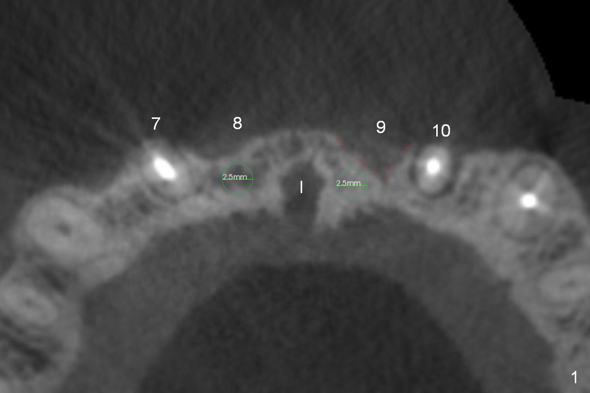
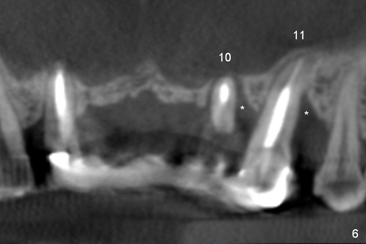
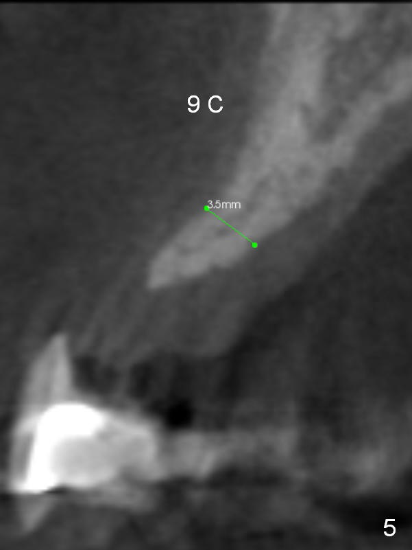
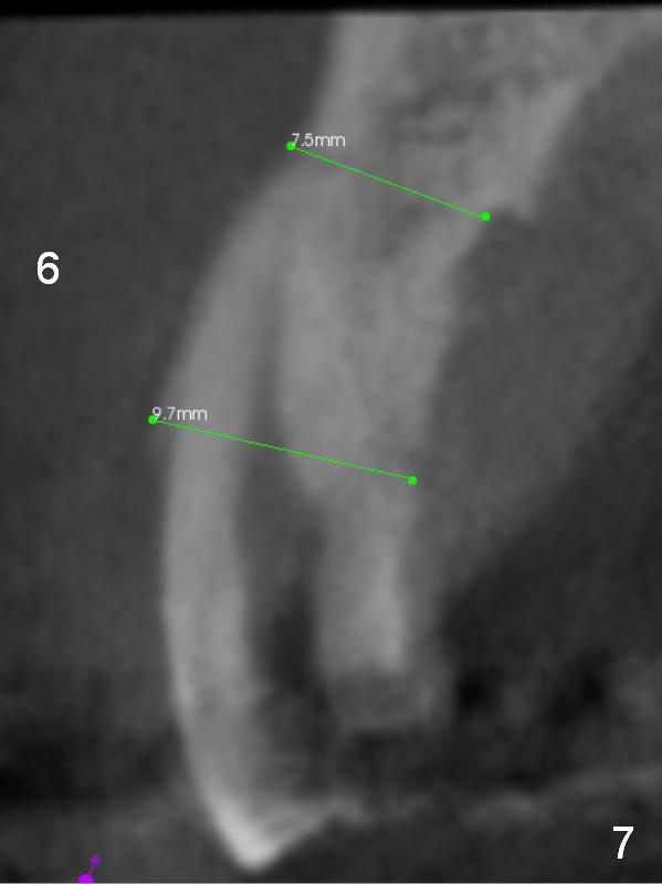
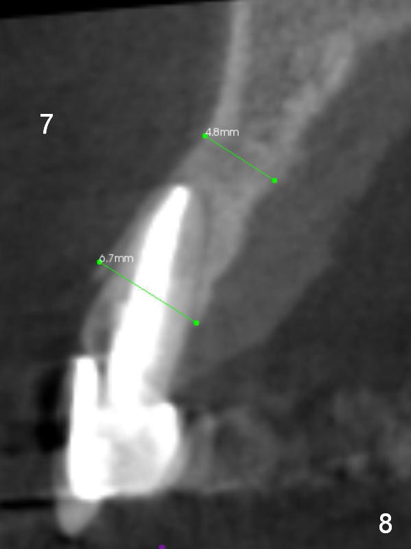
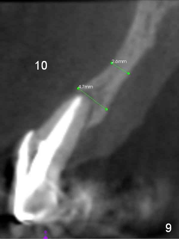
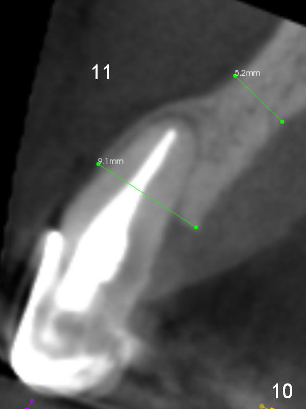
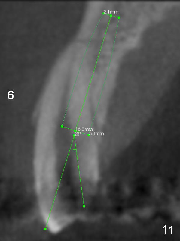
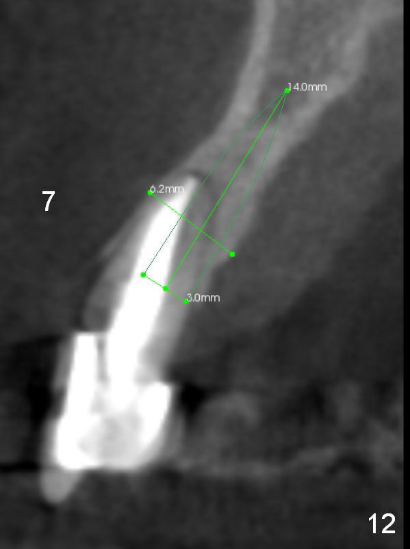
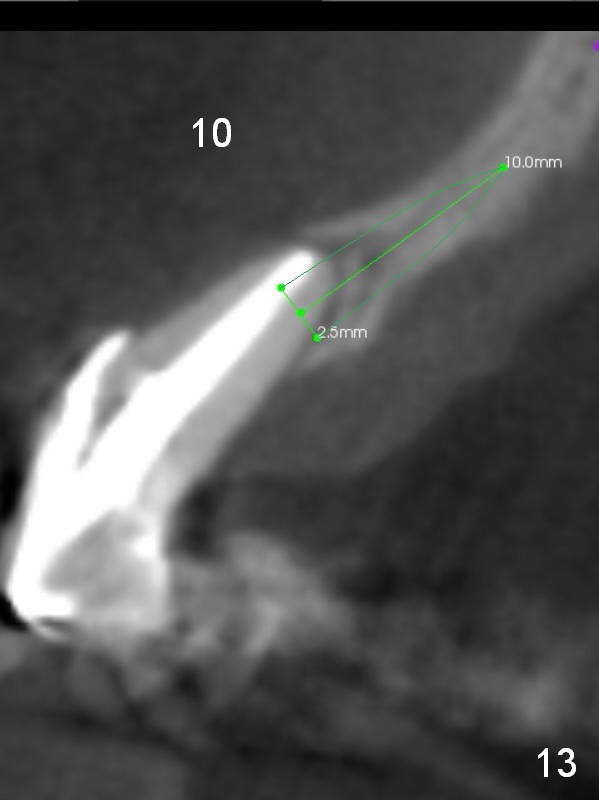
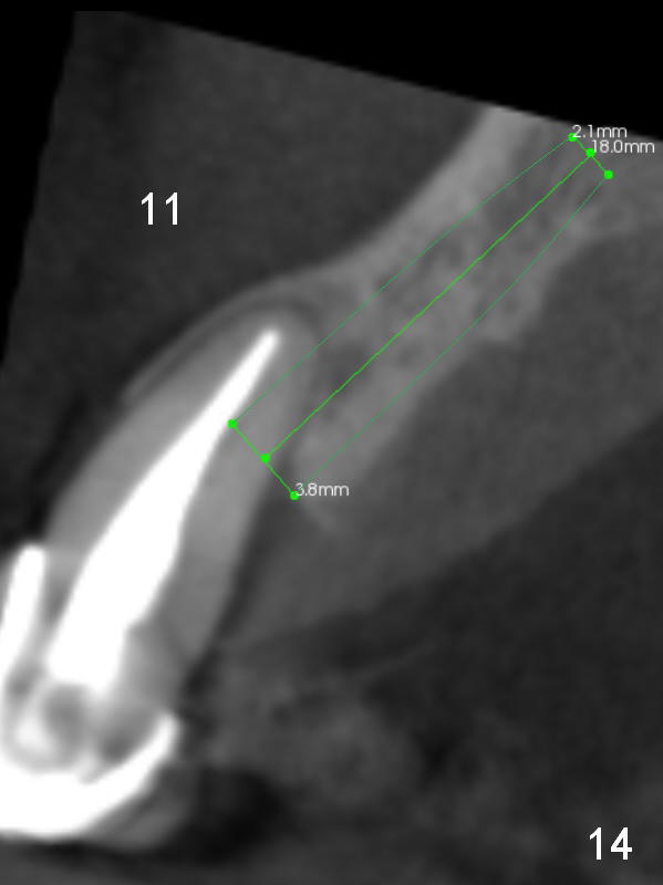
The ridge at #8 and 9 is atrophic (Fig.2-5). In fact it seems more due to inherent bone morphology than post-extraction change. For example, the ridge at #6 and 7 are wider than that at #10 and 11 (Fig.7,8 vs. 9,10), although there are pathologies associated with #10 and 11. It is likely that the left anterior maxilla is congenitally less developed, similar to the fact that cleft lip is predominantly found on the left.
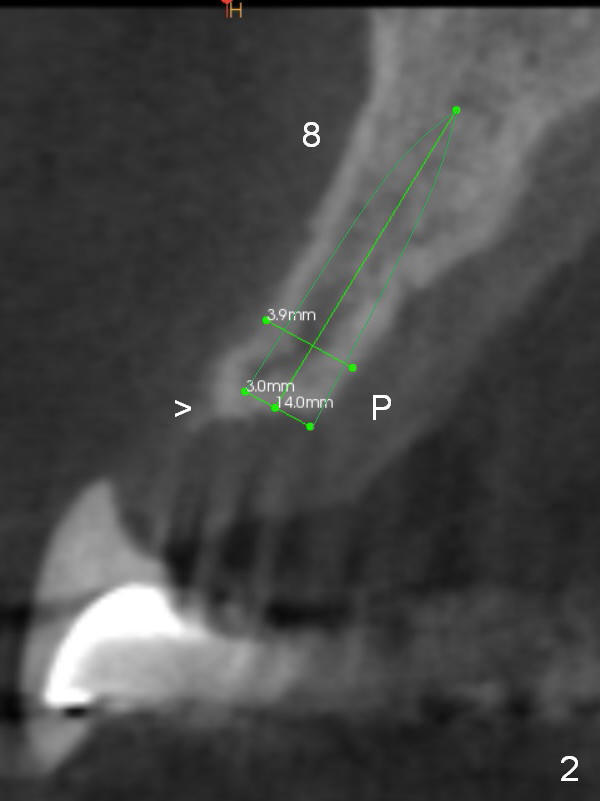
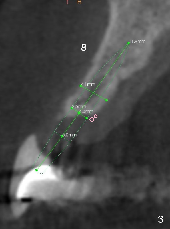
If implants were indicated in the dentulous areas, the ones on the right would be larger than the ones on the left (Fig.11,12 vs. 13,14).
Return to
Full Arch Reconstruction,
IBS,
Atrophic Ridge
Xin Wei, DDS, PhD, MS 1st edition 01/21/2017, last revision 05/24/2019