.jpg)
.jpg)
|
|
|
|
|
|
|
|
|
|
|
Osteotomy in Healed Sites through Surgical Stent
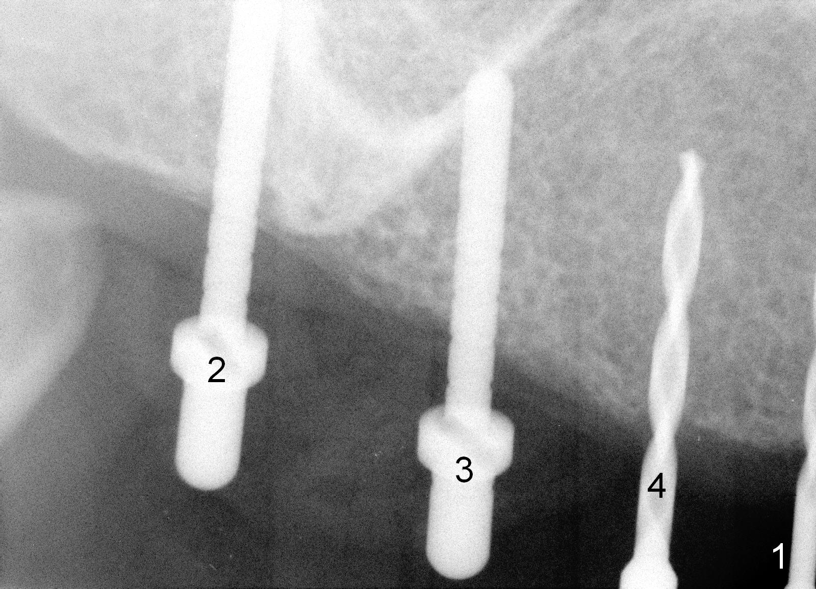
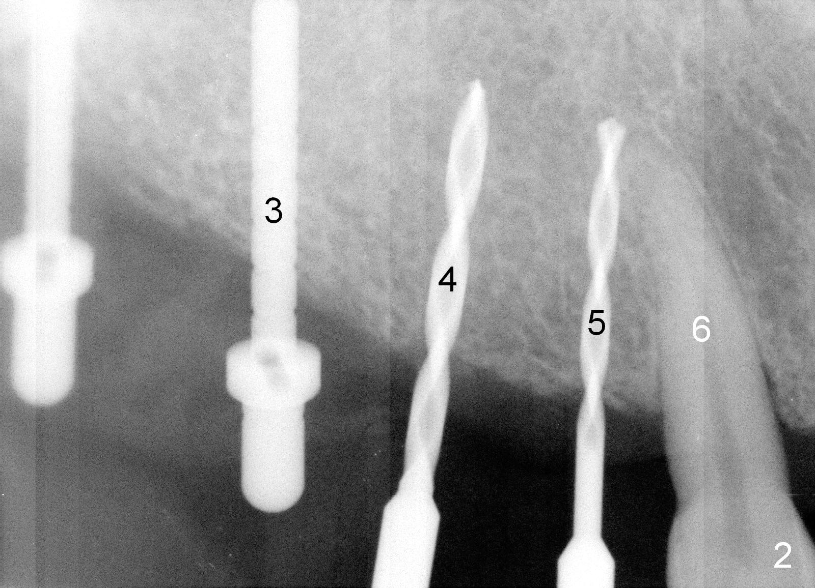
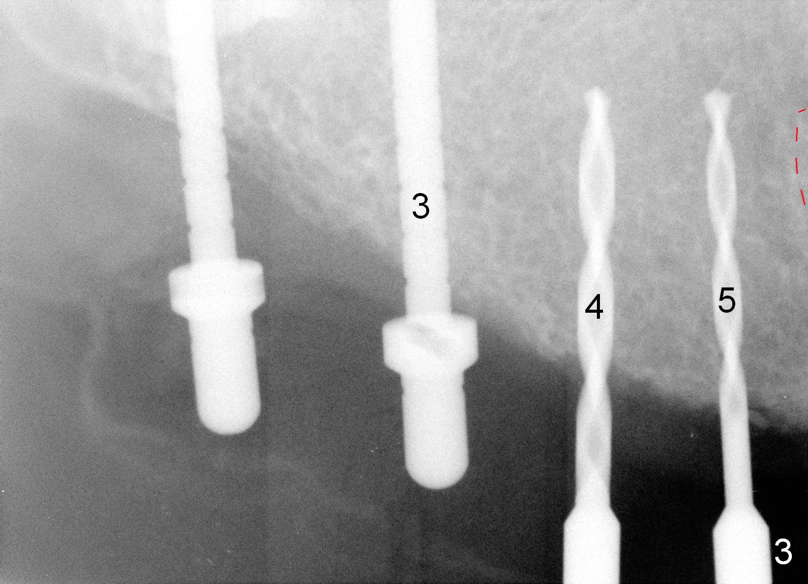
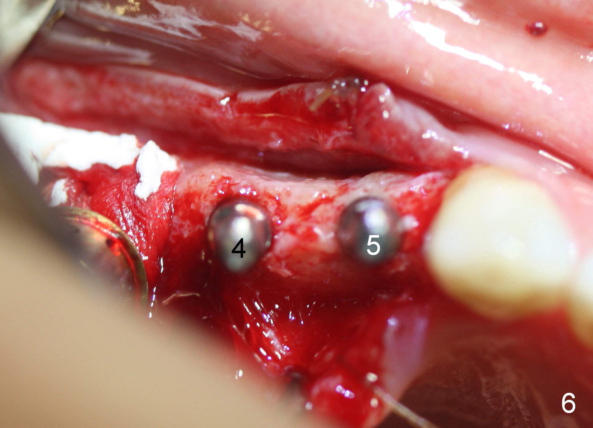
Osteotomy in the upper posteriors starts through a surgical stent for 12 mm. After incision, each osteotomy is examined, adjusted and extended 2 mm shy of intended (Fig.1,2). It appears that the trajectories at #4,5 are off (Fig.2). After re-adjustment, the trajectories are acceptable (Fig.3 (red dashed line; part of the root of the tooth #6)). Four implants are placed basically in accordance with the plan: 5.9x10 mm at #2, 5x14 at 3, and 3x14 mm 1-piece at 4 and 5 (Fig.4,5). Fig.6 shows the narrow ridge at #4 and 5 after implant placement. Although abutments are placed at #2 and 3, an immediate provisional bridge cannot be fabricated because of lack of enough clearance (supraeruption of the opposing dentition).
Drilling through the surgical stent helps establish the initial osteotomy, especially the mesiodistal position.
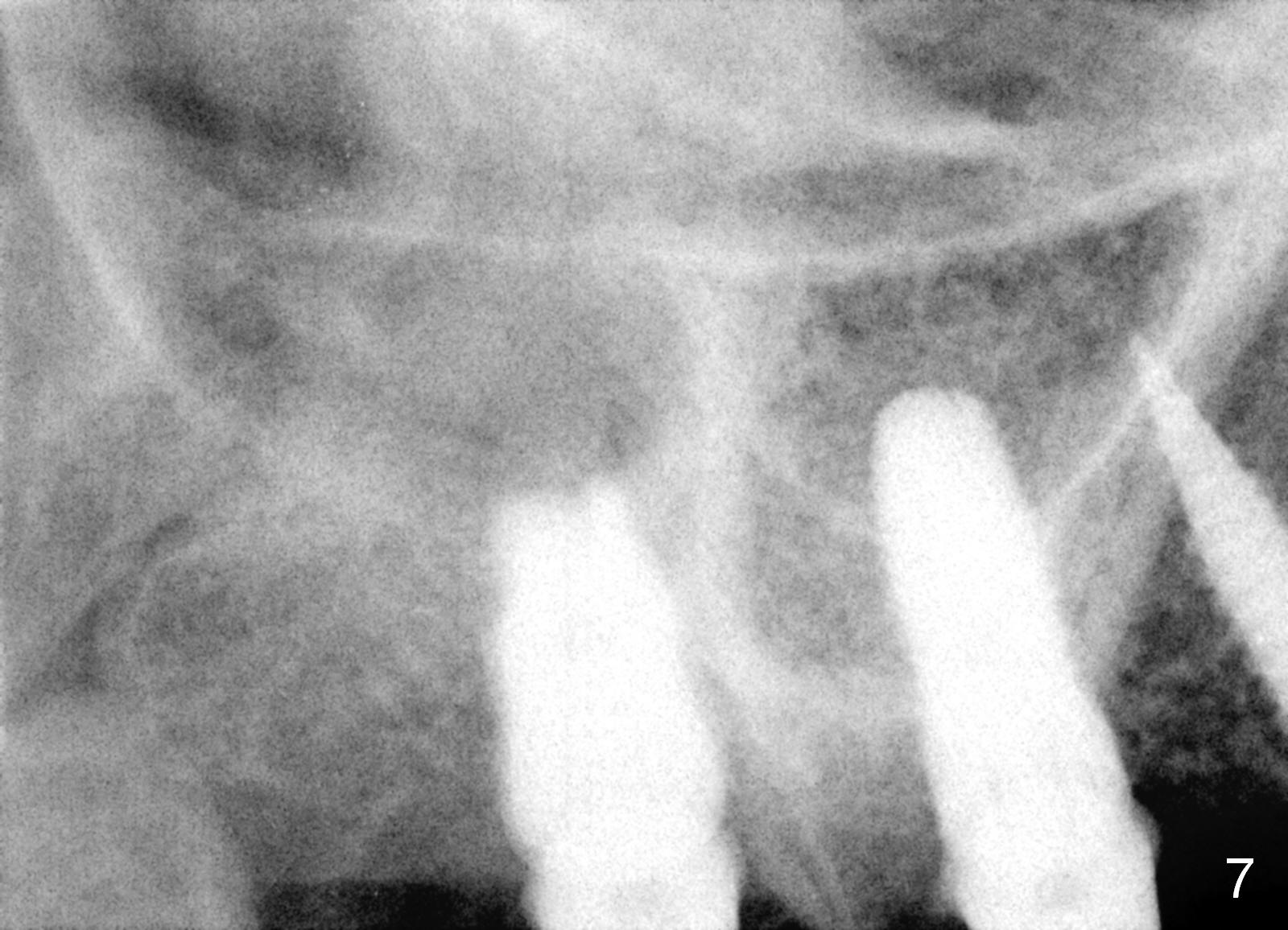
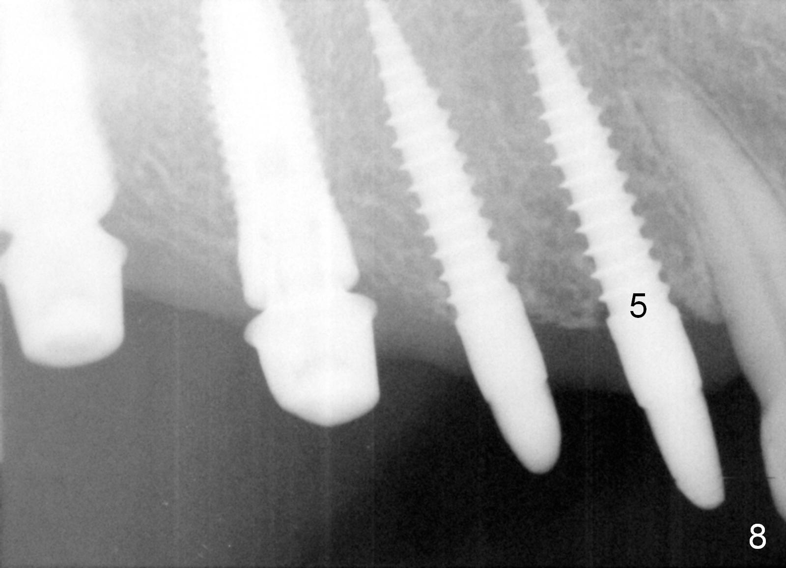
Provisional bridge is fabricated after wound healing. Four months postop, there is minimal bone resorption at the site of #5 (Fig.7,8). Single-unit crowns are fabricated.
Return to Upper Arch Reconstruction with Implants,
Upper Bicuspid Immediate
Implant,
#12-14, Course 1
2
1-Piece
Xin Wei, DDS, PhD, MS 1st edition 10/08/2015, last revision 06/02/2018