,%207.5x4(3),%20more%20bone%20graft.jpg)
|
|
|
|
|
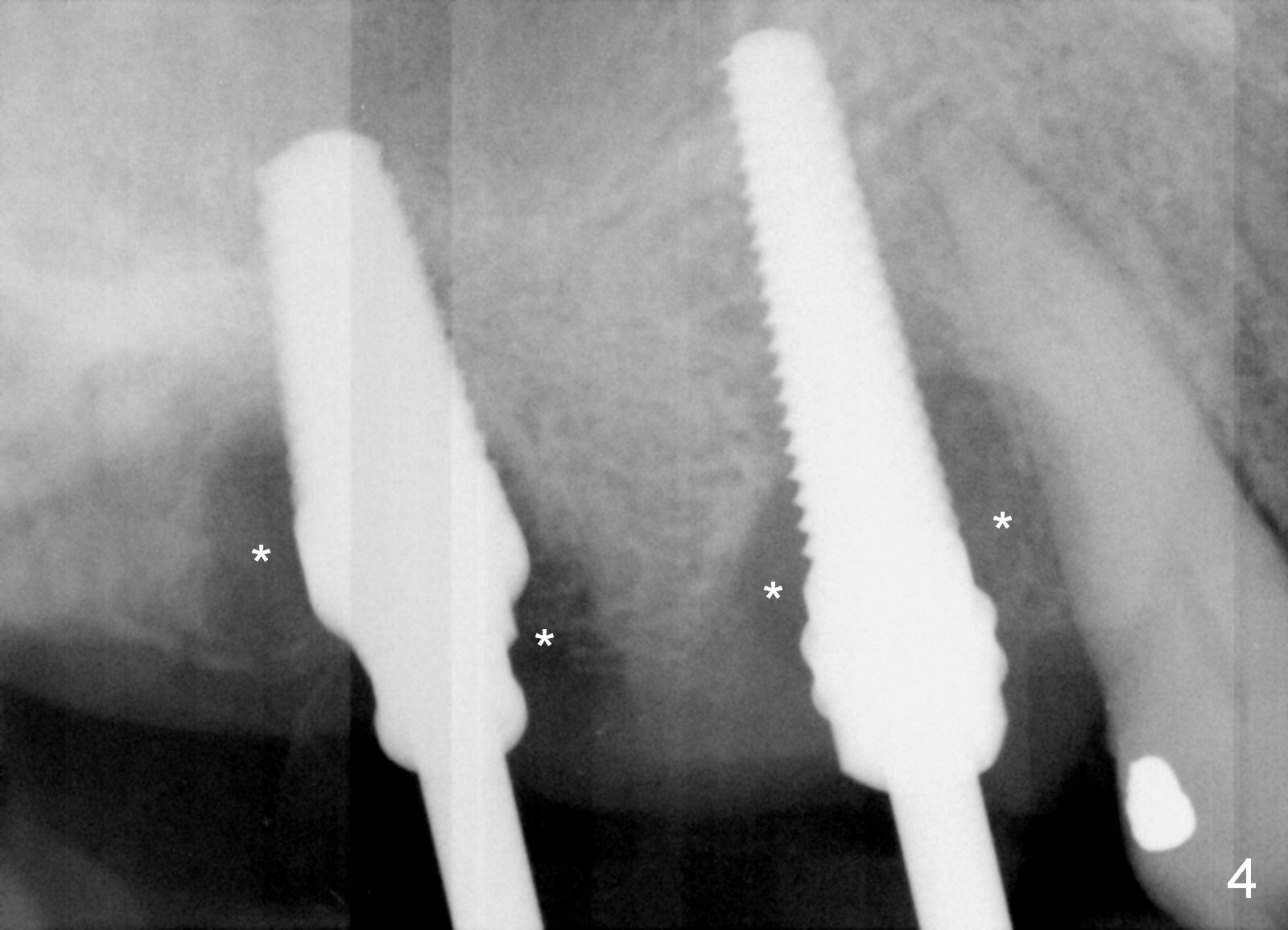
After extraction, the buccal and palatal plates of the teeth #3 and 5 are found basically non-existent. When taps (6x17 mm at #3 and 4.5x20 mm at #5) are placed, large socket defects are evident (Fig.4 *).
When bone graft is placed after implant placement, bone density increases in the defect areas (Fig.8 *).
Two months and a half postop (#3,5 implantation), the patient agrees to have #6-8 to be extracted because of persistent infection. She reports dislodgement of the provisional at #3-5 using water pik. The provisional is found to be mobile. PA shows decrease in bone density around the implant at #5, probably due to proximity to the infection (Fig.14 *).
Xin Wei, DDS, PhD, MS 1st edition 03/08/2016, last revision 05/24/2016