|
|
|
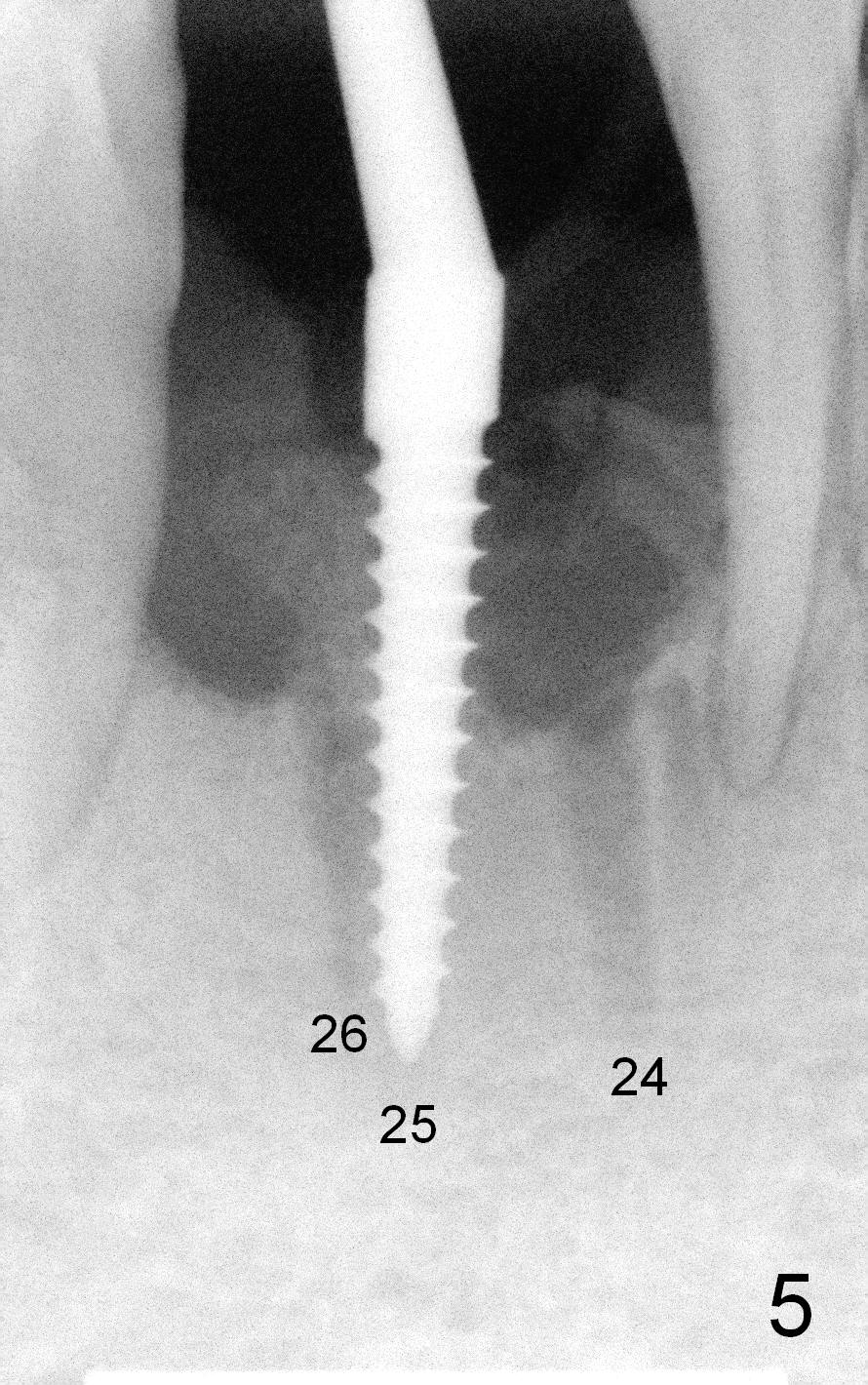
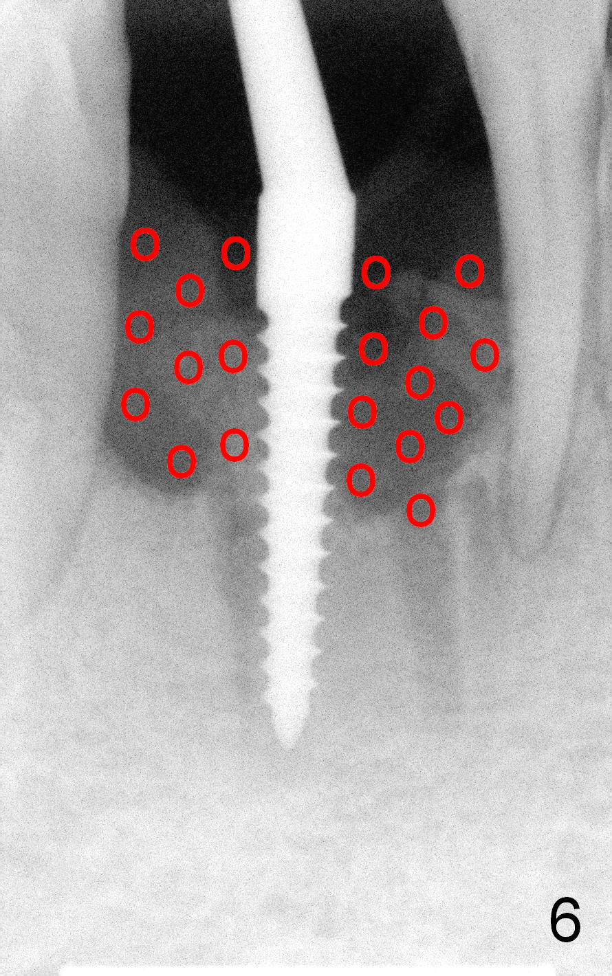
As the osteotomies increase in diameter and depth, the lingual plate starts to be perforated with hemorrhage (Fig.7b). Osteotomy is changed buccal and between the 2 previous ones (Fig.7c pink). When a 3x17 mm angulated (15°) 1-piece implant is placed (Fig.5), the buccal plate is thinner, but not perforated (Fig.7d). No attempt is tried to increase the length of the osteotomy to avoid buccal plate perforation. Bone graft is placed around the implant and over the roots of the neighboring teeth (Fig.6 red circles).
Xin Wei, DDS, PhD, MS 1st edition 03/01/2015, last revision 01/19/2018