
%20dummy.jpg)
.jpg)
|
|
|
|
|
|
|
|
|
|
|
|||
|
|
|
|
|||
|
|
|
||||
Change Position of Osteotomy
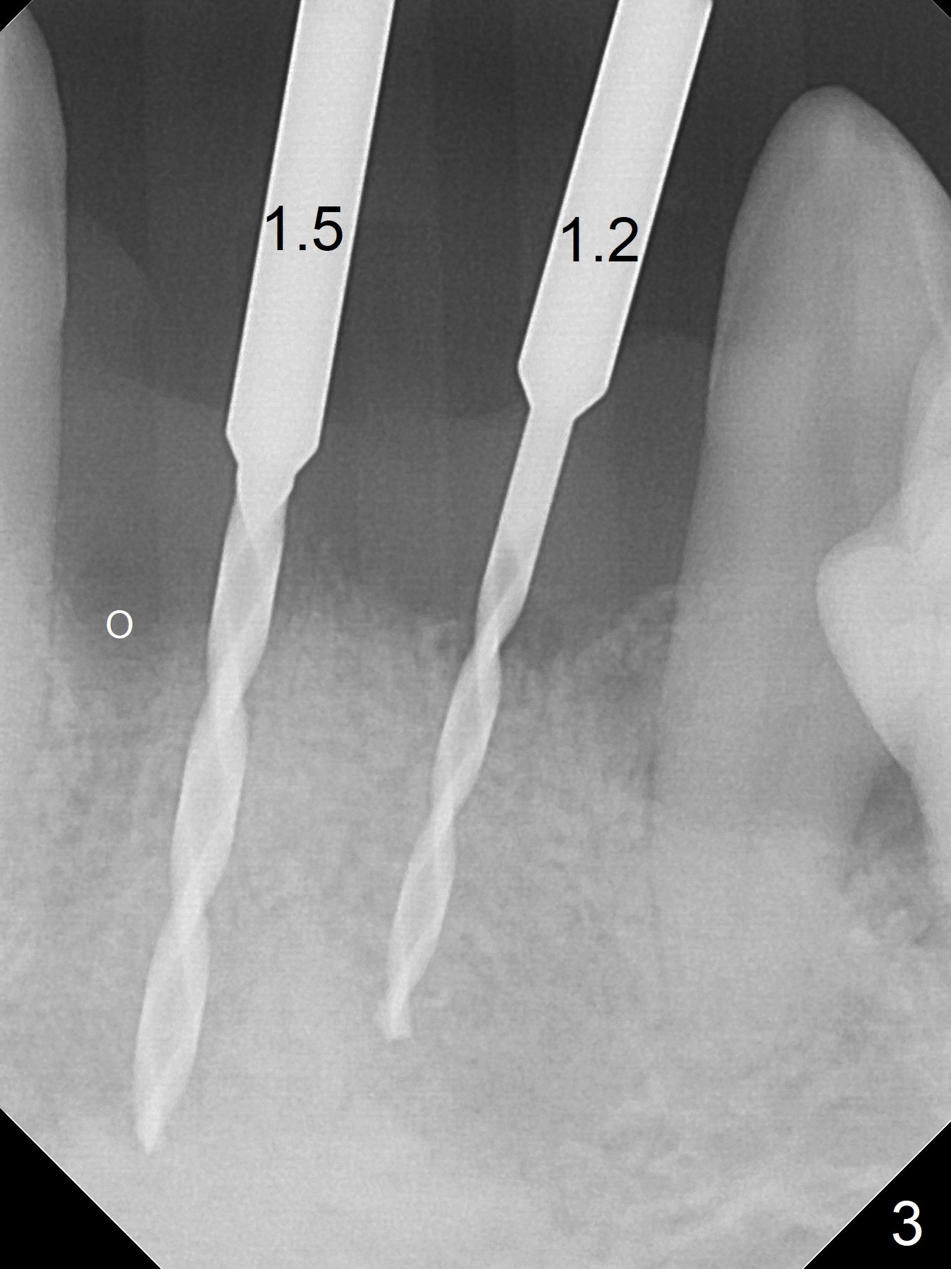
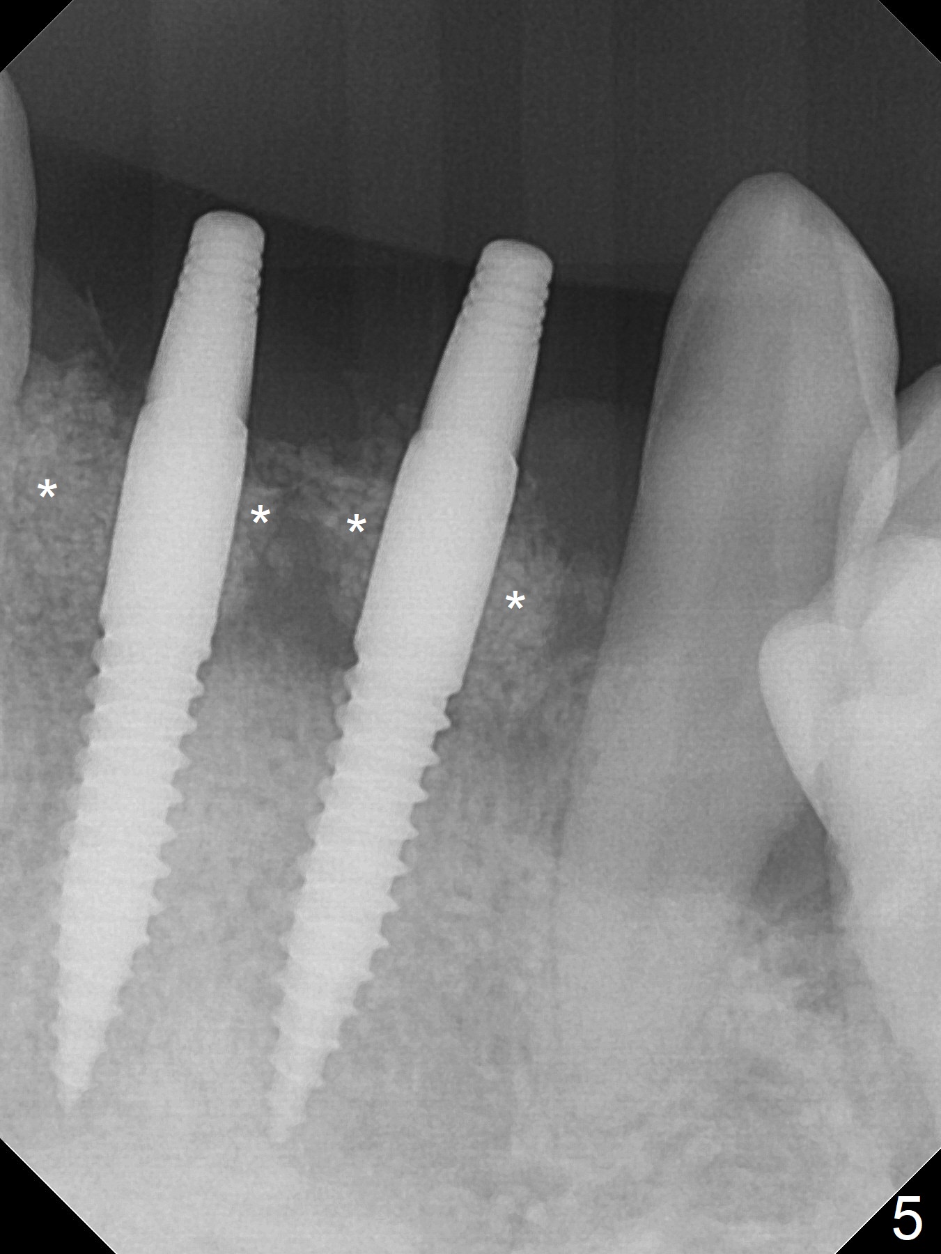
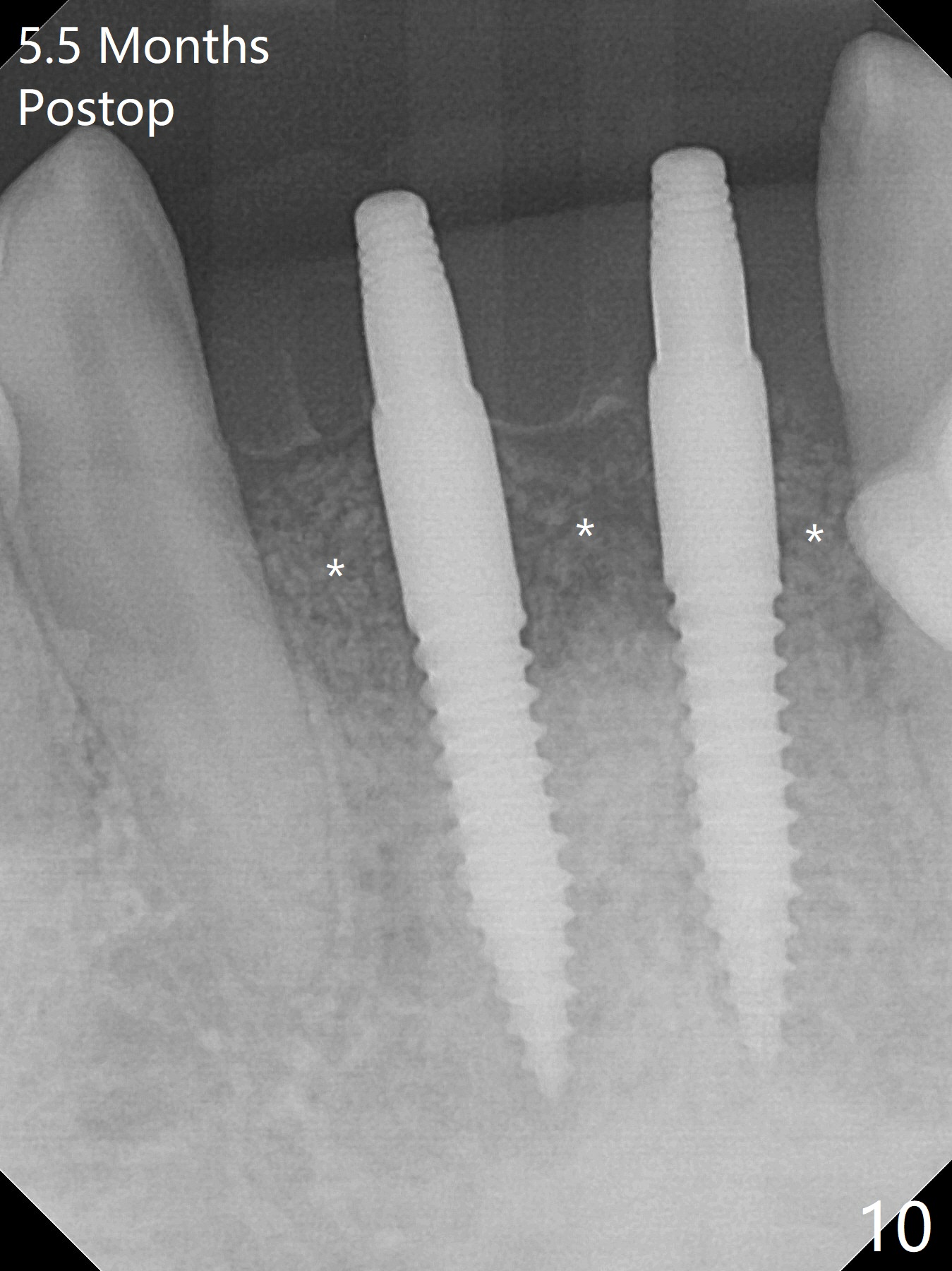
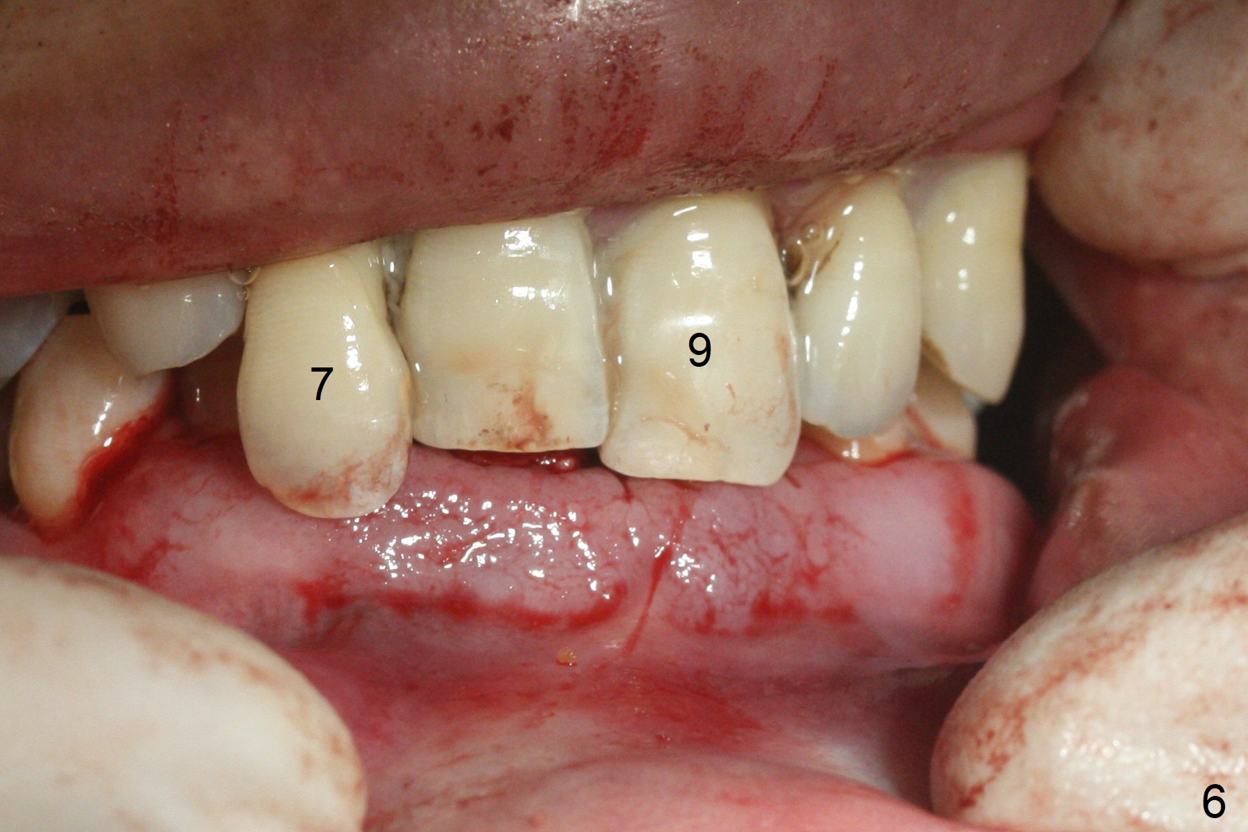
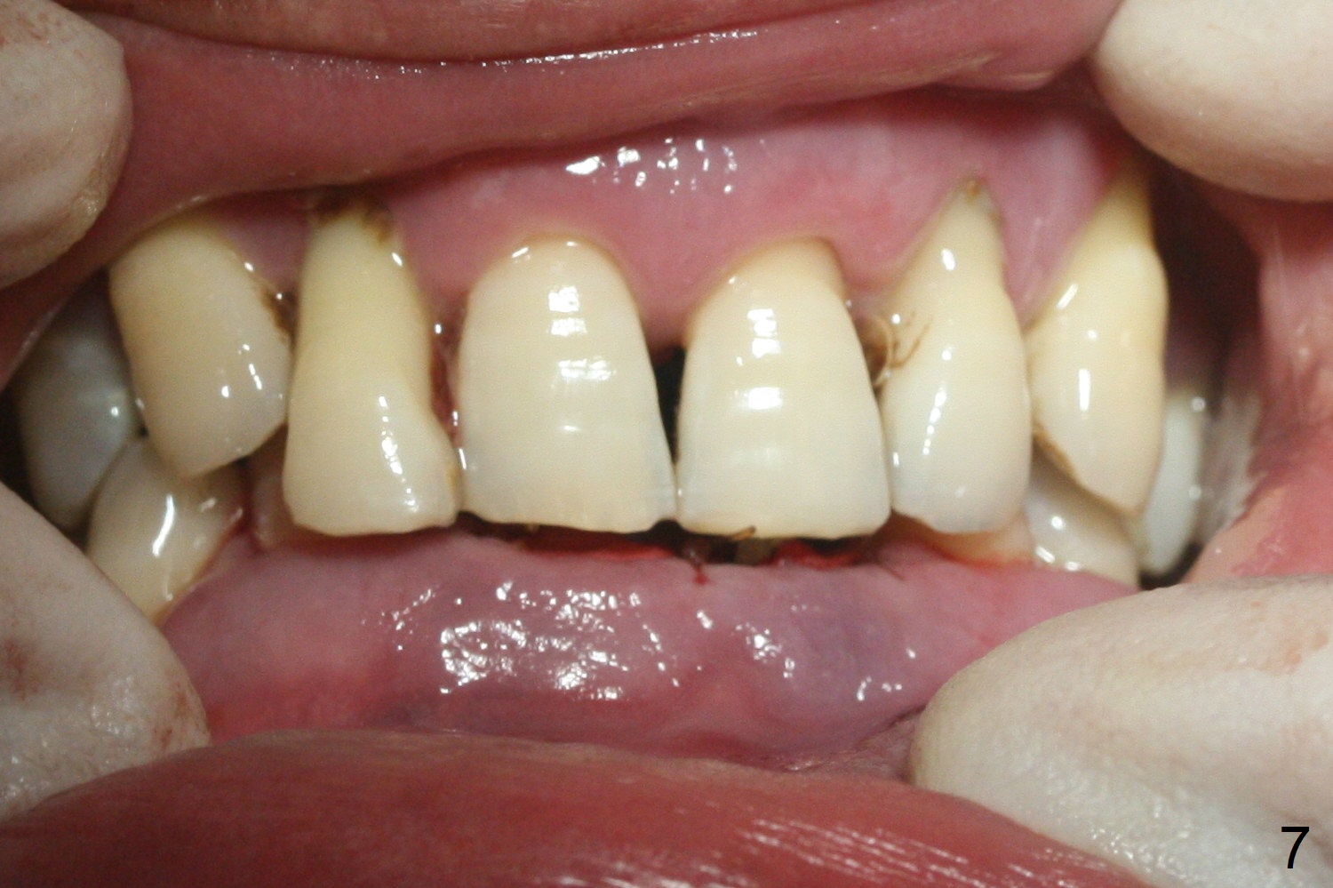
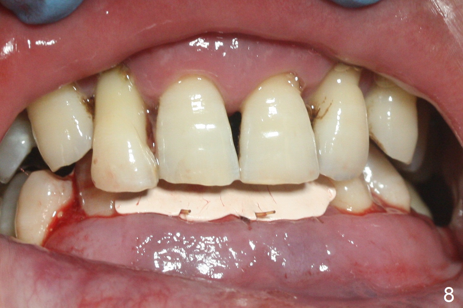
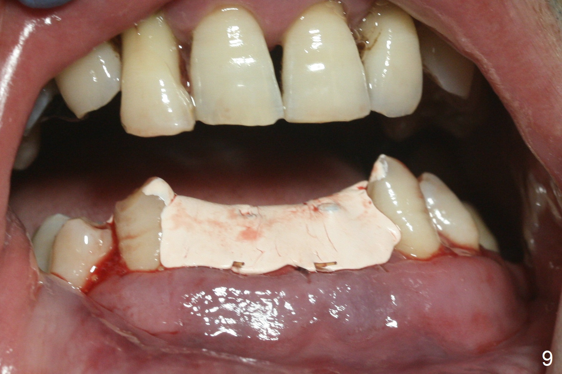
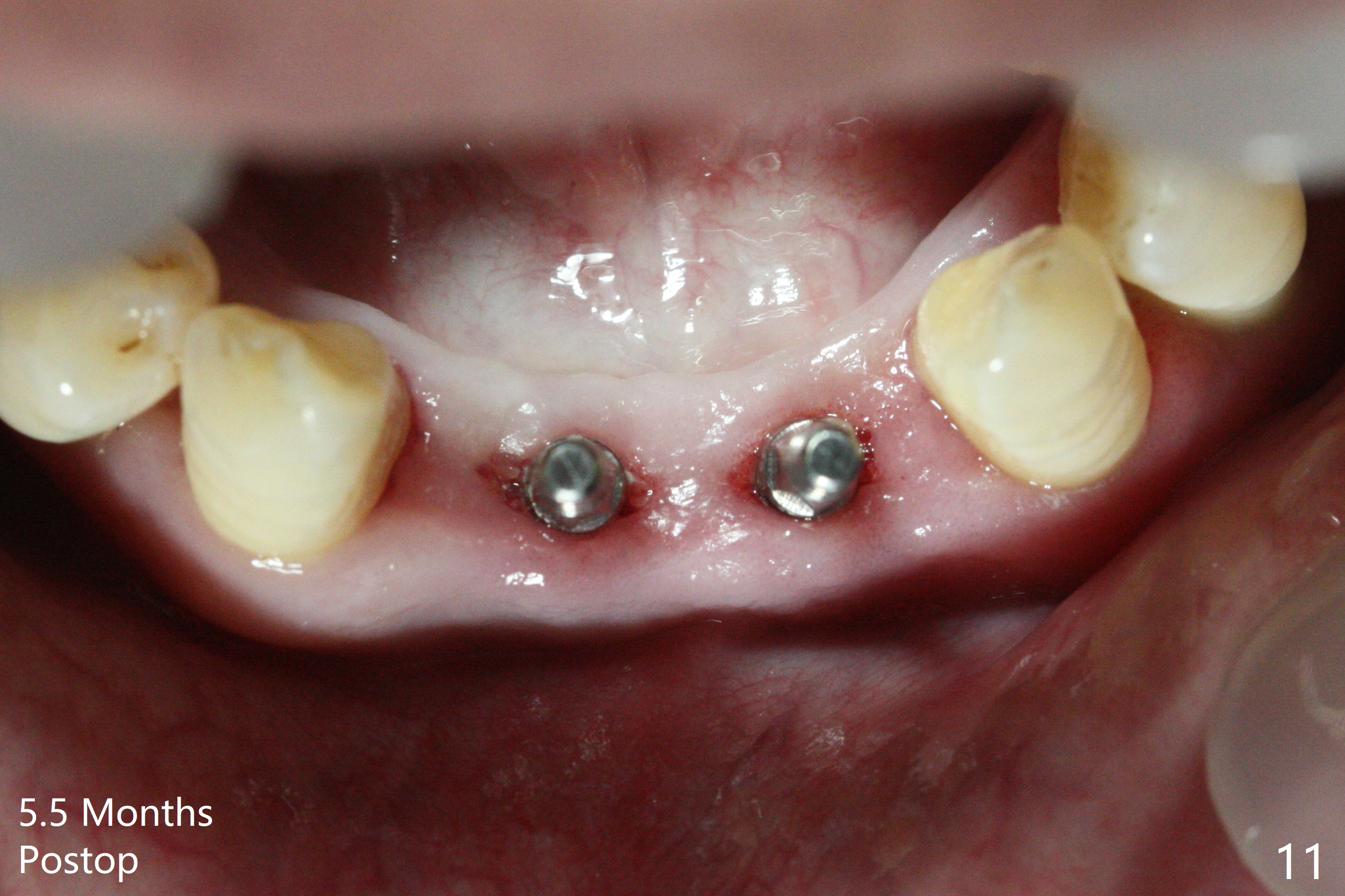
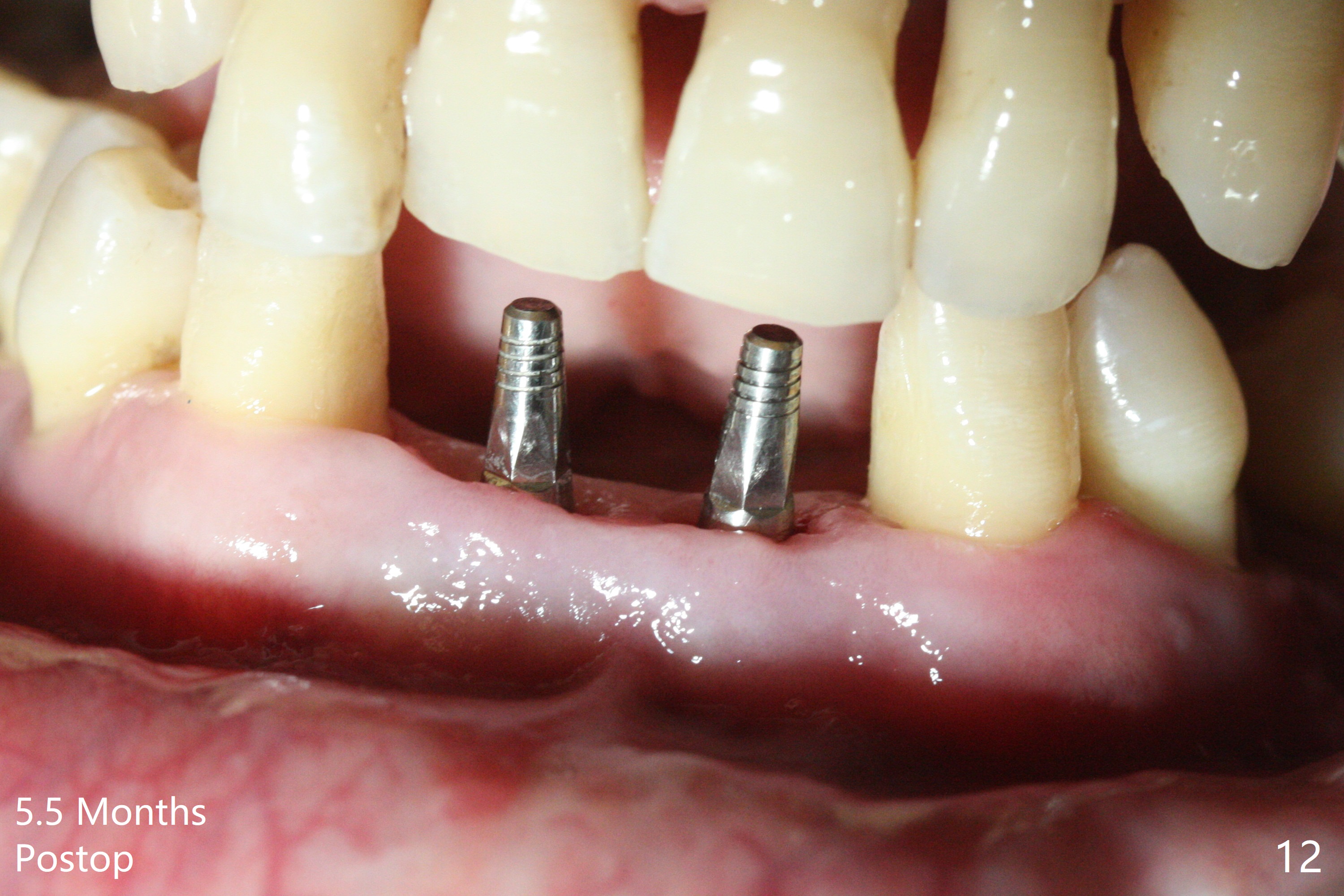
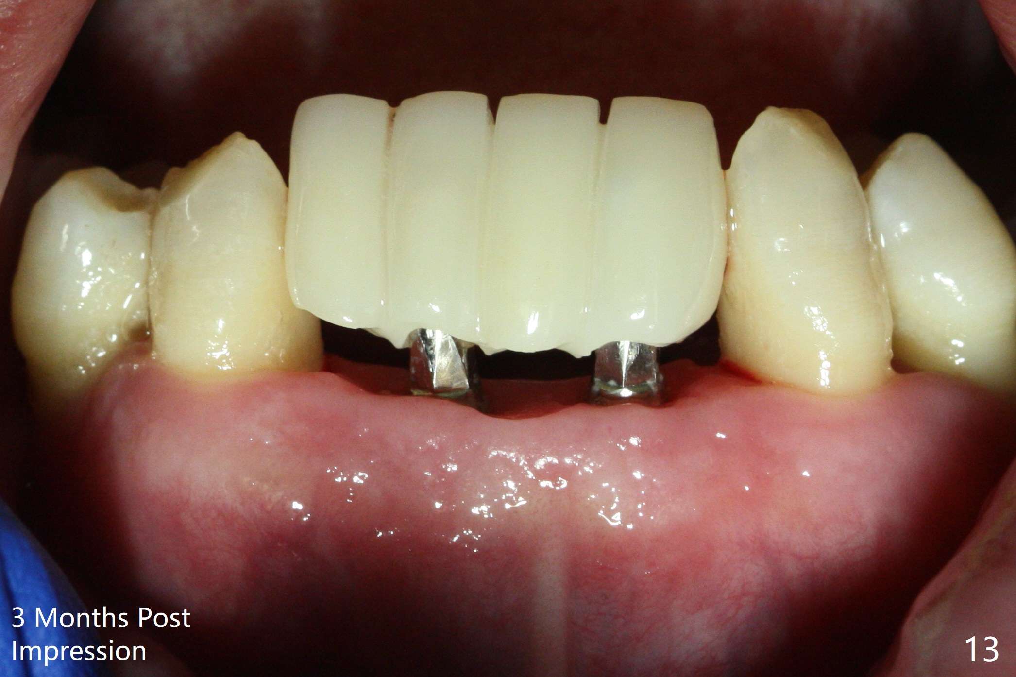
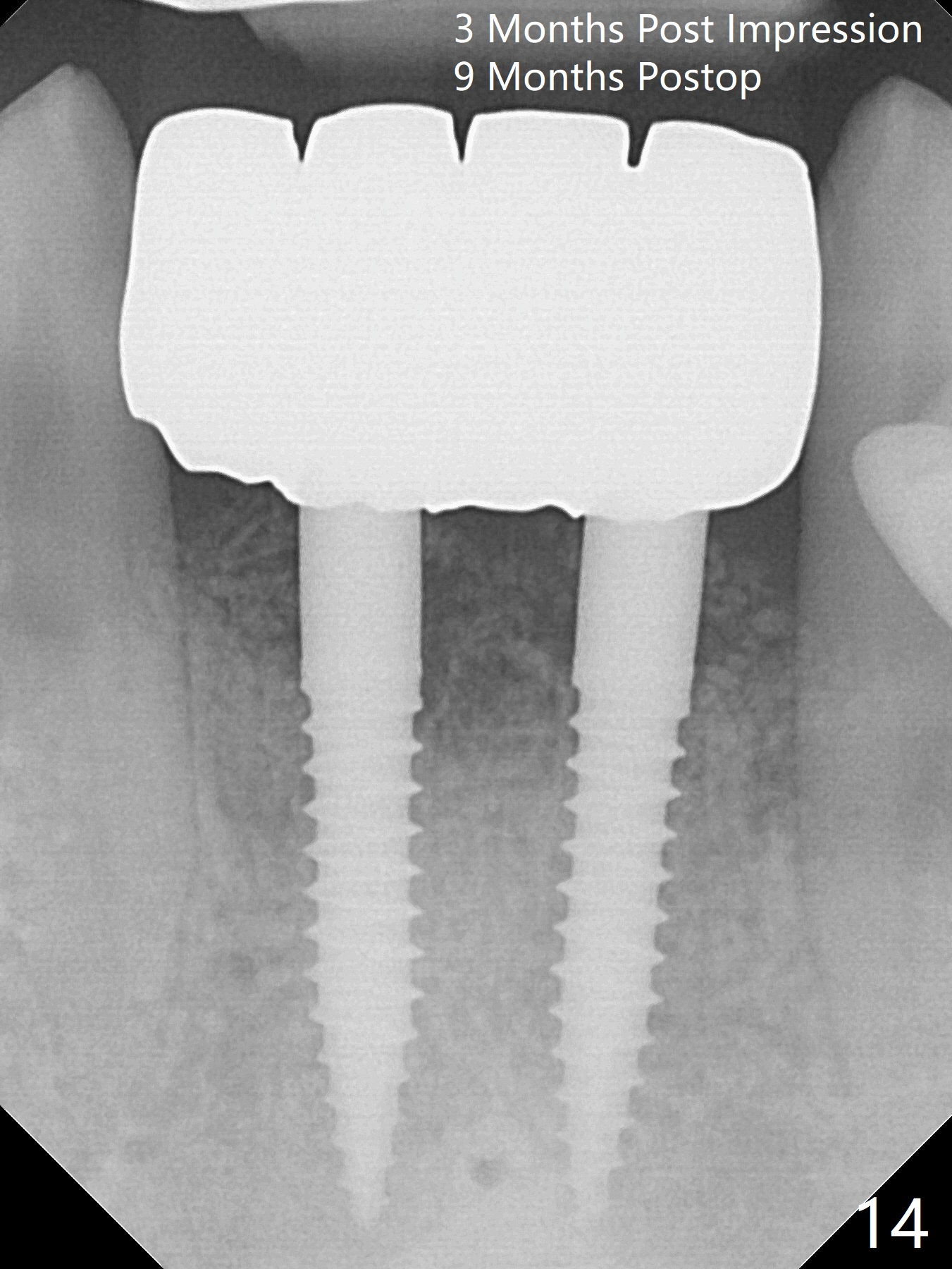
After extraction of 4 of the lower incisors, there are basically 2 sockets (#23/24 and 25/26), separated by the apparently midline bone (Fig.1 red line). In spite of using Lindamann bur to move the 25/26 osteotomy mesially, a 3x14(2) mm dummy implant remains close to the tooth #27 (Fig.2). The terminal branch of the Incisive Canal (<) is located between the lateral and central incisors. A de novo osteotomy (Fig.3 (1.5 mm drill)) is made mesial to the original one (O). While the 3x14(2) mm dummy implant is incompletely placed at #25/26, a 3x14(4) mm 1-piece one is placed at #23/24 (Fig.4). Finally the same implant is placed at #25/26 with placement of mineralized cortical/cancellous bone (Fig.5 *). When the large sockets are sutured, the supraerupted teeth #7-9 touch the lower gingiva (Fig.6). The incisal edge is reduced for clearance (Fig.7). Periodontal dressing is less likely to be dislodged with the incisal edge reduction (Fig.8,9). A provisional FPD is fabricated 1 week postop. Hard (Fig.10) and soft (Fig.11,12) tissues heal 5.5 months postop. The patient returns for crown cementation 3 months post impression (9 months postop, Fig.13,14).
Return to
Lower Incisor Immediate Implant,
IBS,
#3,5,
7,
19/20,
Systemic Diseases
Xin Wei, DDS, PhD, MS 1st edition 08/22/2017, last revision 05/12/2018