mm%20after%20implant%203-4%20more%20turn,%20bone%20graft.jpg)
|
|
|
|
|
|
|
|
|
|
|
|
||
|
|
|||||
Take Pan Early to Determine Osteotomy Depth
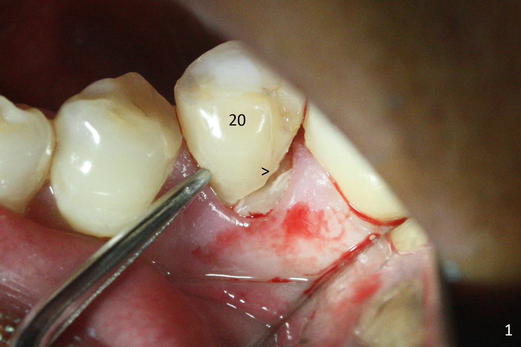
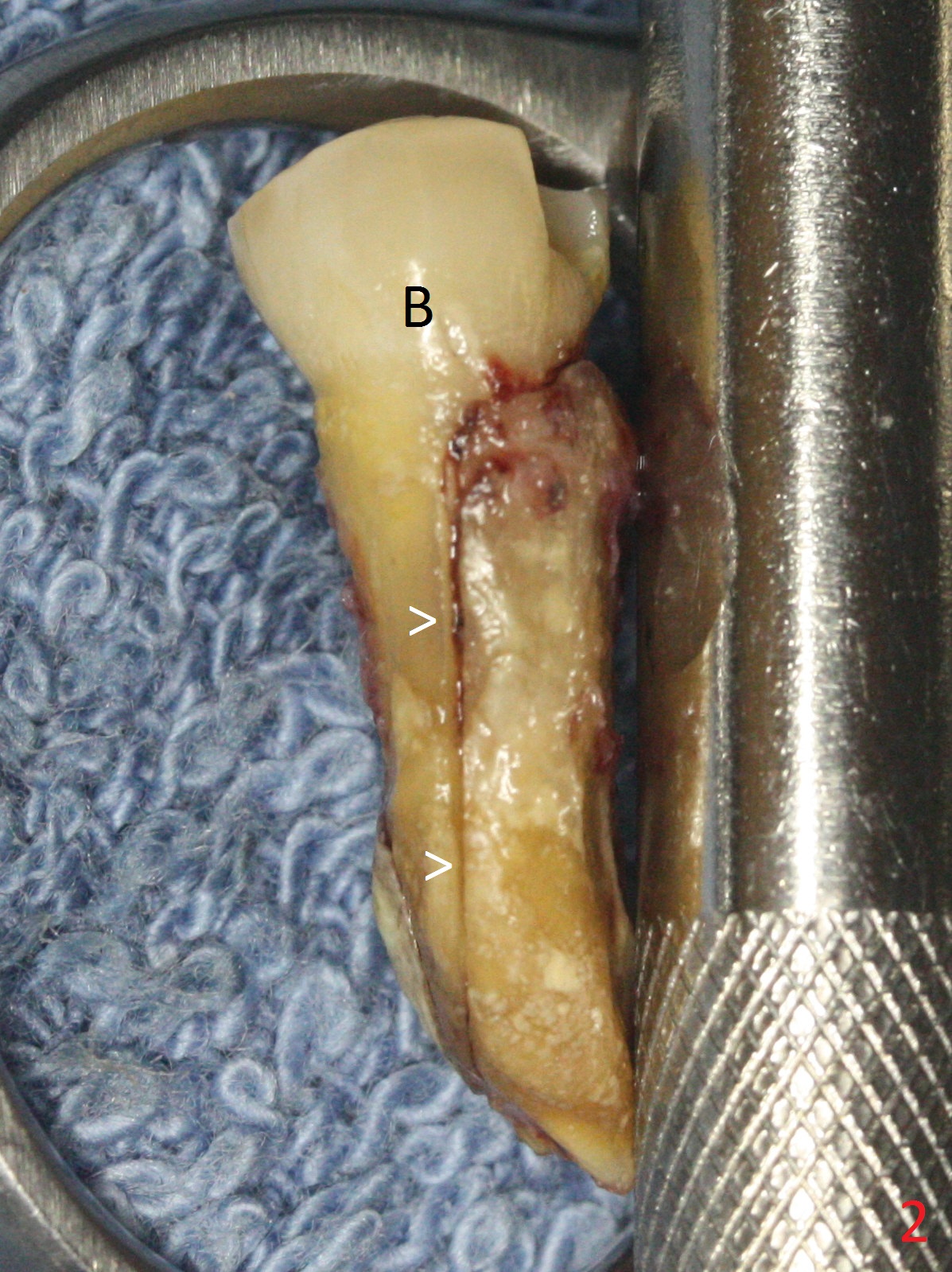
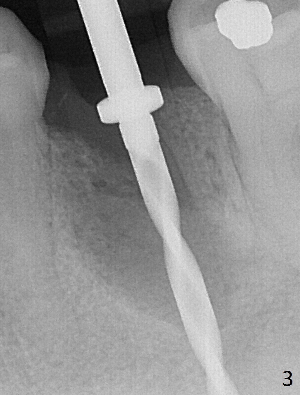
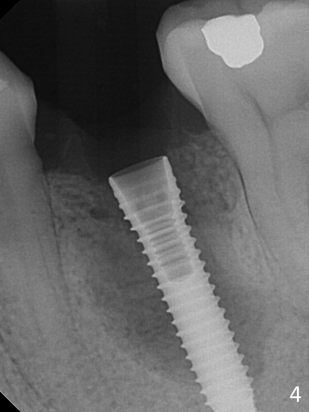
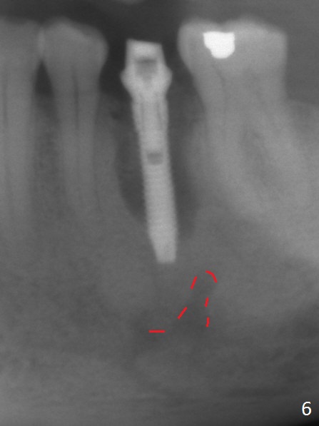
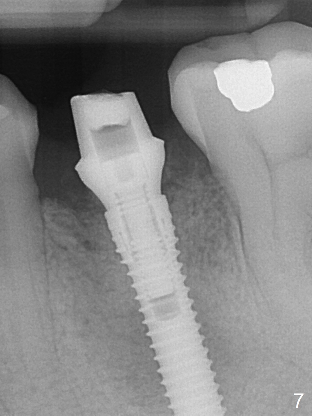
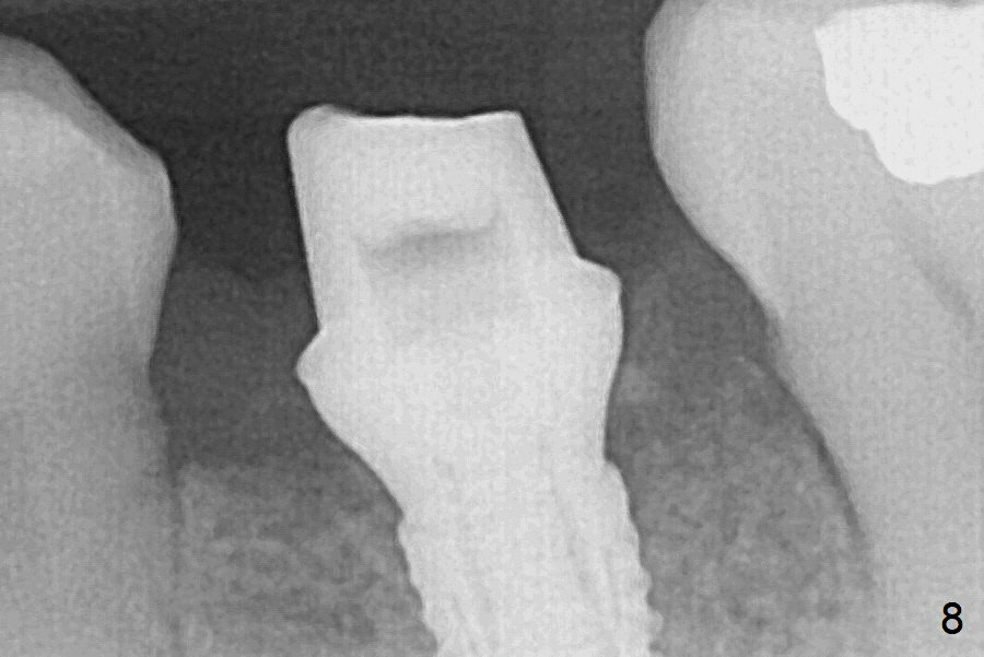
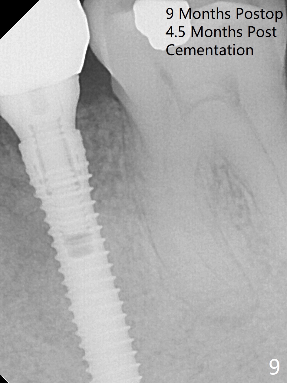
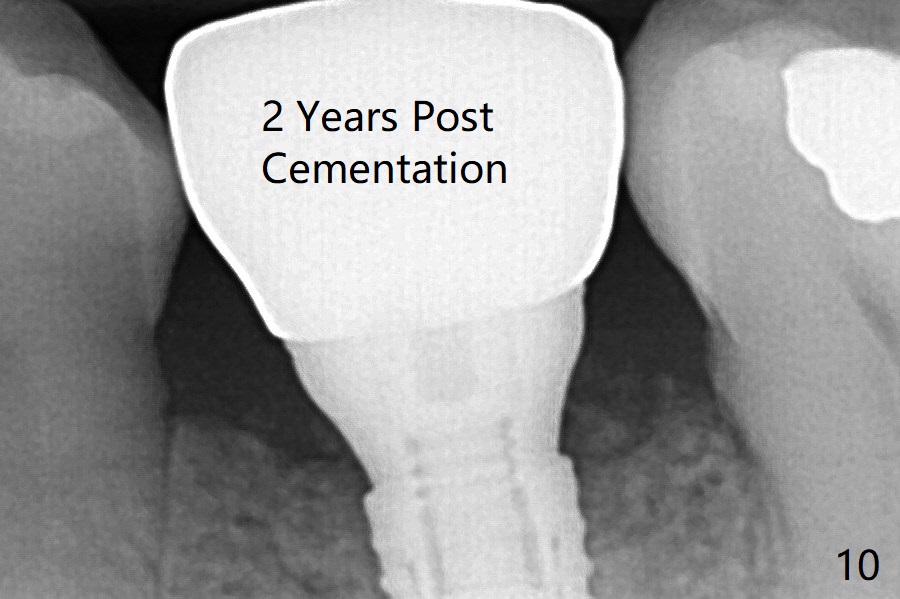
Extraction of the lower left 2nd premolar with vertical fracture (Fig.1,2 >) is easy because of peri-radicular radiolucency. The apical end of the osteotomy is not shown with a 2 mm pilot drill (Fig.3) or a 3.8x18 mm implant (Fig.4) in place. It appears that the implant is not placed deep enough. Following 3-4 more turns of the implant and placement of a 5.5x4(3) mm abutment (Fig.5 A), allograft is placed (*). A postop panoramic X-ray is taken (Fig.6); the osteotomy could have been deepened to reduce the possibility of periimplantitis. Retrospectively, the panoramic X-ray should be taken after use of the pilot drill. The bone around the implant appears to have regenerated 4 months postop (Fig.7,8). Bone density appears to continue increasing 9 months postop (i.e., 4.5 months post cementation, Fig.9). Bone loss is minimal 2 years post cementation (Fig.10).
Return to
Lower
Premolar Immediate Implant, IBS,
14, 29,
18,
Short Delayed
Implant
Xin Wei, DDS, PhD, MS 1st edition 05/08/2017, last revision 09/04/2019